What Imaging Is Best for Surveillance After Muscle-Invasive Bladder Cancer Treatment?

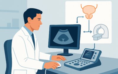
A 68-year-old male is in your urology clinic for his one-year follow-up after a radical cystectomy with ileal conduit creation for muscle-invasive bladder cancer (MIBC). He is asymptomatic and his physical exam is unremarkable. As you plan his ongoing surveillance, you consider the appropriate imaging to screen for distant recurrence, specifically in the thorax, which is the most common site of metastasis. You need to balance the need for detection with the risks of cumulative radiation exposure from repeated scans. This article details the American College of Radiology (ACR) Appropriateness Criteria workflow for this exact decision. For routine thoracic surveillance in this asymptomatic patient, a `Radiography chest` is rated *Usually Appropriate*.
Who Fits This Clinical Scenario for Bladder Cancer Surveillance?
This guidance applies to patients in the post-treatment surveillance phase for muscle-invasive bladder cancer (T2-T4). This includes individuals who have undergone definitive therapy, such as a radical cystectomy with urinary diversion, or those who have completed trimodal therapy (maximal transurethral resection of the bladder tumor followed by chemoradiation) with a complete response. The key element is that the patient is currently asymptomatic and being monitored for potential local, regional, or distant recurrence according to established follow-up protocols.
This workflow is distinct from the evaluation of other patient groups. It does not apply to:
- Patients with non-muscle invasive bladder cancer (NMIBC): These patients follow a different surveillance protocol, which is often more focused on intravesical monitoring with cystoscopy and urine cytology. Systemic imaging is typically reserved for those who develop symptoms or have high-risk features.
- Patients with active symptoms concerning for recurrence: If a patient presents with new flank pain, gross hematuria (from the upper tracts or diversion), palpable masses, or constitutional symptoms like unexplained weight loss, the workup shifts from routine surveillance to a diagnostic evaluation. This often necessitates more advanced imaging, such as contrast-enhanced CT or MRI, to investigate the source of the symptoms.
- Patients currently undergoing initial staging or active treatment: This guidance is for post-treatment follow-up, not the initial workup or monitoring of treatment response.
What Diagnoses Are You Working Up During MIBC Surveillance?
Post-treatment surveillance for MIBC is a systematic search for recurrent disease, which can manifest in several locations. The choice of imaging is tailored to detect the most common patterns of failure for this aggressive malignancy.
The primary concern addressed by thoracic imaging is **distant metastatic disease to the lungs and pleura**. The lungs are the most frequent site of hematogenous spread from bladder cancer. Surveillance imaging aims to detect pulmonary nodules or pleural-based masses at an early, asymptomatic stage when intervention may be more effective.
Another key target is **metastatic disease to thoracic lymph nodes**. While less common than pulmonary parenchymal metastases, involvement of the mediastinal and hilar lymph nodes represents another pathway of distant spread. Chest imaging provides a clear view of the mediastinal contours and can identify suspicious adenopathy.
While not the primary target of a chest radiograph, a comprehensive surveillance strategy also monitors for **upper tract urothelial carcinoma (UTUC)**. Patients with a history of bladder cancer have a “field defect,” meaning their entire urothelium is at increased risk for developing new primary tumors. This is why abdominal and pelvic imaging with CT Urography or MR Urography is also a cornerstone of surveillance, though it is a separate component from thoracic screening.
Finally, surveillance includes monitoring for **local and regional recurrence** in the pelvis (e.g., in the cystectomy bed) or retroperitoneal lymph nodes. This is the domain of cross-sectional imaging of the abdomen and pelvis, such as CT or MRI, which complements thoracic imaging to provide a complete picture of the patient’s disease status.
Why Is a Chest Radiograph Usually Appropriate for Surveillance in This Scenario?
For routine thoracic surveillance in an asymptomatic patient post-treatment for muscle-invasive bladder cancer, the ACR panel designates `Radiography chest` as *Usually Appropriate*. This recommendation is based on a careful balance of diagnostic yield, cost, accessibility, and, most critically, radiation dose.
The rationale is that a chest radiograph serves as an effective, low-risk screening tool for the most common site of distant metastasis—the lungs. Its primary role is to detect asymptomatic pulmonary nodules that would trigger a more definitive diagnostic workup. Given that surveillance occurs at regular intervals over many years, minimizing cumulative radiation exposure is a paramount concern. A chest radiograph delivers a very low radiation dose (adult relative radiation level ☢ <0.1 mSv), making it highly suitable for repeated use.In contrast, other modalities are rated lower for routine screening in this specific context:
- CT chest without IV contrast is rated *May be appropriate*. While a CT scan is undoubtedly more sensitive than a radiograph for detecting small pulmonary nodules, it imparts a substantially higher radiation dose (☢☢☢ 1-10 mSv). Its role is typically reserved for evaluating an indeterminate finding on a chest radiograph or for patients considered to be at a particularly high risk of recurrence where the benefit of higher sensitivity outweighs the radiation risk.
- FDG-PET/CT skull base to mid-thigh is also rated *May be appropriate*. This powerful functional imaging study is not a primary surveillance tool. Its utility lies in problem-solving, such as evaluating equivocal findings on other imaging, staging a confirmed recurrence prior to salvage therapy, or in cases of high clinical suspicion for recurrence despite negative conventional imaging. Its high radiation dose (☢☢☢☢ 10-30 mSv) and cost make it unsuitable for routine screening.
It is crucial to recognize that a comprehensive surveillance strategy is multi-modal. While the chest radiograph addresses the thorax, other studies like `CT abdomen and pelvis with IV contrast` or `MRU without and with IV contrast` are also rated *Usually Appropriate* to monitor for pelvic recurrence and upper tract disease. The overall plan integrates these different studies at specified intervals based on guideline recommendations.
What’s the Next Step After a Surveillance Chest Radiograph?
The results of the surveillance chest radiograph guide the subsequent clinical workflow, determining whether to continue routine monitoring or escalate to a diagnostic evaluation.
**If the chest radiograph is negative or stable:** When the study shows no evidence of metastatic disease, or findings are unchanged from prior examinations, the patient continues with their scheduled surveillance protocol. This typically involves repeating the chest radiograph and other indicated imaging (e.g., abdominal/pelvic CT or MRI) at the next planned interval, which may be every 6 to 12 months in the initial years post-treatment.
**If the chest radiograph is positive for a new or growing nodule:** The presence of a suspicious finding, such as a new pulmonary nodule or enlarging lymph node, immediately shifts the goal from surveillance to diagnosis. The recommended next step is typically a `CT chest without IV contrast` (rated *May be appropriate* for this purpose) to better characterize the lesion’s size, morphology, and number. Depending on the CT findings and the patient’s overall clinical picture, subsequent steps may include a biopsy (percutaneous or bronchoscopic), a PET/CT scan for further staging, or multidisciplinary consultation to plan treatment.
**If the chest radiograph is indeterminate:** Sometimes, findings are equivocal—for example, a tiny, sub-centimeter nodule or subtle opacity. In these cases, management depends on the degree of suspicion. Options include a short-interval follow-up chest radiograph in 3-6 months to assess for stability or growth, or proceeding directly to a chest CT for more definitive characterization, especially in a patient with a high pre-test probability of recurrence.
Pitfalls to Avoid (and When to Get Help)
Navigating post-treatment surveillance for MIBC requires careful attention to detail to avoid common errors that can impact patient outcomes.
One major pitfall is **surveillance fatigue**, leading to inconsistent follow-up. It is critical to adhere to a structured, guideline-based surveillance schedule, as recurrences are often detected in asymptomatic patients. Another common error is **using high-dose imaging for routine screening**. Relying on annual CT scans instead of chest radiographs for thoracic surveillance unnecessarily increases a patient’s lifetime cumulative radiation dose.
A third pitfall is **failing to integrate clinical context**. An “indeterminate” nodule on a chest radiograph in a former smoker with a history of MIBC carries a different weight than the same finding in a non-smoker. Always interpret imaging results in the context of the patient’s full history and risk factors. Finally, avoid **delaying the workup of a new symptom**. The onset of cough, hemoptysis, or chest pain should prompt an immediate diagnostic evaluation, superseding the routine surveillance schedule.
If a new or suspicious finding is identified, especially if it is complex or involves multiple sites, escalation to a multidisciplinary tumor board including urology, medical oncology, radiation oncology, and radiology is the appropriate next step.
Related ACR Topics and Tools
For a comprehensive overview of imaging recommendations across all clinical variants related to this condition, please consult the parent topic article. It provides a breadth of information that complements this in-depth workflow.
- For breadth across all scenarios in Post-Treatment Surveillance of Bladder Cancer, see our parent guide: Post-Treatment Surveillance of Bladder Cancer: ACR Appropriateness Decoded.
To explore adjacent clinical scenarios, refine imaging techniques, or discuss radiation dose with your patients, the following GigHz resources are available:
- ACR Appropriateness Criteria Lookup — for adjacent scenarios
- Imaging Protocol Library — for technique on the recommended study
- Radiation Dose Calculator — for cumulative dose conversations
Frequently Asked Questions
Why not just order a chest CT every time for better sensitivity?
While a chest CT is more sensitive for small lung nodules, it delivers a significantly higher radiation dose than a chest radiograph (1-10 mSv vs. <0.1 mSv). For routine, repeated surveillance in an asymptomatic patient, the American College of Radiology prioritizes the lower radiation dose of a chest radiograph, reserving CT for problem-solving or for patients at very high risk. The goal is to balance detection with minimizing long-term radiation-related risks.
Does this surveillance guidance apply if the patient received trimodal therapy instead of a cystectomy?
Yes, this guidance applies to patients after any definitive treatment for muscle-invasive bladder cancer, including radical cystectomy or trimodal therapy (chemoradiation), provided they had a complete response and are now in the asymptomatic surveillance phase.
How often should surveillance imaging be performed?
The frequency of surveillance imaging is guided by recommendations from organizations like the AUA/SUO and NCCN. Generally, imaging is most frequent in the first 2-3 years after treatment (e.g., every 6-12 months) when the risk of recurrence is highest, and then the interval may be extended. The exact schedule should be tailored to the individual patient’s pathologic stage and risk factors.
What about surveillance of the upper urinary tracts and pelvis?
This article focuses on thoracic surveillance. However, a complete surveillance plan for MIBC is multi-modal. It also includes regular imaging of the abdomen and pelvis to monitor for local recurrence, lymph node metastasis, and new upper tract urothelial tumors. Studies like CT Urography or MR Urography are rated ‘Usually Appropriate’ for this purpose and are performed concurrently with thoracic imaging according to surveillance guidelines.
If my patient had a cystectomy, why do I need to image their upper tracts?
Patients with bladder cancer have a ‘field defect,’ meaning the entire urothelial lining of their urinary system (renal pelvis, ureters, bladder) is at an elevated risk of developing cancer. Even after the bladder is removed, they remain at risk for developing a new primary tumor in the ureters or kidneys (Upper Tract Urothelial Carcinoma or UTUC). Therefore, surveillance must include the upper tracts.
Reviewed by Pouyan Golshani, MD, Interventional Radiologist — May 21, 2026