What Imaging Is Best for Chronic Disequilibrium with Sensory Ataxia in Adults?
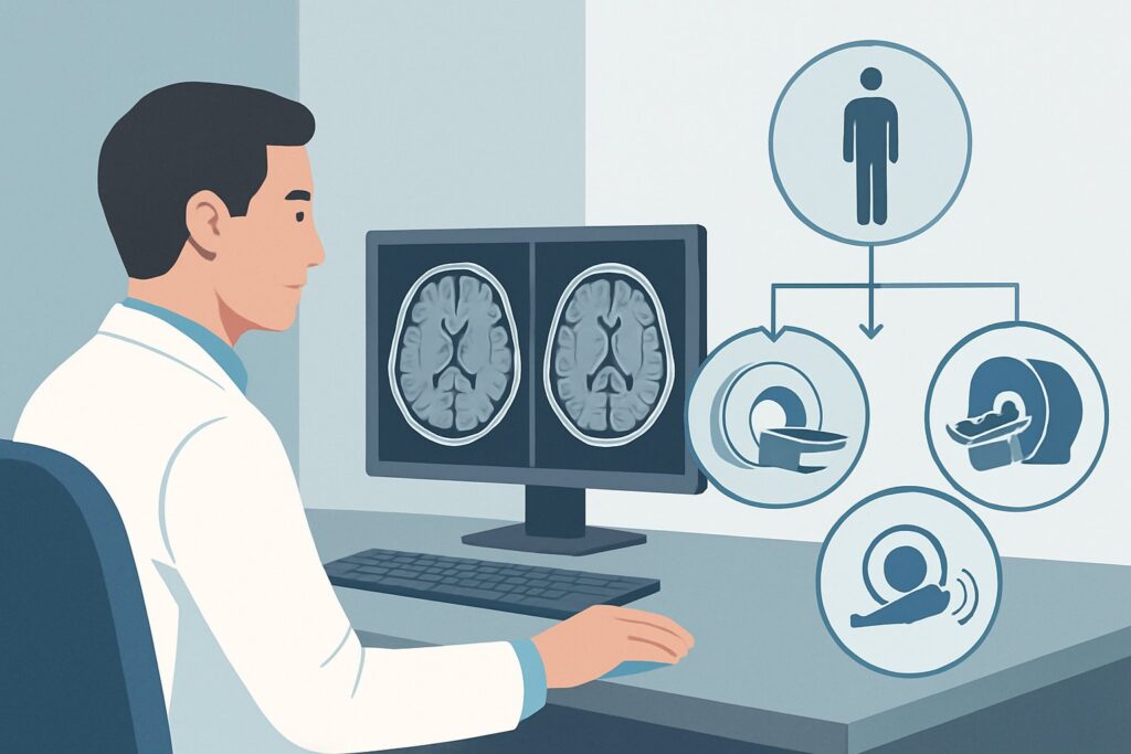
A 62-year-old patient presents to your neurology clinic with a six-month history of worsening balance. He describes it not as a spinning vertigo, but as a profound unsteadiness, particularly when walking in a dimly lit room or closing his eyes to wash his hair in the shower. On examination, he has a wide-based gait and a strongly positive Romberg test, but no nystagmus, dysmetria, or dysarthria. You suspect a lesion affecting the posterior columns of the spinal cord. The immediate clinical question is which imaging study to order first to investigate this sensory ataxia. According to the American College of Radiology (ACR) Appropriateness Criteria, for this specific presentation, `MRI cervical and thoracic spine without and with IV contrast` is rated Usually appropriate.
Who Fits This Clinical Scenario?
This guidance applies specifically to adult patients presenting with chronic disequilibrium where the clinical examination points toward a sensory or proprioceptive ataxia. The key features of this presentation include:
- Chronicity: Symptoms have developed and persisted over weeks to months, rather than appearing acutely.
- Disequilibrium, not vertigo: The patient feels unsteady or “off-balance” but does not experience a true spinning sensation (vertigo).
- Signs of sensory ataxia: The physical exam reveals deficits in proprioception (joint position sense) or vibration sense. A positive Romberg sign—where the patient becomes significantly more unstable when standing with feet together and eyes closed—is the classic finding, indicating a reliance on visual input to maintain balance.
This workflow is distinct from other causes of dizziness. It does not apply to patients with:
- Brief, episodic vertigo triggered by head movements: This presentation suggests benign paroxysmal positional vertigo (BPPV), which is a clinical diagnosis and follows a different imaging pathway if any is needed at all.
- Signs of cerebellar ataxia: If the patient exhibits dysmetria (e.g., past-pointing on finger-to-nose testing), an intention tremor, or dysdiadochokinesia, the workup should target the cerebellum, following the ACR variant for chronic disequilibrium with cerebellar signs.
- Acute, persistent vertigo: A sudden onset of continuous vertigo points toward either a peripheral vestibular issue (like vestibular neuritis) or a central cause (like a posterior circulation stroke), each with its own urgent evaluation protocol.
What Diagnoses Are You Working Up in This Scenario?
The choice of imaging is driven by a differential diagnosis centered on pathologies that disrupt the dorsal columns of the spinal cord, which carry proprioceptive and vibratory information. The goal is to identify a structural cause for the patient’s sensory deficits.
Cervical Spondylotic Myelopathy: This is one of the most common causes of progressive spinal cord dysfunction in older adults. Chronic degenerative changes in the cervical spine, such as disc herniations, osteophytes, and ligamentous hypertrophy, can lead to mechanical compression of the spinal cord, specifically affecting the posterior columns and causing sensory ataxia.
Demyelinating Disease: Multiple sclerosis (MS) can present with plaques in the dorsal columns of the cervical or thoracic spinal cord. An enhancing plaque would indicate active inflammation. While often associated with other neurologic deficits, a “sensory MS” presentation can be dominated by proprioceptive loss.
Spinal Cord Tumors: Though less common, both intrinsic (e.g., ependymoma, astrocytoma) and extrinsic (e.g., meningioma, schwannoma) tumors can compress or invade the spinal cord. Contrast enhancement is critical for characterizing these lesions.
Inflammatory or Infectious Myelitis: Conditions like transverse myelitis, neurosarcoidosis, or even tabes dorsalis (a late manifestation of syphilis) can cause inflammation of the spinal cord. These etiologies often produce characteristic patterns of cord signal abnormality and enhancement on MRI.
Subacute Combined Degeneration: Severe vitamin B12 deficiency classically affects the posterior and lateral columns of the spinal cord. While this is a metabolic diagnosis, MRI can show a characteristic T2-hyperintense signal in the dorsal columns, often described as the “inverted V” sign.
Why Is MRI of the Cervical and Thoracic Spine the Recommended Study?
For a patient with chronic disequilibrium and signs of sensory ataxia, the ACR designates `MRI cervical and thoracic spine without and with IV contrast` as Usually appropriate. This recommendation is based on the modality’s superior ability to visualize the spinal cord parenchyma and surrounding structures, directly addressing the most likely differential diagnoses.
Magnetic Resonance Imaging (MRI) provides excellent soft-tissue contrast, allowing for detailed evaluation of the spinal cord for signs of compression, intrinsic signal abnormality, or abnormal enhancement. The non-contrast sequences (T1 and T2-weighted images) are highly sensitive for detecting structural compression from degenerative disease, disc herniations, or syrinx formation. T2-weighted sequences are particularly crucial for identifying edema or gliosis within the cord (myelomalacia), which appears as a high-signal-intensity focus.
The addition of intravenous (IV) gadolinium-based contrast is essential for this workup. Contrast enhancement highlights areas where the blood-brain barrier (or blood-spinal cord barrier) has broken down, which is a key feature of active inflammatory processes (e.g., MS plaques), infections, and most primary or metastatic spinal tumors. Without contrast, these critical diagnoses could be missed. For this reason, `MRI cervical and thoracic spine without IV contrast` is also rated Usually appropriate, but the addition of contrast provides more diagnostic information for a wider range of pathologies.
Alternative studies are rated lower for good reason. For instance, `MRI head without IV contrast` is rated Usually not appropriate because it completely fails to evaluate the primary area of concern—the spinal cord. Similarly, `CT head without IV contrast` is Usually not appropriate; it offers poor visualization of the spinal cord parenchyma and exposes the patient to ionizing radiation (ACR Relative Radiation Level ☢☢☢, 1-10 mSv) without diagnostic benefit for this specific clinical question. The recommended MRI has a radiation level of 0 mSv.
Once you’ve decided on the recommended study, our protocol guide covers the technical details for the foundational sequences. For a detailed look at the non-contrast cervical spine sequences, see our guide: MRI Cervical Spine Without Contrast.
What’s Next After MRI? Downstream Workflow
The results of the spinal MRI will guide the subsequent clinical pathway. The workflow branches based on whether the findings are positive, negative, or indeterminate.
If the study is positive for significant spinal cord compression: A finding of severe cervical stenosis with cord signal change, a large disc herniation causing myelopathy, or a compressive tumor is an actionable result. The next step is typically a prompt referral to a neurosurgeon or orthopedic spine surgeon for evaluation for potential surgical decompression.
If the study is positive for enhancing or non-compressive lesions: The presence of T2-hyperintense lesions, with or without enhancement, suggests an intrinsic cord pathology like demyelination or inflammation. This should trigger a broader neurologic workup, which may include an MRI of the brain (to look for disseminated lesions consistent with MS), lumbar puncture for cerebrospinal fluid analysis (e.g., oligoclonal bands), and a panel of blood tests for inflammatory, infectious, and metabolic causes (e.g., aquaporin-4 antibodies, vitamin B12, copper).
If the study is negative: A normal cervical and thoracic spine MRI is a crucial finding, as it effectively rules out a structural myelopathy. The diagnostic focus should then shift away from the central nervous system. The next steps would be to investigate peripheral causes of sensory loss, such as a large-fiber peripheral neuropathy. This typically involves nerve conduction studies (NCS) and electromyography (EMG). A comprehensive metabolic and toxic screening should also be revisited.
Pitfalls to Avoid (and When to Get Help)
In working up sensory ataxia, several common pitfalls can delay diagnosis or lead to unnecessary testing. Be mindful of the following:
- Incomplete Imaging: Ordering only a cervical spine MRI when the pathology could be in the thoracic cord. The dorsal columns run the length of the cord, and thoracic lesions are a definite possibility.
- Omitting Contrast: Forgoing IV contrast in the initial study can miss active inflammatory, infectious, or neoplastic disease, potentially requiring the patient to return for a second scan.
- Attribution Error: Mistakenly attributing the patient’s profound ataxia to mild, incidental degenerative changes seen on MRI. The clinical signs of myelopathy should outweigh incidental imaging findings.
- Fixation on the Spine: If the MRI is negative, avoid the trap of repeatedly imaging the spine. The workup must pivot to consider peripheral neuropathies or, rarely, re-evaluating for a subtle cerebellar or parietal lobe process.
If a patient presents with acute or rapidly progressive signs of myelopathy (e.g., new-onset weakness, sensory level, bowel or bladder dysfunction), this constitutes a neurologic emergency. Escalate immediately with urgent imaging and a neurology or neurosurgery consultation.
Related ACR Topics and Tools
This article focuses on a single clinical variant. For a comprehensive overview of imaging for all types of dizziness and ataxia, from BPPV to central vertigo, please see our parent guide. It provides a hub-and-spoke model to help you navigate all the related scenarios.
- For breadth across all scenarios in Dizziness and Ataxia, see our parent guide: Dizziness and Ataxia: ACR Appropriateness Decoded.
- To explore other scenarios and find the right study for your patient, use the ACR Appropriateness Criteria Lookup.
- For detailed procedural techniques on recommended studies, browse our Imaging Protocol Library.
- To discuss radiation exposure from alternative studies like CT, our Radiation Dose Calculator can help frame conversations with patients.
Frequently Asked Questions
Why not start with a brain MRI for chronic disequilibrium?
A brain MRI is the appropriate first step if the clinical signs point to a cerebellar ataxia (e.g., dysmetria, intention tremor). However, in this specific scenario, the signs of sensory ataxia (positive Romberg, impaired proprioception) strongly localize the problem to the spinal cord’s dorsal columns, making a spine MRI the highest-yield initial study.
Is it acceptable to order the MRI without contrast first?
Yes, the ACR rates ‘MRI cervical and thoracic spine without IV contrast’ as ‘Usually appropriate.’ This can be a reasonable strategy if the pre-test probability for a compressive myelopathy from degenerative disease is very high and the suspicion for inflammatory or neoplastic disease is low. However, if the non-contrast study is negative or equivocal, a follow-up study with contrast will likely be needed, so ordering it upfront can be more efficient.
What if my patient has a pacemaker or other contraindication to MRI?
If a patient has an absolute contraindication to MRI, the next best test is a CT myelogram. This involves a lumbar puncture to inject intrathecal contrast followed by a CT scan of the spine. While more invasive and involving radiation, it provides excellent detail of the spinal canal and can identify significant structural compression. However, it provides very little information about intrinsic cord abnormalities.
Does a normal spine MRI completely rule out a spinal cause for sensory ataxia?
A technically adequate, normal spine MRI makes a structural cause like compression, tumor, or significant inflammation highly unlikely. However, it may not show the subtle signal changes of early subacute combined degeneration (B12 deficiency) or other metabolic myelopathies. If clinical suspicion remains high despite a normal MRI, the focus should shift to metabolic testing and electrophysiologic studies (nerve conduction studies/EMG).
Should I order imaging for the lumbar spine as well?
Generally, no. The spinal cord itself (the upper motor neuron structure) ends at the L1-L2 level (the conus medullaris). While lumbar pathology can cause radiculopathy and affect sensation in the legs, it does not cause the truncal and gait ataxia characteristic of a dorsal column myelopathy. The workup for sensory ataxia is focused on the cervical and thoracic cord.
Reviewed by Pouyan Golshani, MD, Interventional Radiologist — May 30, 2026