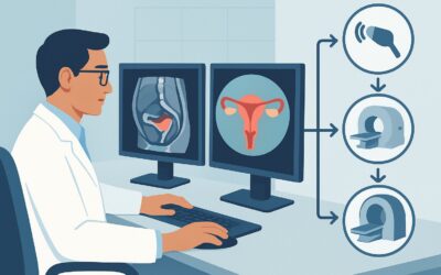
What Imaging Is Best for Early Postpartum Hemorrhage After Vaginal Delivery?

It’s 3 a.m. on the labor and delivery unit. Your patient, a 28-year-old G1P1, is six hours out from an uncomplicated vaginal delivery but continues to have heavier-than-expected bleeding. Uterotonics have been administered and fundal massage is ongoing, but the bleeding persists. You suspect retained products of conception but need to rule out other structural causes. This is a time-sensitive decision: what is the right imaging study to order right now, at the bedside, to guide immediate management? This article provides a focused workflow for this exact scenario, explaining why the American College of Radiology (ACR) rates US duplex Doppler pelvis as Usually Appropriate for initial imaging.
Who Fits This Clinical Scenario for Early Postpartum Hemorrhage?
This guidance applies specifically to a patient presenting with postpartum hemorrhage (PPH) within the first 24 hours of a vaginal delivery. The patient is typically still on the labor and delivery or postpartum unit, and while there is concern for ongoing blood loss, they are sufficiently stable for a diagnostic imaging study. The primary clinical question is to identify an underlying structural cause, such as retained tissue, that is amenable to intervention.
This workflow is NOT intended for:
- Patients with PPH after a Cesarean delivery: The differential diagnosis and potential for surgical complications like bladder flap hematomas or uterine dehiscence are different. This presentation is covered in a separate ACR variant.
- Patients with late PPH: Bleeding that begins more than 24 hours after delivery has a different differential, including subinvolution of the placental site or infection, and is also addressed in a separate variant.
- Hemodynamically unstable patients: If a patient is in shock, hypotensive, or has signs of massive hemorrhage, imaging should not delay definitive management. These patients require immediate resuscitation and escalation to the operating room for examination under anesthesia, potential surgical intervention, or to interventional radiology for embolization.
What Diagnoses Are You Working Up in This Scenario?
In early PPH, the clinical workup is often guided by the “four T’s” mnemonic. Imaging is primarily used to evaluate for structural causes related to Tissue and Trauma.
Tissue (Retained Products of Conception): This is a primary concern and a key target for imaging. Retained placental fragments or membranes can prevent the uterus from contracting effectively, leading to persistent bleeding. While blood clots can mimic retained tissue, the presence of a distinct, echogenic, and often vascularized mass within the endometrial cavity on ultrasound is highly suggestive of Retained Products of Conception (RPOC).
Tone (Uterine Atony): This is the most common cause of PPH. While largely a clinical diagnosis based on a soft, “boggy” uterus on physical exam, ultrasound can provide supporting evidence. Findings may include a large, fluid-filled endometrial cavity with blood and clot, but the primary role of imaging in this context is to exclude other concurrent causes, like RPOC, that may be contributing to the atony.
Trauma (Lacerations and Hematomas): Significant cervical or high vaginal lacerations are typically diagnosed on physical examination. However, if bleeding persists from an unknown source, ultrasound can be invaluable for identifying occult pelvic hematomas. A large or expanding hematoma in the broad ligament or retroperitoneum may not be apparent on a standard pelvic exam but can be readily visualized with ultrasound.
Thrombin (Coagulopathy): This is a systemic issue diagnosed with laboratory tests. Imaging does not diagnose coagulopathy but can identify its consequences, such as extensive hematoma formation, which might appear disproportionate to the suspected injury.
Why Is Pelvic Ultrasound the Recommended First Step for This Presentation?
For a patient with early postpartum hemorrhage after a vaginal delivery, the ACR designates US duplex Doppler pelvis, US pelvis transabdominal, and US pelvis transvaginal as Usually Appropriate. These studies are the cornerstone of initial imaging for several critical reasons.
First and foremost, ultrasound involves no ionizing radiation (0 mSv) and does not require IV contrast, making it the safest option for a postpartum patient. Its portability is a major advantage, allowing for a rapid bedside assessment without needing to transport a potentially unstable patient to a radiology suite.
The diagnostic strength of ultrasound lies in its ability to directly visualize the uterine cavity. A combined transabdominal and transvaginal approach provides a comprehensive evaluation. The transabdominal view gives a global picture of the uterus and surrounding pelvis, while the transvaginal view offers high-resolution detail of the endometrium. Adding Duplex Doppler is crucial; it assesses blood flow within any identified intrauterine mass. Vascularity within an echogenic mass significantly increases the likelihood of RPOC, helping to distinguish it from an avascular blood clot.
Why Alternatives Are Rated Lower
- CT abdomen and pelvis with IV contrast: Rated as May be appropriate, this study is not a first-line choice. It exposes the patient to ionizing radiation (☢☢☢ 1-10 mSv) and requires IV contrast. Its role is reserved for situations where ultrasound is inconclusive or there is a high suspicion of a vascular injury, such as a pseudoaneurysm or arteriovenous fistula, which are more common after instrumentation or surgery.
- MRI pelvis without and with IV contrast: Rated as Usually not appropriate in the acute setting. MRI is time-consuming, less accessible, and offers no significant advantage over ultrasound for answering the primary question of RPOC. It may have a role in complex, subacute cases or for problem-solving an indeterminate ultrasound finding in a stable patient, but it is not suited for the initial, urgent workup of early PPH.
What’s Next After Pelvic Ultrasound? Downstream Workflow
The results of the pelvic ultrasound will directly guide your next steps in management. The workflow typically branches based on the key findings.
- Positive for Retained Products of Conception (RPOC): If ultrasound identifies a vascular, echogenic mass consistent with RPOC, the next step is typically uterine evacuation. This is most often performed via suction D&C (Dilation and Curettage). The imaging provides a clear indication for the procedure and helps the obstetrician anticipate the location and extent of the retained tissue.
- Negative for RPOC, Hematoma, or other pathology: If the ultrasound is normal and shows an empty endometrial cavity, the diagnosis of uterine atony is presumed. Management should be focused on continued medical therapy with uterotonics (oxytocin, methergine, misoprostol, etc.) and other supportive measures. If bleeding persists despite maximal medical therapy, further investigation for coagulopathy or an unidentified traumatic injury may be warranted.
- Indeterminate or Equivocal Findings: Sometimes, it can be difficult to distinguish a large blood clot from avascular RPOC. If the patient is stable, a period of observation with ongoing medical management may be appropriate. If bleeding continues and the diagnosis remains unclear, proceeding to an examination under anesthesia with possible D&C may be necessary for both diagnostic and therapeutic purposes. In rare, complex cases where a vascular abnormality is suspected, a CT Angiography (CTA) may be considered if the patient is stable enough for the study.
Pitfalls to Avoid (and When to Get Help)
Navigating this clinical scenario requires avoiding several common pitfalls to ensure timely and effective care.
- Delaying imaging for an unstable patient: Ultrasound is fast, but it should never delay resuscitation and transfer to the operating room for a patient in hemorrhagic shock.
- Relying solely on a transabdominal scan: A transabdominal-only ultrasound may miss small but significant retained products. A transvaginal scan is essential for detailed evaluation of the endometrial cavity.
- Forgetting to order Doppler: An ultrasound without Doppler cannot reliably differentiate vascular retained tissue from avascular clot, which is the key distinction needed to guide management.
- Ordering CT as a first-line test: Jumping to CT exposes the patient to unnecessary radiation and contrast when a safer, faster, and often more definitive test (ultrasound) is available.
If bleeding is massive and refractory to all initial measures, or if a complex vascular injury is identified on imaging, immediate escalation is critical. This involves activating your institution’s massive transfusion protocol and consulting with interventional radiology for potential uterine artery embolization, as well as surgical colleagues for possible hysterectomy.
Related ACR Topics and Tools
For a comprehensive overview of all clinical variants related to Postpartum Hemorrhage, please see our parent guide: Postpartum Hemorrhage: ACR Appropriateness Decoded.
To explore other clinical scenarios or refine your imaging orders, the following GigHz tools are available:
- ACR Appropriateness Criteria Lookup — for adjacent scenarios
- Imaging Protocol Library — for technique on the recommended study
- Radiation Dose Calculator — for cumulative dose conversations
Frequently Asked Questions
Why is Duplex Doppler so important for this specific ultrasound?
Duplex Doppler is critical because it assesses blood flow. Retained products of conception (RPOC) are placental tissue and will typically demonstrate internal vascularity. A simple blood clot, which can look similar on grayscale imaging, will be avascular. This distinction is key to confirming RPOC and justifying a procedure like a D&C.
What if the patient is too unstable to even get a bedside ultrasound?
If a patient is hemodynamically unstable with massive postpartum hemorrhage, clinical priorities shift from diagnosis to resuscitation and intervention. Imaging should not delay treatment. The patient should be taken directly to the operating room for examination under anesthesia, manual evacuation of the uterus, and potential surgical intervention. The algorithm ‘resuscitate first, diagnose second’ applies here.
Can ultrasound reliably diagnose uterine atony?
Uterine atony is primarily a clinical diagnosis based on finding a soft, boggy, and poorly contracted uterus on physical exam. While ultrasound can show supportive findings like a large, distended uterus with a fluid-filled endometrial cavity, its main role in this setting is to rule out other causes of bleeding, especially retained products of conception.
Is there any role for MRI in early postpartum hemorrhage?
In the acute setting (the first 24 hours), MRI is rated as *Usually not appropriate* by the ACR. It is too slow, not readily available at the bedside, and provides little additional information over ultrasound for the most common causes. Its role is limited to complex, stable cases, often in the subacute or late postpartum period, to evaluate for conditions like placenta accreta spectrum disorders if they were not diagnosed prenatally.
My patient had a vaginal delivery but also had an episiotomy. Does that change the imaging choice?
No, the initial imaging choice remains pelvic ultrasound. An episiotomy is a form of controlled trauma, and if a hematoma is forming at the repair site or extending into the pelvis, ultrasound is an excellent first-line modality to assess its size and extent without using radiation.
Reviewed by Pouyan Golshani, MD, Interventional Radiologist — May 29, 2026