What Imaging Is Best for High-Risk Transfeminine Breast Cancer Screening with Minimal Hormone Use?

It’s a busy afternoon in your primary care clinic. Your next patient is a 28-year-old transfeminine individual here for a routine physical. They have not started gender-affirming hormone therapy. During the family history review, they mention their mother was diagnosed with breast cancer at age 45, and a paternal aunt had ovarian cancer. You recognize these as significant risk factors that place the patient in a higher-than-average risk category, prompting the question of when and how to initiate breast cancer screening. This scenario differs from routine screening guidelines for both cisgender men and women, and requires a specific, evidence-based approach.
For this precise clinical presentation, the American College of Radiology (ACR) Appropriateness Criteria rate Digital breast tomosynthesis screening as May be appropriate. This article provides a detailed clinical workflow for this specific scenario, outlining the rationale, downstream steps, and common pitfalls.
Who Fits This Clinical Scenario?
This guidance applies to a very specific patient population: transfeminine (male-to-female) individuals who are 25 to 30 years of age or older and have higher-than-average risk for breast cancer, but have not used gender-affirming hormones or have used them for less than five years. The presence of one or more high-risk factors is the key determinant for initiating screening in this group.
Inclusion criteria for “higher-than-average risk” include:
- A personal history of breast cancer.
- Chest wall irradiation between the ages of 10 and 30.
- A known personal or first-degree relative’s genetic predisposition to breast cancer (e.g., BRCA1, BRCA2 mutations).
- A significant family history of breast or ovarian cancer, even without known genetic testing.
This workflow is not appropriate for individuals who fall into other clinical categories. For instance, a transfeminine patient with a similar risk profile but who has been on gender-affirming hormones for five or more years has a different set of recommendations due to hormone-induced breast tissue development. Similarly, a transfeminine individual with no significant risk factors (average risk) would not typically begin screening at this age. This guidance also does not apply to transmasculine patients, who have separate screening considerations based on their surgical history and anatomy.
What Diagnoses Are You Working Up in This Scenario?
The primary goal of initiating screening in this high-risk population is the early detection of breast malignancy. While the absolute risk of breast cancer in individuals assigned male at birth is low, certain genetic predispositions and risk factors significantly elevate this risk, making screening a necessary consideration.
Invasive Ductal Carcinoma (IDC): This is the most common type of breast cancer in all populations, including cisgender men and transfeminine individuals. Screening aims to detect these lesions when they are small and have not spread to lymph nodes, offering the best prognosis. The patient’s genetic or family history directly increases the lifetime risk for this diagnosis.
Ductal Carcinoma In Situ (DCIS): A non-invasive, or pre-invasive, form of breast cancer, DCIS is often detected on mammography as microcalcifications. Identifying and treating DCIS can prevent its progression to invasive cancer. Digital breast tomosynthesis is particularly effective at characterizing calcifications and architectural distortion that may represent DCIS.
Benign Breast Changes: Even without hormone use, some breast tissue is present. Conditions like gynecomastia (benign proliferation of glandular tissue) are common. While not a target of screening, it is a key differential for a palpable lump and can affect the interpretation of screening images. The radiologist must differentiate benign changes from suspicious findings that warrant further investigation.
Why Is Digital Breast Tomosynthesis Screening the Recommended Study for This Presentation?
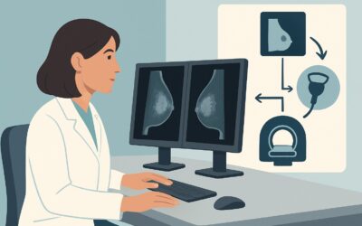
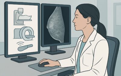
The ACR rates both Digital Breast Tomosynthesis (DBT) and standard 2D Mammography as May be appropriate for this scenario. However, DBT is often preferred in practice as it provides a quasi-three-dimensional view of the breast, which can be particularly advantageous.
DBT acquires multiple low-dose images from different angles as the x-ray tube moves in an arc. These images are then reconstructed into thin one-millimeter slices. This technique helps to reduce the effect of overlapping breast tissue, which can sometimes hide a small cancer or create a “summation artifact” that mimics a mass on standard 2D mammography. This can lead to improved cancer detection rates and a reduction in recall rates for non-cancerous findings.
The rationale for choosing mammographic techniques over other modalities for screening is rooted in their proven ability to detect microcalcifications, an early sign of DCIS, which other modalities may miss.
Alternative Studies and Their Ratings:
- Breast Ultrasound (US): Rated as Usually not appropriate for screening in this context. While excellent for evaluating palpable lumps or clarifying findings from a mammogram (a diagnostic role), ultrasound is not a suitable primary screening tool. It has poor sensitivity for microcalcifications and can result in a high number of false positives for benign cysts and nodules.
- Breast MRI with and without IV Contrast: Also rated as Usually not appropriate for screening in this specific scenario. While breast MRI is the most sensitive test for detecting invasive breast cancer and is used for screening in some very high-risk cisgender women, its role here is not established. The potential for high false-positive rates, leading to unnecessary biopsies and patient anxiety, along with its higher cost and need for IV contrast, makes it less suitable for initial screening in this population.
Both DBT and standard mammography involve a low level of ionizing radiation (ACR Relative Radiation Level ☢☢, corresponding to 0.1-1 mSv). This dose is considered safe and the benefits of early cancer detection in a high-risk individual are felt to outweigh the minimal risk.
When ordering, specifying “screening mammogram with tomosynthesis” is sufficient. For detailed technical specifications, refer to our complete guide on the Screening Mammography (with DBT) protocol.
What’s Next After Digital Breast Tomosynthesis Screening? Downstream Workflow
The results of the screening DBT will be categorized using the Breast Imaging Reporting and Data System (BI-RADS) score, which dictates the next steps in the clinical workflow.
- BI-RADS 0: Incomplete. This means additional imaging is needed for a final assessment. The patient will be called back for diagnostic mammography (which may include magnification or spot compression views) and/or a targeted breast ultrasound to clarify the initial finding.
- BI-RADS 1 (Negative) or 2 (Benign Finding): The exam is normal. The patient should continue annual screening mammography due to their high-risk status. The exact interval should be determined in consultation with breast imaging specialists and based on the specific risk factors.
- BI-RADS 3: Probably Benign. This indicates a finding that has a very high probability of being benign (<2% risk of malignancy). The standard recommendation is a short-interval follow-up, typically a repeat mammogram in six months, to ensure stability.
- BI-RADS 4 (Suspicious) or 5 (Highly Suggestive of Malignancy): These findings warrant a biopsy for definitive pathologic diagnosis. The patient should be referred promptly to a breast surgeon or breast radiologist for a tissue sampling procedure, most commonly a core needle biopsy.
If the patient later begins long-term gender-affirming hormone therapy, their screening plan will need to be re-evaluated, as this will align them with a different ACR Appropriateness Criteria scenario that accounts for hormone-induced breast development.
Pitfalls to Avoid (and When to Get Help)
Navigating breast cancer screening in high-risk transgender individuals requires careful attention to detail. Here are several common pitfalls to avoid:
- Underestimating Risk: Failing to take a thorough family history or recognize risk factors like chest irradiation can lead to a delay in initiating appropriate screening.
- Applying Cisgender Male Guidelines: A high-risk transfeminine individual, even without hormone use, has different screening needs than an average-risk cisgender man due to their elevated risk profile.
- Using Ultrasound for Primary Screening: Do not order a breast ultrasound as the first and only screening test. It is a diagnostic tool, not a screening tool, and can miss non-palpable, calcified lesions like DCIS.
- Ignoring a Palpable Lump: If the patient presents with a palpable breast lump, the workflow shifts from screening to diagnostic. This requires an immediate diagnostic mammogram and targeted ultrasound, regardless of recent screening results.
If you are uncertain about a patient’s specific risk level or the appropriate screening interval, a consultation with a breast imaging specialist or a genetics counselor is the appropriate next step.
Related ACR Topics and Tools
This article covers one specific scenario in transgender breast cancer screening. For a comprehensive overview of all related patient presentations, from average-risk individuals to those on long-term hormone therapy and transmasculine patients, please see our parent guide. Additional GigHz tools can help you navigate imaging decisions and patient conversations.
- For breadth across all scenarios in Transgender Breast Cancer Screening, see our parent guide: Transgender Breast Cancer Screening: ACR Appropriateness Decoded.
- To look up other clinical scenarios, use the ACR Appropriateness Criteria Lookup.
- For details on imaging technique, consult the Imaging Protocol Library.
- To discuss radiation exposure with your patients, the Radiation Dose Calculator can help frame the conversation.
Frequently Asked Questions
Why is screening initiated at age 25-30 in this scenario, and not earlier?
The age recommendation balances the benefit of early detection against the risks of radiation exposure over a lifetime. For most high-risk groups, including those with BRCA mutations, major guidelines recommend starting screening between ages 25 and 30. Starting earlier may not yield significant benefits and would increase cumulative radiation dose. This decision is based on risk-modeling data from high-risk cisgender populations, which is the best available evidence applied to this scenario.
How does this recommendation change if this patient starts gender-affirming hormone therapy?
If a transfeminine patient begins and continues gender-affirming hormone therapy (typically estrogen) for five or more years, their breast tissue develops, and their screening recommendations change. They would then fall under a different ACR scenario, which generally aligns more closely with screening guidelines for high-risk cisgender women, often involving annual mammography and consideration of supplemental screening with MRI.
Why is breast MRI ‘Usually not appropriate’ for screening here when it’s used for high-risk cisgender women?
Breast MRI is extremely sensitive but has lower specificity, leading to a higher rate of false positives. In cisgender women with dense breasts and very high risk, this trade-off is often accepted. For a transfeminine patient with minimal or no hormone-induced breast development, the breast tissue volume is typically small. In this context, the high false-positive rate of MRI is less acceptable, and mammography (especially DBT) is considered a more balanced initial screening tool. MRI is reserved for diagnostic problem-solving or for different risk profiles.
What specific genetic factors place a patient in this ‘higher-than-average risk’ category?
A known pathogenic or likely pathogenic variant in a cancer susceptibility gene, such as BRCA1 or BRCA2, is the most well-known factor. Other genes like PALB2, CHEK2, and ATM also confer increased risk. A patient with a first-degree relative (parent, sibling, child) who has a known mutation but has not undergone testing themselves is also considered high-risk. Genetic counseling is recommended for any patient with a significant personal or family history to clarify their specific risk.
Is there a difference in the types of breast cancer seen in transfeminine individuals?
Current data suggests that the types of breast cancer that develop in transfeminine individuals are similar to those seen in both cisgender men and women, with invasive ductal carcinoma being the most common. Cancers are often estrogen-receptor (ER) positive, which is consistent with cancers found in cisgender men and the majority of postmenopausal cisgender women.
Reviewed by Pouyan Golshani, MD, Interventional Radiologist — May 29, 2026