What Imaging Is Best for Low Back Pain After Minor Trauma or in an Elderly Patient?
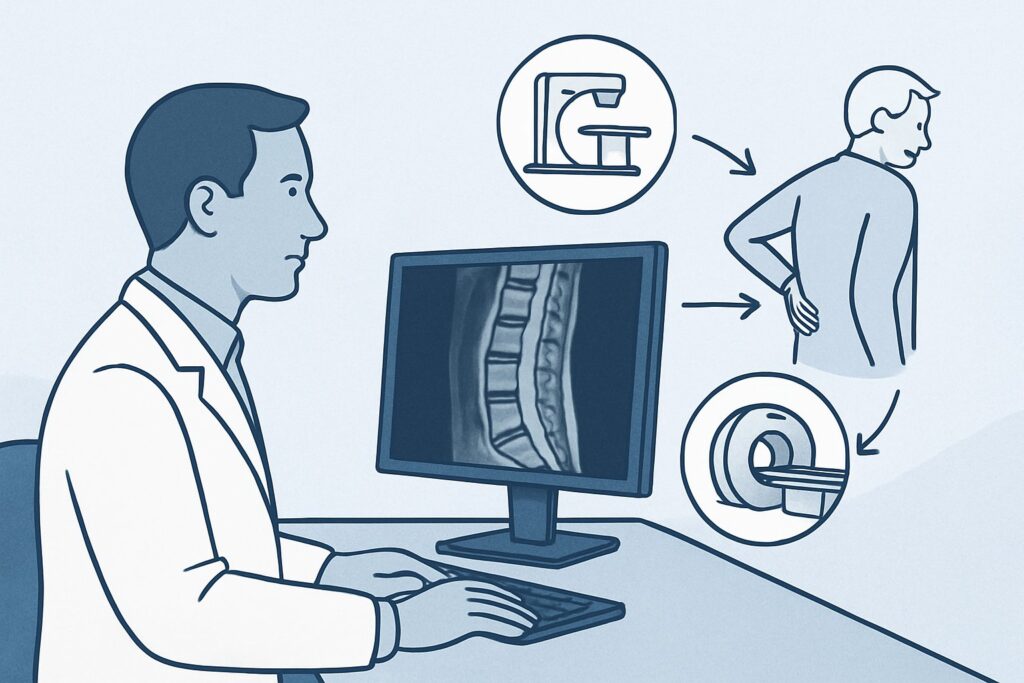
An 82-year-old female with known osteoporosis stumbles over a rug in her living room. While she doesn’t fall completely, she twists and catches herself, immediately feeling a sharp, new pain in her mid-lumbar spine. The pain persists over the next two days, making it difficult to stand up straight. As her physician, you are concerned about a potential fracture given her age and bone density. This article details the appropriate initial imaging workflow for this specific clinical scenario: low back pain in an individual with risk factors for vertebral fracture, such as an elderly patient, a person with osteoporosis, one on chronic steroids, or someone who has sustained low-velocity trauma. Based on the American College of Radiology (ACR) Appropriateness Criteria, the initial study of choice, Radiography lumbar spine, is rated Usually appropriate.
Who Fits This Clinical Scenario?
This guidance applies to a specific subset of patients presenting with low back pain, with or without radiculopathy, who have risk factors that increase the suspicion for a vertebral fracture. The key inclusion criteria are the presence of new or worsening low back pain coupled with one or more of the following:
- Elderly individual: Generally defined as age 65 or older, where bone density naturally decreases.
- Known osteoporosis or osteopenia: A documented diagnosis that makes the spine susceptible to fracture from minimal stress.
- Chronic steroid use: Long-term use of glucocorticoids is a well-established cause of secondary osteoporosis and increased fracture risk.
- Low-velocity trauma: This includes events like a fall from a standing height, a simple slip or twist, or even strenuous lifting in a susceptible individual.
It is crucial to distinguish this scenario from others. This workflow does not apply to patients with high-velocity trauma (e.g., motor vehicle collision, fall from a significant height), as they require a different trauma-focused imaging protocol. It also does not apply to patients with “red flag” symptoms suggesting cancer, infection, or cauda equina syndrome (e.g., unexplained weight loss, fever, new bowel/bladder incontinence), which necessitate a more urgent and often different imaging pathway. Finally, this is distinct from the workup for a younger, healthier patient with acute back pain and no risk factors, where imaging is often not indicated initially.
What Diagnoses Are You Working Up in This Scenario?
The primary clinical question driving the imaging decision in this context is the presence or absence of an acute or subacute vertebral compression fracture. The patient’s risk factors—age, osteoporosis, steroid use, or a traumatic event—significantly elevate this possibility above other causes of back pain.
Vertebral Compression Fracture (VCF): This is the most consequential diagnosis to identify or exclude. A VCF occurs when a vertebral body collapses, which can cause significant pain, deformity (kyphosis), and functional limitation. In patients with compromised bone quality, this can happen with minimal or no trauma. Identifying a fracture is critical as it changes management from symptomatic care to specific treatments for the fracture and underlying osteoporosis.
Exacerbation of Degenerative Disease: While patients in this demographic almost universally have underlying degenerative disc disease, spondylosis, or facet arthropathy, the new, acute nature of the pain following a minor traumatic event suggests a new process. Imaging helps determine if a new fracture is superimposed on these chronic changes, which can be a common diagnostic challenge.
Pathologic Fracture from Malignancy: Less commonly, a fracture may be the first presentation of an underlying malignancy, such as multiple myeloma or metastatic disease, that has weakened the bone. While not the primary suspicion without other red flags, imaging can sometimes reveal lytic or blastic lesions that would prompt a different, more urgent workup.
Why Is Lumbar Spine Radiography the Recommended Initial Study?
For this specific presentation, the ACR designates Radiography lumbar spine as Usually appropriate. The rationale is grounded in its ability to effectively answer the primary clinical question—is there a fracture?—in a rapid, accessible, and low-radiation manner.
Plain radiographs (X-rays) provide excellent visualization of bony structures and are highly effective at detecting most vertebral compression fractures. They can clearly show loss of vertebral body height, cortical disruption, and changes in spinal alignment like kyphosis. As the first-line study, radiography quickly triages patients, confirming a fracture in many cases and guiding immediate management without the delay or expense of more advanced imaging.
While other modalities are also highly rated, they are typically not the first choice.
- MRI lumbar spine without IV contrast is also rated Usually appropriate. MRI is superior for detecting marrow edema, which can identify an occult fracture not visible on X-ray and help determine the age of a fracture. However, it is more expensive, less available, and takes longer to perform, making it a better second-line test if radiographs are negative but clinical suspicion remains high.
- CT lumbar spine without IV contrast is another Usually appropriate option. CT provides superior bony detail compared to radiography and is excellent for characterizing complex fracture patterns or assessing the posterior elements. Its primary drawback is the significantly higher radiation dose (☢☢☢ 1-10 mSv) compared to the benefit it offers for an initial screen. It is often reserved for pre-procedural planning (e.g., for kyphoplasty) or if radiography is equivocal.
Studies like CT lumbar spine with IV contrast are rated Usually not appropriate for this initial evaluation. Intravenous contrast adds no value to the primary goal of assessing for a simple compression fracture and introduces risks associated with the contrast agent itself. The focus here is on bony integrity, not soft-tissue infection or tumor enhancement, which would be indications for contrast.
What’s Next After Lumbar Spine Radiography? Downstream Workflow
The results of the initial lumbar radiograph directly guide the subsequent clinical pathway. The decision tree branches based on whether the findings are positive, negative, or indeterminate.
If the radiograph is positive for a compression fracture: The immediate next steps involve pain management, bracing if indicated, and initiating or optimizing treatment for osteoporosis. A key downstream decision is whether the patient is a candidate for vertebral augmentation procedures like vertebroplasty or kyphoplasty. This often requires further imaging. An MRI without contrast may be ordered to assess for marrow edema, which confirms the fracture is acute or subacute and therefore more likely to benefit from intervention. A CT without contrast may be used to better define the fracture anatomy and assess for any retropulsion of bone fragments into the spinal canal.
If the radiograph is negative: For a patient with persistent, functionally limiting pain despite a negative X-ray, the workup should not stop. There is a significant possibility of an occult fracture (one not visible on plain films). The appropriate next step is an MRI lumbar spine without IV contrast. The high sensitivity of MRI for bone marrow edema will definitively rule in or rule out an occult fracture. If MRI is also negative, the diagnosis is more likely a severe musculoskeletal strain or an exacerbation of degenerative disease, and management can proceed with conservative, non-surgical care.
If the radiograph is indeterminate: Sometimes, X-rays show old, healed fractures or severe degenerative changes that make it difficult to identify an acute process. In this situation, similar to a negative result with high clinical suspicion, an MRI without contrast is the best next step to differentiate an acute-on-chronic process by looking for edema.
Pitfalls to Avoid (and When to Get Help)
In managing this patient population, several common pitfalls can delay diagnosis or lead to suboptimal outcomes. Be mindful of the following:
- Dismissing persistent pain after a negative X-ray: In a high-risk patient (e.g., elderly with osteoporosis), a negative radiograph is not sufficient to rule out a fracture. Persisting with a diagnosis of “muscular strain” without considering follow-up MRI can miss an occult fracture.
- Attributing symptoms solely to chronic degenerative changes: It is easy to look at an X-ray showing severe spondylosis and assume it is the cause of the patient’s new, acute pain. Always correlate the chronic findings with the acute history to avoid missing a superimposed fracture.
- Prematurely ordering advanced imaging: Starting with a CT or MRI without first obtaining a radiograph can lead to unnecessary radiation (CT) or healthcare costs and delays (MRI) when a simple X-ray would have answered the primary clinical question.
If a patient develops progressive or severe neurologic deficits, such as new, significant leg weakness, saddle anesthesia, or bowel/bladder dysfunction, this constitutes a clinical emergency. Escalate immediately for an emergent MRI of the lumbar spine to evaluate for severe spinal canal compromise or cauda equina syndrome.
Related ACR Topics and Tools
The ACR Appropriateness Criteria are a powerful resource for ensuring evidence-based imaging decisions. For a comprehensive overview of all low back pain scenarios, from acute uncomplicated pain to suspected cancer, please consult our parent guide. For tools to help implement these guidelines in your practice, see the resources below.
- For breadth across all scenarios in Low Back Pain, see our parent guide: Low Back Pain: ACR Appropriateness Decoded.
- To look up other clinical scenarios, visit the ACR Appropriateness Criteria Lookup.
- For details on imaging techniques, explore the Imaging Protocol Library.
- To discuss radiation exposure with patients, use the Radiation Dose Calculator.
Frequently Asked Questions
Why not just get an MRI first for an elderly patient with back pain?
While an MRI is highly sensitive, a lumbar spine radiograph (X-ray) is the recommended initial test because it is faster, more widely available, and lower cost, and it effectively answers the most immediate question: is there an obvious vertebral compression fracture? If the X-ray is positive, management can begin immediately. If it’s negative but suspicion remains high, an MRI is the appropriate next step to look for an occult fracture.
Does the presence of radiculopathy change the initial imaging choice from an X-ray?
Not for this specific scenario. The ACR criteria group patients ‘with or without radiculopathy’ together when risk factors for fracture are present. The primary concern is still ruling out a fracture, which an X-ray does well. If the X-ray is negative and radicular symptoms are the dominant issue, an MRI would be the logical next step to evaluate for disc herniation or nerve root compression.
How does ‘low-velocity trauma’ differ from other trauma in terms of imaging workup?
Low-velocity trauma (e.g., a fall from standing height) in a patient with risk factors like osteoporosis primarily raises suspicion for an isolated vertebral compression fracture. High-velocity trauma (e.g., a car accident) requires a much more extensive workup, often with CT scans of the entire spine and other body regions, to look for unstable, complex fractures and associated injuries.
If the X-ray shows a compression fracture, is a CT or MRI always needed next?
Not always. If the fracture is clearly identified and the clinical plan is conservative management (pain control, osteoporosis treatment), no further imaging may be needed immediately. However, if the patient has severe, unremitting pain, neurologic symptoms, or is being considered for a procedure like kyphoplasty, an MRI or CT is typically required to assess the fracture’s age and stability.
Does chronic steroid use alone, without trauma, warrant an X-ray for new back pain?
Yes. Chronic steroid use is a major risk factor for osteoporosis and fragility fractures. In a patient on long-term steroids, new, significant low back pain should be considered a potential fracture until proven otherwise, even without a specific traumatic event. A lumbar spine radiograph is the appropriate initial imaging step.
Reviewed by Pouyan Golshani, MD, Interventional Radiologist — June 27, 2026