What Imaging Is Best for Suspected Breast Cancer Recurrence After Lumpectomy?
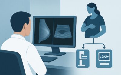
A 58-year-old woman, eight years status post-lumpectomy and radiation for early-stage breast cancer, presents to your clinic with a new concern. Over the past month, she has noticed a firm, non-tender nodule near her surgical scar. On examination, you confirm a 1.5 cm palpable abnormality. The clinical question is immediate: Is this benign post-treatment change, or is it a local recurrence? Deciding on the most effective initial imaging study is critical for guiding the subsequent diagnostic pathway. This article provides a detailed workflow for this specific scenario, focusing on the American College of Radiology (ACR) Appropriateness Criteria. For a patient with a history of breast-conserving therapy (BCT) presenting with symptoms or signs of a potential local recurrence, the ACR rates breast ultrasound as a Usually Appropriate first-line imaging examination.
Who Fits This Clinical Scenario?
This guidance applies specifically to patients with a history of breast-conserving therapy (BCT)—which includes lumpectomy with or without radiation—who now present with new signs or symptoms suggestive of a local recurrence. This presentation is distinct from routine, asymptomatic surveillance.
Inclusion criteria for this workflow:
- A prior diagnosis of invasive breast cancer or ductal carcinoma in situ (DCIS).
- Previous treatment with BCT (lumpectomy).
- New clinical findings, such as a palpable lump, focal pain, skin changes (dimpling, erythema, thickening), or suspicious nipple discharge.
It is crucial to distinguish this scenario from similar but distinct clinical situations that require different imaging strategies. This workflow does not apply to:
- Asymptomatic Surveillance: Patients undergoing routine annual screening without any new symptoms or physical exam findings. This falls under the ACR’s surveillance guidelines, which typically involve annual mammography.
- Post-Mastectomy Patients: The evaluation of the chest wall after a mastectomy is a different clinical problem with a distinct imaging approach, often involving ultrasound or MRI.
- Newly Diagnosed Breast Cancer: This workflow is for suspected recurrence, not the initial workup of a new primary breast cancer in a treatment-naïve patient.
What Diagnoses Are You Working Up in This Scenario?
When a patient with a history of BCT presents with a new palpable abnormality, the differential diagnosis is centered on distinguishing recurrent malignancy from expected or benign post-treatment changes. Imaging is essential for this differentiation.
Local Recurrence of Breast Cancer
This is the primary concern. A recurrence can manifest as a new mass, architectural distortion, or suspicious microcalcifications at or near the original tumor bed. The goal of imaging is to identify features suspicious for malignancy that warrant a biopsy for definitive pathologic diagnosis.
Fat Necrosis
A very common and benign consequence of surgery and radiation therapy. Damaged fatty tissue can form firm, irregular masses that are often clinically indistinguishable from a recurrent tumor. On imaging, fat necrosis has a variable appearance but often demonstrates characteristic features like oil cysts that confirm its benign nature.
Post-Surgical Scarring (Fibrosis)
Fibrosis in the lumpectomy bed is an expected finding. However, evolving or thickening scar tissue can sometimes create a palpable finding that raises clinical concern. Imaging helps characterize the stability and features of the scar to differentiate it from a developing recurrence.
Suture Granuloma
A benign inflammatory reaction to retained suture material. This can present as a firm, palpable nodule, often directly along the surgical scar line. Ultrasound is particularly effective at identifying the suture material within the nodule, which can suggest the diagnosis.
Why Are Ultrasound and Diagnostic Mammography the Recommended Studies?
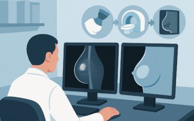
For a patient with a new palpable finding in a previously treated breast, the ACR designates both breast ultrasound and diagnostic mammography (including digital breast tomosynthesis, or DBT) as Usually Appropriate. In practice, these two studies are almost always performed together as a comprehensive diagnostic evaluation.
Breast Ultrasound (US) is the ideal first step for characterizing any palpable abnormality. Its high spatial resolution is excellent for assessing mass margins, shape, and internal characteristics (cystic vs. solid). In the post-treatment breast, where scar tissue and architectural distortion can obscure findings on mammography, ultrasound provides a targeted, real-time evaluation of the precise area of concern. It is also the primary modality for guiding a percutaneous biopsy if a suspicious solid lesion is identified. Importantly, ultrasound uses no ionizing radiation (0 mSv).
Diagnostic Mammography / Tomosynthesis provides the essential global view of the entire breast and axilla. While the palpable finding is the primary target, a recurrence may manifest elsewhere in the breast as non-palpable microcalcifications or a developing asymmetry. Tomosynthesis (3D mammography) is particularly valuable in this setting, as it reduces the effect of overlapping tissue and post-surgical distortion, improving the detection of subtle abnormalities. This modality involves a low dose of ionizing radiation (☢☢ 0.1-1 mSv).
The combination of these two modalities provides a powerful diagnostic pairing: mammography for the global survey and detection of calcifications, and ultrasound for the targeted characterization of the palpable finding and biopsy guidance.
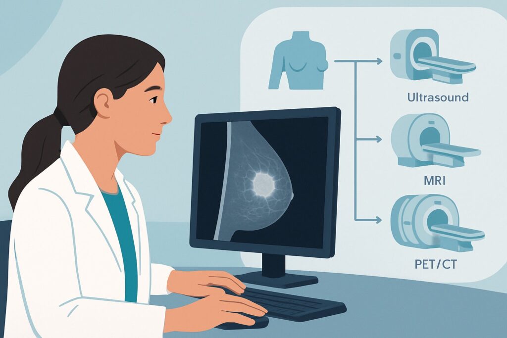
Why are other studies rated lower for this initial workup?
- Breast MRI with and without IV Contrast is rated May be appropriate. While highly sensitive for detecting invasive cancer, its specificity can be lower in the post-treatment setting. Benign enhancing findings, such as granulation tissue and inflammation, are common after BCT and can lead to false-positive results. MRI is typically reserved as a problem-solving tool if mammography and ultrasound are inconclusive or to evaluate the extent of disease if a recurrence is biopsy-proven.
- FDG-PET/CT is rated Usually not appropriate for evaluating a suspected local recurrence. This is a systemic staging tool designed to detect distant metastatic disease. It has poor sensitivity for small or low-grade local recurrences and involves a significant radiation dose (☢☢☢☢ 10-30 mSv). Its use is not indicated for the initial workup of a focal breast symptom.
What’s Next After Imaging? Downstream Workflow
The results of the diagnostic mammogram and targeted ultrasound will dictate the next steps in the patient’s management. The workflow typically follows one of three paths.
If the finding is suspicious for malignancy (BI-RADS 4 or 5):
The immediate next step is an image-guided core needle biopsy, most commonly performed under ultrasound guidance for palpable or sonographically visible lesions. The tissue diagnosis from pathology will confirm or exclude recurrence and provide crucial information (e.g., hormone receptor status) to guide treatment planning.
If the finding is definitively benign (BI-RADS 2):
If imaging clearly identifies a benign cause for the palpable finding, such as a simple cyst or classic fat necrosis, the patient can be reassured. The recommendation is typically a return to routine annual surveillance imaging. No further immediate workup is needed for that specific finding.
If the finding is probably benign (BI-RADS 3) or imaging is negative (BI-RADS 1):
If a palpable finding corresponds to a “probably benign” lesion on imaging (e.g., a stable-appearing fibrotic scar), the standard recommendation is short-interval follow-up imaging, usually in 6 months, to ensure stability. If imaging is entirely negative despite a persistent and distinct palpable abnormality, clinical correlation is paramount. A surgical consultation for consideration of excisional biopsy may be warranted, as a small percentage of cancers can be mammographically and sonographically occult.
Pitfalls to Avoid (and When to Get Help)
Navigating the workup of a suspected breast cancer recurrence requires careful attention to clinical and imaging correlation. Here are a few common pitfalls to avoid:
- Dismissing a palpable finding with negative imaging: A persistent, distinct palpable mass that is not explained by imaging findings (a “discordant” case) should not be ignored. Escalate with a referral to a breast surgeon for consideration of excisional biopsy.
- Attributing all changes to scarring: While post-treatment fibrosis is common, do not assume a new or evolving palpable abnormality is scar tissue without a thorough imaging workup. Recurrences often arise within or adjacent to the surgical bed.
- Ordering MRI as the first-line test: Jumping to breast MRI without initial mammography and ultrasound can lead to unnecessary biopsies of benign post-treatment enhancement and misses the crucial information provided by mammography (e.g., calcifications).
If the clinical and imaging findings are discordant or the diagnosis remains uncertain after initial studies, consultation with a breast radiologist or breast surgeon is the appropriate next step.
Related ACR Topics and Tools
For a comprehensive overview of all clinical scenarios related to imaging of invasive breast cancer, from initial diagnosis to surveillance, please see our parent topic guide. The tools below can help you navigate other clinical questions and discuss imaging choices with your patients.
- For breadth across all scenarios in Imaging of Invasive Breast Cancer, see our parent guide: Imaging of Invasive Breast Cancer: ACR Appropriateness Decoded.
- ACR Appropriateness Criteria Lookup — for adjacent scenarios
- Imaging Protocol Library — for technique on the recommended study
- Radiation Dose Calculator — for cumulative dose conversations
Frequently Asked Questions
Should I order a diagnostic mammogram or an ultrasound first for a palpable lump after BCT?
In practice, both are considered ‘Usually Appropriate’ and are typically ordered and performed together during the same diagnostic visit. The mammogram provides a global view of the breast, while the ultrasound provides a targeted evaluation of the palpable abnormality. Ordering a ‘diagnostic mammogram with ultrasound of the palpable area of concern’ is standard practice.
If the patient had dense breasts at her original diagnosis, should I skip mammography and go straight to MRI?
No. Even in dense breasts, diagnostic mammography (especially with tomosynthesis) is crucial for detecting suspicious microcalcifications, which can be a sign of recurrence and are not well-visualized on MRI. The standard first-line approach remains diagnostic mammography and targeted ultrasound. MRI is reserved for problem-solving if these initial studies are inconclusive.
How long after treatment can fat necrosis develop and cause a palpable lump?
Fat necrosis is a dynamic process that can evolve over time. While it often develops within the first year after treatment, it can appear or change months or even years later, frequently mimicking a recurrence. This is why any new, persistent palpable finding warrants a full diagnostic imaging workup, regardless of how long it has been since treatment ended.
What if the palpable lump is right on the surgical scar? Is it still suspicious?
Yes. While it could be benign scar tissue or a suture granuloma, the surgical bed is the most common site for a local recurrence. Any new or changing abnormality in or near the scar requires the same thorough diagnostic workup with mammography and ultrasound as a lump elsewhere in the breast.
Does this guidance change if the patient’s original cancer was invasive lobular carcinoma (ILC)?
The initial imaging approach (diagnostic mammogram and ultrasound) remains the same. However, it’s important to remember that ILC can be more subtle on imaging and may present as architectural distortion or an area of shadowing rather than a distinct mass. If initial imaging is negative or equivocal but clinical suspicion remains high for an ILC recurrence, there may be a lower threshold to proceed to breast MRI for further evaluation.
Reviewed by Pouyan Golshani, MD, Interventional Radiologist — May 30, 2026