What Imaging Is Next for a Palpable Breast Mass with a Benign Mammogram?

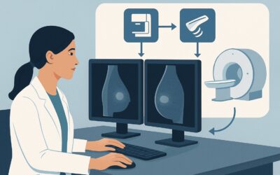
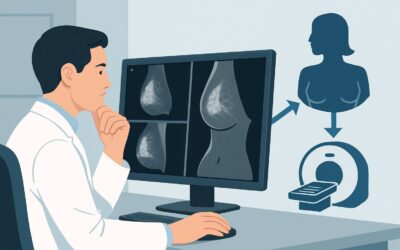
A 48-year-old female presents for follow-up. Her screening mammogram from last month was reported as benign, BI-RADS 2. However, during a self-exam last week, she felt a new, firm lump in her left breast that corresponds to the area of a benign calcification on the mammogram. She is anxious, and on clinical exam, you can confirm a vague, mobile, 1.5 cm palpable area. The mammogram report is reassuring, but the patient’s palpable finding creates a clinical dilemma. This situation, a palpable-mammographic discordance, requires a specific next step to ensure a subtle lesion is not missed. This article details the American College of Radiology (ACR) recommended workflow for this exact scenario. For this presentation, a targeted breast ultrasound is rated as Usually Appropriate.
Who Fits This Clinical Scenario?
This guidance applies to a specific and common clinical situation: an adult female, 40 years of age or older, who presents with a palpable breast mass, and whose recent diagnostic mammogram (including tomosynthesis) shows benign findings (BI-RADS 2) at the precise location of the palpable concern. The key element is the discordance between a benign imaging report and a persistent physical finding.
This workflow is distinct from other related scenarios. This article does not apply if:
- The mammogram is negative (BI-RADS 1): A negative mammogram with a palpable lump is a different type of discordance that also requires further workup, but it represents a separate ACR variant.
- The mammogram is suspicious (BI-RADS 4 or 5): If the mammogram identifies a suspicious lesion corresponding to the palpable mass, the next step is typically image-guided biopsy, not further diagnostic imaging.
- The patient is younger than 40: For women under 30, ultrasound is often the initial imaging modality. For women between 30 and 39, the initial workup may vary.
Correctly identifying your patient within this specific scenario—a benign mammogram at the site of a palpable mass in a woman 40 or older—is crucial for applying the appropriate next step.
What Diagnoses Are You Working Up in This Scenario?
The primary goal of ordering the next imaging study is to resolve the discordance between the physical exam and the benign mammogram. The differential diagnosis ranges from common benign entities to the rare but critical possibility of a mammographically occult cancer.
Benign Cyst or Fibroglandular Tissue: This is the most common explanation. A simple or complicated cyst may not be apparent on a mammogram, especially in dense breast tissue, but is easily characterized by ultrasound. Similarly, what the patient feels may be a focal area of normal, dense fibroglandular tissue that presents no suspicious mammographic features.
Fibroadenoma or Other Benign Solid Mass: A fibroadenoma, a common benign tumor, can be isodense to the surrounding breast parenchyma, making it difficult to distinguish on mammography. Ultrasound is highly effective at identifying and characterizing these well-circumscribed solid masses, which often have classic benign features.
Mammographically Occult Malignancy: This is the “can’t-miss” diagnosis driving the workup. While uncommon, some breast cancers do not have typical mammographic signs like spiculated masses or suspicious calcifications. Invasive lobular carcinoma, in particular, is known for its subtle presentation, sometimes appearing only as an area of architectural distortion or being completely occult on mammography. These cancers are often detectable on ultrasound as a hypoechoic, irregular, or shadowing mass.
Why Is Breast Ultrasound the Recommended Study for This Presentation?
When a patient has a palpable mass but a benign mammogram, the ACR designates a targeted breast ultrasound (US) as Usually Appropriate. The rationale is centered on ultrasound’s unique ability to investigate a specific area of concern with high-resolution imaging, independent of breast density.
Ultrasound excels at tissue characterization. It can definitively distinguish a simple cyst (which requires no further workup) from a complex cyst or a solid mass. For solid masses, sonographic features such as shape, orientation, margins, and posterior acoustic features can help determine the likelihood of malignancy and guide the decision for biopsy. In the context of a benign mammogram, ultrasound serves as the essential problem-solving tool to find a sonographic correlate for the palpable finding.
Alternative studies are rated lower for this specific clinical question:
- MRI breast without and with IV contrast is rated Usually Not Appropriate. While highly sensitive, breast MRI has a higher rate of false positives in this setting, which could lead to unnecessary anxiety and biopsies. It is generally reserved for high-risk screening or as a problem-solving tool if both mammography and ultrasound are negative but clinical suspicion remains high.
- Image-guided core biopsy is also rated Usually Not Appropriate as the next step. A biopsy cannot be performed without an imaging target. The purpose of the ultrasound is to identify a targetable lesion corresponding to the palpable mass. If a suspicious sonographic correlate is found, biopsy then becomes the appropriate subsequent step.
From a safety perspective, ultrasound is an ideal choice. It involves no ionizing radiation (Relative Radiation Level: O) and does not require intravenous contrast, making it a safe, non-invasive, and readily available examination.
What’s Next After Breast Ultrasound? Downstream Workflow
The results of the targeted breast ultrasound will direct the subsequent steps in the patient’s care. The workflow branches based on whether a sonographic correlate for the palpable mass is identified.
If a suspicious sonographic correlate is found (BI-RADS 4 or 5): The palpable-mammographic discordance is resolved. The next step is an ultrasound-guided core needle biopsy of the identified lesion to obtain a pathologic diagnosis.
If a benign or probably benign sonographic correlate is found (BI-RADS 2 or 3): If ultrasound identifies a classic simple cyst (BI-RADS 2) or a classic fibroadenoma (BI-RADS 3) that fully explains the palpable finding, the discordance is resolved. For a BI-RADS 2 finding, the patient can return to routine screening. For a BI-RADS 3 finding, the standard of care is short-term imaging follow-up, typically with ultrasound in 6 months, to ensure stability.
If no sonographic correlate is found (BI-RADS 1): This is a challenging situation where both mammogram and targeted ultrasound are negative in the area of a palpable mass. This is termed “triple negative” (negative clinical exam, negative mammogram, negative ultrasound) is not correct here, as the clinical exam is positive. This is a persistent imaging-palpable discordance. Management may involve a discussion between the radiologist and the referring clinician. Options include short-term clinical follow-up (e.g., re-examination in 3-6 months) or, if clinical suspicion is high, consideration of breast MRI or even surgical consultation for excisional biopsy.
Pitfalls to Avoid (and When to Get Help)
Navigating this scenario requires careful correlation between clinical and imaging findings. Here are a few common pitfalls to avoid:
- Dismissing the Palpable Finding: Do not assume a benign mammogram definitively excludes malignancy in the presence of a persistent palpable mass. The discordance itself is an indication for further evaluation.
- Accepting an Incomplete Ultrasound: Ensure the ultrasound report explicitly states whether a sonographic correlate for the palpable finding was identified. A generic “benign breast ultrasound” is insufficient if it doesn’t address the specific area of clinical concern.
- Not Correlating Location: The radiologist must be clearly informed of the exact location of the palpable mass (e.g., using a clock-face position and distance from the nipple) to ensure the ultrasound is truly targeted to the correct area.
If both mammography and targeted ultrasound are negative but the palpable mass is persistent, growing, or highly suspicious on clinical exam, this warrants escalation. A discussion with the breast radiologist is the critical next step to decide between close clinical follow-up, breast MRI, or surgical consultation.
Related ACR Topics and Tools
For a comprehensive overview of all clinical variants related to palpable breast masses, from initial workup to post-biopsy management, please see our parent guide. It provides a breadth of information that complements this in-depth article.
- For breadth across all scenarios in Palpable Breast Masses, see our parent guide: Palpable Breast Masses: ACR Appropriateness Decoded.
- To explore other clinical scenarios, use the Imaging Appropriateness Selector.
- For details on imaging techniques, visit the Imaging Protocol Library.
- To discuss cumulative radiation exposure with patients, consult the Radiation Dose Calculator.
Frequently Asked Questions
Why not just repeat the mammogram in 6 months if it was BI-RADS 2?
A BI-RADS 2 (Benign) mammogram indicates no evidence of malignancy, so repeating it in the short term is not indicated. The clinical problem is the palpable finding that the mammogram does not explain. Ultrasound is the appropriate modality to investigate this specific palpable area, as it can detect mammographically occult lesions like cysts or some cancers.
What if the patient has extremely dense breasts? Is ultrasound still the best next step?
Yes, in fact, ultrasound is particularly valuable in women with dense breasts. Mammographic sensitivity is reduced in dense tissue, which can obscure underlying masses. Ultrasound’s effectiveness is not limited by breast density, making it the ideal tool to evaluate a palpable finding in this context.
If the ultrasound is also negative (BI-RADS 1), can I confidently tell the patient she does not have cancer?
When both a high-quality mammogram and a targeted ultrasound are negative at the site of a palpable mass, the likelihood of malignancy is very low. However, it does not fall to zero. This situation of persistent palpable-imaging discordance requires careful clinical judgment. The next step is often close clinical follow-up, but if the palpable finding is highly suspicious, a discussion with the radiologist about proceeding to breast MRI or surgical consultation is warranted.
Should I order a ‘whole breast’ ultrasound or a ‘targeted’ ultrasound?
For this scenario, you should order a targeted (or limited) breast ultrasound. The goal is to interrogate the specific area corresponding to the palpable mass. A whole breast screening ultrasound is a different examination used for supplemental screening in women with dense breasts and is not the indicated study for evaluating a focal problem.
Does it matter if the BI-RADS 2 finding (e.g., a benign calcification) is in the same spot as the lump?
Yes, the location is critical. The scenario assumes the benign mammographic finding is at the site of the palpable mass. This still constitutes discordance because a benign entity like a simple calcification should not be palpable. The ultrasound is needed to determine if there is another, non-calcified lesion in that exact location (e.g., a cyst, fibroadenoma, or occult cancer) that is responsible for the palpable finding.
Reviewed by Pouyan Golshani, MD, Interventional Radiologist — May 26, 2026