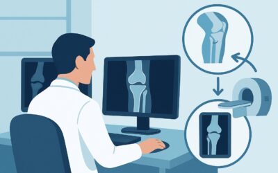
What Imaging Is Best for Suspected Erosive Osteoarthritis When Radiographs Are Normal?

A 55-year-old woman presents to your clinic with six months of worsening pain, stiffness, and swelling in the joints of her fingers, particularly the ones closest to her nails. The symptoms are worse in the morning. On exam, you note tender, mildly swollen distal interphalangeal (DIP) joints. You suspect erosive osteoarthritis (EOA), but the hand radiographs you ordered are read as normal, with no visible erosions or significant joint space narrowing. You are now faced with a common clinical dilemma: what is the most appropriate next imaging study to confirm your suspicion, rule out mimics, and guide management? This article provides a detailed workflow for this specific scenario, based on the American College of Radiology (ACR) Appropriateness Criteria. For this presentation, US area of interest is rated May be appropriate.
Who Fits This Clinical Scenario for Suspected Erosive Osteoarthritis?
This guidance is intended for a specific patient population: those with chronic extremity joint pain where erosive osteoarthritis is the leading diagnosis, but initial radiographs are either normal or inconclusive. The classic presentation involves middle-aged to older adults, most commonly postmenopausal women, with symptoms localized to the small joints of the hands (particularly the DIP and proximal interphalangeal [PIP] joints) or feet.
Inclusion criteria for this workflow are:
- Chronic symptoms (weeks to months) of joint pain, stiffness, or swelling.
- Clinical suspicion for EOA based on demographics and joint distribution.
- Initial radiographs that do not show definitive features of EOA (e.g., central “gull-wing” erosions, ankylosis).
It is crucial to distinguish this scenario from similar presentations that require a different diagnostic approach. This workflow does not apply if:
- Inflammatory arthritis is the primary suspicion. If the patient is seropositive (e.g., positive rheumatoid factor or anti-CCP) or has a joint pattern more typical for rheumatoid arthritis (e.g., symmetric wrist and metacarpophalangeal [MCP] joint involvement), a different ACR variant for suspected inflammatory arthritis should be consulted.
- Crystalline arthropathy is suspected. A patient presenting with acute, severe monoarticular pain and swelling, especially in the great toe or knee, should be evaluated according to the ACR guidelines for suspected gout or pseudogout.
What Diagnoses Are You Working Up When Radiographs Are Inconclusive?
When initial X-rays are unrevealing in a patient with suspected EOA, advanced imaging is used to investigate a narrow but important differential. The goal is to identify early inflammatory and structural changes that radiographs cannot detect.
Erosive Osteoarthritis (EOA)
This is the primary diagnosis under consideration. EOA is an aggressive, inflammatory subset of osteoarthritis characterized by synovial inflammation and central bony erosions, which can lead to significant functional impairment. In its early stages, the hallmark “gull-wing” erosions may not yet be visible on radiographs, but the underlying synovitis and subtle cortical breaks can be detected with more sensitive imaging modalities like ultrasound.
Psoriatic Arthritis (PsA)
PsA is a major mimic of EOA, as it frequently affects the DIP joints and can be seronegative. Differentiating the two is critical due to vastly different treatment pathways. Advanced imaging can help identify features more characteristic of PsA, such as enthesitis (inflammation where tendons and ligaments attach to bone), dactylitis (“sausage digit”), and a different pattern of bone erosion and proliferation.
Seronegative Rheumatoid Arthritis (RA)
While less commonly DIP-predominant, seronegative RA remains on the differential. RA typically causes marginal erosions (at the “bare areas” of the joint where synovium contacts bone), in contrast to the central erosions of EOA. Identifying the location of early erosions and the specific character of synovial proliferation on imaging can help distinguish RA from EOA.
Non-Erosive Inflammatory Osteoarthritis
Some patients may have a significant inflammatory component to their osteoarthritis without developing the destructive erosions that define EOA. Imaging can confirm the presence of synovitis and joint effusion, establishing an inflammatory phenotype that may respond to anti-inflammatory treatments, even in the absence of erosions.
Why Is Ultrasound the Next Step for Suspected Erosive OA with Normal Radiographs?
In this scenario, where radiographs are normal or inconclusive, the ACR rates US area of interest as May be appropriate. This rating signifies that ultrasound is a valuable tool that can provide diagnostic clarity when clinical suspicion is high but first-line imaging is negative. It offers a unique combination of high-resolution soft tissue visualization, dynamic assessment, and lack of ionizing radiation.
Ultrasound excels at detecting the key pathological features of early EOA and its mimics. It is highly sensitive for identifying synovitis, joint effusions, and tenosynovitis. Using power Doppler, the operator can visualize and quantify synovial hyperemia, providing a direct measure of active inflammation. Most importantly, ultrasound can detect subtle bone erosions and cortical irregularities long before they become apparent on a plain radiograph. This capability is essential for confirming an erosive process and differentiating it from non-erosive inflammatory OA.
The ACR rates alternative advanced imaging modalities lower for this specific clinical question:
- MRI area of interest without IV contrast is rated Usually not appropriate. While MRI provides excellent anatomical detail, the assessment of active synovitis—a critical feature for differentiating inflammatory arthritides—is significantly limited without the use of gadolinium-based contrast.
- Bone scan whole body is also rated Usually not appropriate. A bone scan is a sensitive but highly non-specific test. It may show increased radiotracer uptake in inflamed joints but cannot distinguish between EOA, PsA, or RA, nor can it directly visualize the synovium, erosions, or tendons.
The primary advantages of ultrasound in this context are its safety profile (0 mSv radiation dose), cost-effectiveness, and ability to perform a dynamic, multi-joint examination in a single session. When ordering, it is helpful to specify the joints of highest concern and to request evaluation for synovitis, erosions, and enthesitis with power Doppler assessment.
What’s Next After Ultrasound? Downstream Workflow
The results of the ultrasound will guide your subsequent management and diagnostic decisions. The workflow typically branches based on whether key inflammatory or erosive features are identified.
If the ultrasound is positive for EOA:
Findings consistent with EOA include synovitis (especially with a positive power Doppler signal) and central erosions in a DIP/PIP distribution. A positive study confirms the diagnosis, allowing you to initiate appropriate management, which may include NSAIDs, intra-articular injections, and hand therapy. It also sets a baseline for monitoring disease progression.
If the ultrasound is negative:
A completely normal ultrasound, showing no synovitis, erosions, or effusion in a symptomatic patient, makes an inflammatory or erosive arthritis less likely. In this case, the diagnosis may be non-inflammatory osteoarthritis. Management would focus on analgesia and supportive care. Re-evaluation may be necessary if symptoms progress.
If the ultrasound is indeterminate or suggests a mimic:
The study might reveal findings that are ambiguous or more suggestive of an alternative diagnosis like PsA (e.g., prominent enthesitis) or RA (e.g., marginal erosions at the MCPs). In this situation, the ACR rates MRI area of interest without and with IV contrast as May be appropriate. A contrast-enhanced MRI can provide a more comprehensive assessment of synovitis, bone marrow edema, and erosions, helping to clarify the diagnosis and guide referral to a rheumatologist.
Pitfalls to Avoid (and When to Get Help)
Navigating the workup for suspected EOA requires careful attention to clinical and imaging details. Here are a few common pitfalls to avoid:
- Over-relying on negative serologies: EOA, PsA, and a subset of RA are all seronegative. Do not dismiss a diagnosis of inflammatory arthritis solely because RF and anti-CCP are negative.
- Not specifying the clinical question: When ordering an ultrasound, clearly state that you are evaluating for erosive osteoarthritis versus other inflammatory arthritides. This helps the sonographer and radiologist focus their search on key discriminating features.
- Ignoring the contralateral hand: If a patient has unilateral symptoms, examining the asymptomatic contralateral joints can sometimes reveal subclinical disease, strengthening the diagnosis of a systemic process.
- Misinterpreting osteophytes as erosions: Standard osteophytes are a feature of non-inflammatory OA. Differentiating these from the central, often “gull-wing” shaped erosions of EOA is critical.
If the diagnosis remains unclear after ultrasound, or if the findings suggest a complex inflammatory condition like PsA or RA, referral to a rheumatologist for further evaluation and management is the appropriate next step.
Related ACR Topics and Tools
For a comprehensive overview of imaging for all related conditions, and for tools to help you implement these guidelines, the following resources are available. For breadth across all scenarios in Chronic Extremity Joint Pain-Suspected Inflammatory Arthritis, Crystalline Arthritis, or Erosive Osteoarthritis, see our parent guide: Chronic Extremity Joint Pain-Suspected Inflammatory Arthritis, Crystalline Arthritis, or Erosive Osteoarthritis: ACR Appropriateness Decoded.
- ACR Appropriateness Criteria Lookup: For exploring adjacent clinical scenarios and alternative imaging recommendations.
- Imaging Protocol Library: For detailed technical guidance on performing the recommended studies.
- Radiation Dose Calculator: For discussing cumulative radiation exposure with patients when considering studies like CT or bone scans in other contexts.
Frequently Asked Questions
Why is ultrasound rated ‘May be appropriate’ instead of ‘Usually appropriate’ for this scenario?
The ‘May be appropriate’ rating reflects that ultrasound is a valuable second-line test in a specific context: when clinical suspicion for erosive osteoarthritis is high but initial radiographs are unrevealing. It is not considered a routine first-line test for all cases of chronic joint pain. Its utility is highest when the pre-test probability is moderate to high and the results will directly influence management, such as differentiating EOA from a mimic like psoriatic arthritis.
If I order an ultrasound, what specific findings should I ask the radiologist to look for?
When ordering, it’s helpful to specify the clinical question. Ask the radiologist to evaluate for: 1) Synovitis and its activity using power Doppler. 2) Bony erosions, noting their location (central vs. marginal). 3) Enthesitis, especially if psoriatic arthritis is on the differential. 4) Tenosynovitis. This level of detail helps tailor the exam to answer the specific clinical question.
Is a contrast-enhanced MRI ever the right first step after a normal radiograph?
According to the ACR criteria for this specific scenario, contrast-enhanced MRI is also rated ‘May be appropriate,’ making it a reasonable alternative to ultrasound. However, ultrasound is often preferred initially due to its lower cost, lack of radiation, and ability to perform dynamic imaging. MRI is typically reserved for cases where ultrasound is inconclusive or when a more global assessment of the joint, including bone marrow edema, is required.
What if the patient has a pacemaker or other contraindication to MRI?
If a patient has a contraindication to MRI, ultrasound becomes an even more critical tool. Ultrasound does not involve magnetic fields or ionizing radiation and is safe for all patients. It provides excellent visualization of the synovium, erosions, and tendons, making it the best alternative to MRI for assessing inflammatory arthritis in this setting.
Can I use CT instead of ultrasound or MRI to look for erosions?
For this clinical scenario, all variants of CT (without contrast, with contrast, and both) are rated ‘Usually not appropriate’ by the ACR. While CT is excellent for visualizing bone and can detect erosions with high resolution, it does not visualize the synovium, effusions, or tendons well. Therefore, it cannot assess for active inflammation, which is a key part of the diagnostic question. It also involves ionizing radiation, which is avoided by both ultrasound and MRI.
Reviewed by Pouyan Golshani, MD, Interventional Radiologist — May 30, 2026