What Imaging Is Best for UGIB When Endoscopy Fails to Control Bleeding?
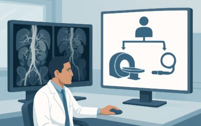
It’s 2 AM in the intensive care unit. Your patient, a 68-year-old man with a history of NSAID use, was admitted with hematemesis. Gastroenterology performed an endoscopy, identifying a large, bleeding duodenal ulcer. Despite attempts at clipping and epinephrine injection, the bleeding continues, and the patient’s hemoglobin is dropping. The GI team states they cannot achieve durable hemostasis endoscopically. You need to act quickly to localize and control the hemorrhage. This situation calls for a shift from endoscopic therapy to interventional radiology or surgery, guided by precise imaging. This article details the American College of Radiology (ACR) Appropriateness Criteria workflow for this exact scenario: a confirmed nonvariceal upper gastrointestinal bleed where endoscopic treatment has failed or is not possible. For this presentation, the ACR rates `Arteriography visceral` as Usually Appropriate.
Who Fits This Clinical Scenario?
This guidance is for a specific, high-acuity patient population. The inclusion criteria are precise: an adult patient with a confirmed nonvariceal upper gastrointestinal (UGI) bleed where the source has been identified on endoscopy, but bleeding persists. This includes cases where endoscopic therapy was attempted but failed to achieve hemostasis, or where the lesion’s location or characteristics made endoscopic treatment impossible from the outset.
This workflow is not for patients in whom:
- No endoscopy has been performed. If UGI bleeding is only suspected clinically without endoscopic confirmation, the initial imaging workup follows a different path. See the ACR variant for suspected UGI bleeding without endoscopy.
- Endoscopy confirmed bleeding but found no clear source. An occult or obscure bleed requires a different diagnostic algorithm, often involving studies designed to survey a wider area.
- Endoscopy was negative. If a thorough endoscopic evaluation is negative despite clinical signs of bleeding, the focus shifts to obscure GI bleeding, which may originate beyond the reach of the endoscope.
- The bleeding is from esophageal or gastric varices. Variceal bleeding is a distinct pathophysiology related to portal hypertension and is managed with a completely different treatment algorithm (e.g., TIPS procedure).
Correctly identifying your patient’s scenario is critical to selecting the most effective and efficient imaging study.
What Diagnoses Are You Working Up in This Scenario?
In this context, endoscopy has already provided the “what” (e.g., an ulcer, a lesion), but imaging is needed to define the “where” of the arterial supply for intervention. The differential diagnosis is less about identifying the lesion type and more about characterizing its vascular supply and planning for hemostasis.
The most common underlying cause is peptic ulcer disease (PUD), particularly posterior duodenal ulcers that can erode into the gastroduodenal artery or its branches. These vessels are large and can cause life-threatening hemorrhage that is difficult to control with endoscopic clips or cautery alone due to the brisk flow and vessel size.
Another important consideration is a Dieulafoy’s lesion. This is a large, aberrant submucosal artery that erodes through the overlying mucosa without a primary ulcer. Because the vessel itself is the problem, endoscopic therapy can be challenging, and re-bleeding is common. Arteriography is ideal for identifying this specific vascular anomaly.
Less common but critically important is an aortoenteric fistula, especially in patients with a history of aortic aneurysm repair. While endoscopy may identify bleeding in the duodenum, it often cannot visualize the fistula itself. CTA and subsequent arteriography are essential to make this diagnosis, which is a true surgical emergency.
Other potential sources include bleeding from tumors (which may have a rich, neovascular supply resistant to endoscopic control) or angiodysplasia. While often associated with lower GI bleeding, these vascular malformations can occur in the upper GI tract and may require embolization for definitive treatment.
Why Is Visceral Arteriography the Recommended Study for This Presentation?
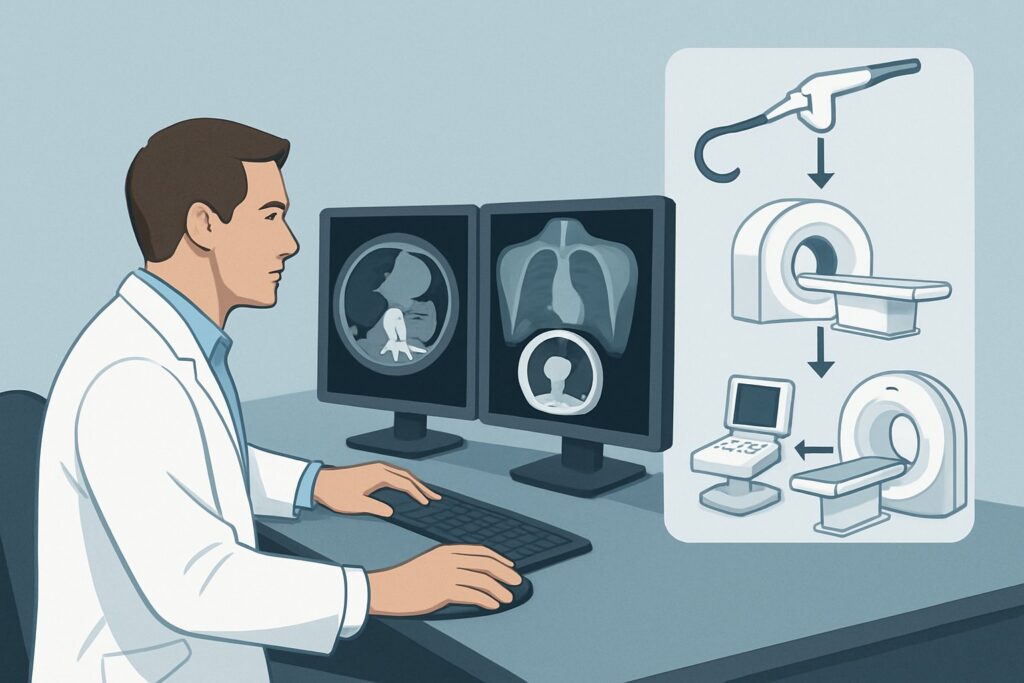
When endoscopic treatment fails, the clinical goal shifts from simple diagnosis to active intervention. This is why the ACR designates `Arteriography visceral` as Usually Appropriate. It is uniquely positioned as both a diagnostic and a therapeutic procedure. During the study, an interventional radiologist can precisely map the celiac and superior mesenteric arteries, identify the exact point of active contrast extravasation, and immediately proceed with transcatheter arterial embolization to stop the bleeding. This single-session approach is crucial for a hemodynamically unstable patient.
`CTA abdomen and pelvis without and with IV contrast` is also rated Usually Appropriate. It serves as a highly sensitive, non-invasive roadmap. A multiphase CTA can pinpoint the bleeding source with excellent spatial resolution, guiding the subsequent arteriogram. This is particularly useful in stable patients or when the interventional radiology suite is not immediately available. However, it is a purely diagnostic study and will always require a subsequent procedure (arteriography or surgery) for treatment. The radiation dose is also higher (☢☢☢☢ 10-30 mSv) compared to the typical diagnostic portion of an arteriogram (☢☢☢ 1-10 mSv), though the therapeutic portion of arteriography can increase the total dose.
Why are other studies rated lower?
- RBC scan abdomen and pelvis is Usually Not Appropriate. A tagged red blood cell scan is far too slow and lacks the spatial resolution needed in this acute setting. It can detect slower bleeding rates but cannot precisely localize the source vessel for embolization, making it unhelpful when a source has already been seen endoscopically.
- Fluoroscopy upper GI series is also Usually Not Appropriate. A barium study has no role in evaluating active, life-threatening hemorrhage. It cannot identify a vascular source and the presence of barium in the GI tract would interfere with subsequent endoscopy, angiography, or surgery.
The choice between proceeding directly to arteriography versus obtaining a preliminary CTA often depends on institutional protocols, patient stability, and interventional radiology availability. In an unstable patient, proceeding directly to the angiography suite is often the most expedient path to hemostasis.
What’s Next After Visceral Arteriography? Downstream Workflow
The results of the angiogram dictate the immediate next steps in patient management. The workflow is a clear decision tree focused on achieving and maintaining hemostasis.
If the arteriogram is positive and embolization is successful: The patient should be transferred back to an intensive care setting for close hemodynamic monitoring. This includes frequent vital signs, serial hemoglobin checks, and monitoring for any signs of re-bleeding. The underlying cause of the bleed (e.g., H. pylori, NSAID use) must be addressed to prevent recurrence. The interventional radiologist will also monitor for rare complications of embolization, such as non-target embolization or bowel ischemia.
If the arteriogram is positive but embolization is technically unsuccessful: This is a critical event. The interventional radiologist may have been unable to selectively catheterize the bleeding vessel, or the vascular anatomy may be unfavorable for safe embolization. An immediate surgical consultation is mandatory. The angiographic images are invaluable to the surgeon, providing a precise map of the bleeding point to guide the operation.
If the arteriogram is negative: This typically means the bleeding was intermittent and had temporarily stopped at the time of the study. In a stable patient, this may lead to a period of observation and medical management. In a patient with continued signs of blood loss, provocative arteriography—using vasodilators or anticoagulants to induce bleeding under controlled conditions—may be considered, though this carries risks. Alternatively, a repeat endoscopy in 24-48 hours may be planned.
Pitfalls to Avoid (and When to Get Help)
In this high-stakes clinical scenario, several common pitfalls can compromise patient outcomes.
- Delaying the Call: Time is critical. Once endoscopic failure is established in a patient with ongoing bleeding, the call to interventional radiology should be made immediately. Delaying for another trial of medical management can lead to further decompensation.
- Inadequate Communication: The interventional radiologist needs to know the precise endoscopic findings. Relay the location of the lesion (e.g., “posterior duodenal bulb,” “gastric body, lesser curvature”) to help them focus their angiographic search.
- Ignoring Renal Function: Both CTA and arteriography require iodinated contrast, which can be nephrotoxic. In patients with pre-existing chronic kidney disease, a careful risk-benefit discussion is needed, and pre-procedural hydration should be administered if clinically feasible.
- Neglecting Coagulopathy: Before an invasive procedure like arteriography, any significant coagulopathy should be assessed and corrected if possible.
If the patient becomes profoundly hemodynamically unstable (e.g., requiring massive transfusion protocol), escalate immediately. This often involves a simultaneous consultation with both interventional radiology and general/trauma surgery, as the patient may be too unstable for transport to the angiography suite and may require an emergency laparotomy.
Related ACR Topics and Tools
This article covers one specific variant within the broader topic of nonvariceal upper gastrointestinal bleeding. For a comprehensive overview of all related clinical scenarios, from initial suspicion to post-procedural follow-up, please consult the parent topic guide. The following GigHz tools can also support your clinical decision-making:
- For breadth across all scenarios in Nonvariceal Upper Gastrointestinal Bleeding, see our parent guide: Nonvariceal Upper Gastrointestinal Bleeding: ACR Appropriateness Decoded.
- ACR Appropriateness Criteria Lookup — for adjacent scenarios and other clinical questions.
- Imaging Protocol Library — for detailed technical parameters of CTA and other imaging studies.
- Radiation Dose Calculator — for discussing cumulative radiation exposure with patients and families.
Frequently Asked Questions
Why not go directly to surgery if endoscopy fails to control the bleeding?
Catheter-directed embolization via arteriography is a highly effective, less invasive alternative to open surgery. It avoids the morbidity and mortality associated with a major laparotomy in a critically ill patient. Surgery is typically reserved for cases where embolization fails, is not anatomically feasible, or if the patient is too unstable for transport to an angiography suite.
What is the minimum bleeding rate required for detection on CTA versus arteriography?
While figures vary, conventional angiography is generally thought to be able to detect active bleeding at rates as low as 0.5 mL/min. Multidetector CTA has improved sensitivity and may detect bleeding at rates of 0.3 to 0.5 mL/min. Both are highly sensitive for the brisk, arterial bleeding typical in this clinical scenario.
Is a CTA always necessary before visceral arteriography?
No, it is not always necessary. In a hemodynamically unstable patient with a known bleeding source from endoscopy, proceeding directly to the angiography suite for diagnosis and treatment is often the fastest and most appropriate course. A pre-procedure CTA is most valuable in stable patients to provide a ‘roadmap’ for the interventionalist, potentially shortening the subsequent angiography procedure time.
What are the major risks of visceral arteriography and embolization?
The primary risks include those common to any arterial catheterization, such as bleeding or hematoma at the access site, vessel dissection, or pseudoaneurysm. Specific to embolization, the most significant risk is non-target embolization, which could lead to ischemia or infarction of healthy tissue, such as a portion of the bowel or liver. This risk is minimized by experienced interventional radiologists using superselective catheterization techniques.
How does profound hemodynamic instability affect the choice between CTA and direct-to-angio?
Profound instability is a key decision point. A patient requiring massive transfusion and high-dose vasopressors may be too unstable for the time and transport required for a CTA. In this case, the decision is often between going directly to the angiography suite (if it’s immediately available and the patient can be stabilized for the procedure) or going directly to the operating room for surgical control of the hemorrhage. This decision requires rapid coordination between the ICU, GI, interventional radiology, and surgical teams.
Reviewed by Pouyan Golshani, MD, Interventional Radiologist — May 30, 2026