What Imaging Should You Order for a Child with Acute Ataxia After Trauma?
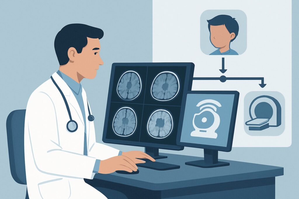
A 7-year-old presents to the emergency department on a Tuesday afternoon. Yesterday, he fell from the monkey bars at a playground. While he seemed fine initially, this morning his parents noticed he was stumbling, unable to walk in a straight line, and complaining of a headache. In the exam room, his gait is wide-based and unsteady. You suspect an acute cerebellar process, and the recent trauma looms large in the differential. The immediate question is which imaging study will provide the clearest, safest, and most definitive answer. This article details the clinical workflow for this specific scenario, guided by the American College of Radiology (ACR) Appropriateness Criteria, which rate an MRI head without IV contrast as Usually Appropriate.
Who Fits This Clinical Scenario for Post-Traumatic Ataxia?
This guidance applies to a specific patient presentation: a child (from infancy through adolescence) who develops acute ataxia following a known or suspected traumatic event. The key features are the new onset of motor incoordination (e.g., unsteady gait, clumsiness, dysmetria) and a clear temporal link to a head or neck injury, which can range from minor falls to more significant accidents.
This workflow is distinct from other causes of pediatric ataxia. It is crucial to differentiate this scenario from presentations that require a different diagnostic approach:
- Acute Ataxia Without Trauma: If there is no history of recent injury, the differential diagnosis shifts towards infectious, post-infectious, toxic-metabolic, and neoplastic causes. This patient presentation is covered in a separate ACR variant.
- Chronic or Progressive Ataxia: Ataxia that develops insidiously over weeks to months suggests a different set of underlying conditions, such as neurodegenerative disorders, inherited metabolic diseases, or slow-growing tumors.
- Recurrent Ataxia: Episodic ataxia with periods of normal neurologic function between attacks points toward channelopathies or metabolic disorders, which have their own dedicated workup.
- Known Toxin Ingestion: If ataxia develops after a known ingestion (e.g., alcohol, anticonvulsants, benzodiazepines), imaging may be deferred in favor of toxicological screening and observation, unless the clinical picture is atypical or fails to resolve as expected.
This article focuses exclusively on the initial imaging decision for the child with new-onset ataxia directly following a traumatic incident.
What Diagnoses Are You Working Up in This Scenario?
In the setting of acute post-traumatic ataxia, the imaging workup is focused on identifying structural injuries to the cerebellum and surrounding posterior fossa structures. The differential diagnosis is weighted heavily toward direct and indirect consequences of the trauma.
A primary concern is a cerebellar contusion or hemorrhage. The cerebellum is vulnerable to injury from direct impact or contrecoup forces. A contusion (bruising of the brain parenchyma) or an intraparenchymal hemorrhage can directly disrupt cerebellar function, leading to ataxia. These injuries need to be identified promptly to guide management and neurosurgical consultation.
Another critical diagnosis is a posterior fossa epidural or subdural hematoma. While less common than supratentorial hematomas, a bleed in the confined space of the posterior fossa can rapidly compress the cerebellum and brainstem. This is a neurosurgical emergency, and ataxia may be the primary presenting sign before signs of brainstem compression, like cranial nerve palsies or altered consciousness, develop.
Consideration must also be given to vertebral artery dissection with subsequent cerebellar infarction. Traumatic injury, particularly involving hyperextension or rotational forces to the neck (common in falls and motor vehicle accidents), can cause an intimal tear in a vertebral artery. This can lead to thromboembolism and a stroke in the cerebellum or brainstem. While less common than contusion, it is a consequential diagnosis that requires specific management to prevent further neurologic injury.
Finally, the trauma may be coincidental. The fall could have been caused by pre-existing ataxia from an underlying posterior fossa mass, such as a medulloblastoma or ependymoma. The trauma may have caused hemorrhage into the tumor, leading to an acute worsening of symptoms. Imaging must be able to differentiate traumatic injury from an underlying neoplasm.
Why Is MRI Head Without IV Contrast the Recommended Study for This Presentation?
The ACR rates MRI head without IV contrast as Usually Appropriate for a child with acute post-traumatic ataxia because it offers the highest diagnostic yield for the most likely pathologies while avoiding ionizing radiation.
The superior soft-tissue contrast of Magnetic Resonance Imaging (MRI) is its key advantage. It can exquisitely delineate the cerebellar gray and white matter, making it highly sensitive for detecting subtle parenchymal contusions, cytotoxic edema from early ischemia, and non-hemorrhagic axonal injury. It can also characterize blood products, helping to determine the age of any hemorrhage. This level of detail is often crucial for differentiating a small contusion from an early infarct or a small tumor.
While MRI is the preferred study, it’s important to understand the role of alternatives:
- CT head without IV contrast is also rated Usually Appropriate. Computed Tomography (CT) is fast, widely available, and excellent for detecting acute hemorrhage and skull fractures. In a clinically unstable patient or in a facility where MRI is not immediately accessible, a non-contrast head CT is an excellent and often necessary first step. However, its utility can be limited by beam-hardening artifacts in the posterior fossa, which can obscure the cerebellum and brainstem. Furthermore, CT is less sensitive than MRI for detecting non-hemorrhagic injuries like early-stage infarction or subtle contusions.
- CTA head and neck with IV contrast is rated May be appropriate. This study is not a first-line screening tool but becomes valuable when there is a high clinical suspicion for vascular injury, such as a vertebral artery dissection. Clinical clues might include neck pain, a significant mechanism of injury involving cervical spine torsion, or evolving neurologic signs. The decision to proceed to CT Angiography (CTA) is often made after initial non-contrast imaging or based on a strong pre-test probability of vascular injury.
The choice between MRI and CT often balances diagnostic precision against practical constraints. However, the radiation burden is a significant factor in pediatric imaging. MRI uses no ionizing radiation (relative radiation level of O 0 mSv), aligning with the ALARA (As Low As Reasonably Achievable) principle. A pediatric head CT, while utilizing low-dose protocols, still imparts a dose of 0.3-3 mSv (☢☢☢). For a stable child where the primary goal is to evaluate the brain parenchyma for injury, MRI is the superior choice.
What’s Next After MRI Head Without IV Contrast? Downstream Workflow
The results of the initial MRI will guide the subsequent clinical pathway. The downstream workflow depends on whether the findings are positive, negative, or indeterminate.
If the MRI is positive for traumatic injury:
A finding of cerebellar contusion, hemorrhage, or a posterior fossa hematoma requires immediate action. This typically involves admission to the hospital for close neurologic monitoring. A neurosurgical consultation is essential to determine if surgical intervention, such as evacuation of a hematoma, is necessary. The subsequent management is supportive, focusing on managing intracranial pressure and monitoring for any clinical deterioration.
If the MRI is positive for cerebellar infarction:
Identifying an ischemic stroke is a critical finding that triggers a different workflow. The patient requires admission, often to a pediatric intensive care unit, and consultation with pediatric neurology and potentially hematology. The immediate next step is to identify the cause, which in this context is most likely a vertebral artery dissection. An MRA or CTA of the head and neck, if not already performed, would be indicated to confirm the vascular injury. Management will focus on antithrombotic therapy to prevent stroke extension or recurrence.
If the MRI is negative:
A normal MRI is highly reassuring and effectively rules out significant structural traumatic injury. At this point, the differential shifts. The ataxia may be due to a concussion (post-concussive syndrome), which is a clinical diagnosis of exclusion. Alternatively, if symptoms persist or evolve, clinicians should reconsider the initial history. Could there be a post-infectious cause (e.g., acute post-infectious cerebellitis) where the trauma was coincidental? Or could a toxin ingestion have been missed? The next steps are typically clinical observation and, if the ataxia does not resolve, a broader workup for non-traumatic causes, which may align with the ACR scenario for Acute ataxia, no history of recent trauma.
Pitfalls to Avoid (and When to Get Help)
Navigating the workup for post-traumatic ataxia in a child requires careful consideration to avoid common diagnostic errors.
- Attribution Error: Do not automatically attribute all symptoms to a “concussion” without first ruling out structural injury. Ataxia is a specific localizing sign that warrants a high index of suspicion for a posterior fossa lesion.
- Ignoring the Neck: Remember that head trauma often involves neck trauma. If the mechanism of injury involved significant flexion, extension, or rotation, maintain a high suspicion for vertebral artery dissection, even if the initial non-contrast MRI is normal.
- Underestimating Posterior Fossa Hematomas: A small hematoma in the posterior fossa can have much more significant clinical consequences than a similarly sized bleed in the supratentorial compartment due to the tight space and proximity to the brainstem.
- Delaying Imaging: Acute ataxia is a red-flag neurologic sign. While a stable child can await an MRI, any sign of deterioration, such as decreasing level of consciousness or new cranial nerve deficits, should prompt an immediate CT scan and escalation of care.
If the clinical picture is worsening or the imaging findings are complex, immediate consultation with pediatric neurology and neurosurgery is the appropriate next step.
Related ACR Topics and Tools
This article covers one specific clinical variant within the broader topic of pediatric ataxia. For a comprehensive overview of all related scenarios and to explore adjacent diagnostic questions, the following resources are valuable.
- For breadth across all scenarios in Ataxia-Child, see our parent guide: Ataxia-Child: ACR Appropriateness Decoded.
- To explore other clinical presentations, use the ACR Appropriateness Criteria Lookup.
- For details on imaging techniques, consult the Imaging Protocol Library.
- To discuss radiation exposure with families, the Radiation Dose Calculator can help contextualize pediatric doses.
Frequently Asked Questions
Why is MRI without contrast preferred over MRI with contrast for the initial study?
For acute trauma, the primary concerns are hemorrhage, contusion, and ischemia. A non-contrast MRI is highly sensitive for these findings. Intravenous contrast is generally not needed to identify these acute traumatic injuries and would add time, cost, and exposure to a gadolinium-based agent. Contrast is typically reserved for cases where there is a concern for an underlying tumor, infection, or specific vascular pathology that is not clear on the initial non-contrast images.
If my hospital’s MRI scanner is unavailable at night, should I wait until morning or get a CT scan now?
This decision depends on the patient’s clinical stability. If the child is clinically stable with no signs of deterioration, it may be reasonable to admit for observation and obtain an MRI in the morning. However, if there are any ‘red flag’ signs—such as worsening ataxia, vomiting, altered mental status, or new focal deficits—an immediate non-contrast head CT is warranted to rule out a life-threatening hemorrhage or hydrocephalus that requires urgent intervention.
Does a normal head CT completely rule out a significant injury in this scenario?
No. While a normal non-contrast head CT is very good at ruling out acute hemorrhage, significant skull fracture, and large territorial strokes, it can miss smaller posterior fossa contusions, non-hemorrhagic axonal injuries, and early ischemic changes from a vertebral artery dissection. If clinical suspicion for an injury remains high despite a negative CT, proceeding to an MRI is the appropriate next step.
What if the trauma was minor, like a simple fall from standing height?
The mechanism of injury helps stratify risk, but the presence of a new, objective neurologic sign like ataxia is the most important factor. Even seemingly minor trauma can cause significant injury, including vertebral artery dissection or hemorrhage, especially if there is an underlying predisposition. Any new-onset ataxia following a traumatic event, regardless of perceived severity, warrants a thorough evaluation and generally proceeds with the imaging workflow described.
Should I also order imaging of the cervical spine?
Dedicated cervical spine imaging is not routinely required for isolated ataxia but should be strongly considered if there is neck pain, tenderness on examination, torticollis, or a high-risk mechanism of injury. In such cases, CT or MRI of the cervical spine may be appropriate to evaluate for fracture or ligamentous injury. If vertebral artery dissection is the primary concern, a CTA or MRA of the neck would be the study of choice.
Reviewed by Pouyan Golshani, MD, Interventional Radiologist — June 27, 2026