Should You Order a CT Scan for a Child with Intermediate-Risk Minor Head Trauma?
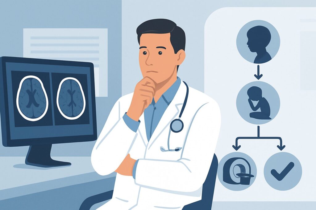
It’s a busy shift in the pediatric emergency department when you see a 6-year-old who fell off the monkey bars two hours ago. He cried immediately, had one episode of non-bilious vomiting in the car, and now has a mild headache. On exam, he has a frontal scalp hematoma but is otherwise alert, interactive, and has a normal neurologic exam. His Glasgow Coma Scale (GCS) is 15. Based on the Pediatric Emergency Care Applied Research Network (PECARN) criteria, he falls into the intermediate-risk group for a clinically important brain injury. The decision now is whether to proceed with imaging or continue observation. This article details the American College of Radiology (ACR) Appropriateness Criteria for this specific scenario, where a CT head without IV contrast is rated as May be appropriate.
Who Fits This Clinical Scenario?
This guidance applies specifically to children with minor acute blunt head trauma who are classified as having an intermediate risk for a clinically important traumatic brain injury (ciTBI) according to the validated PECARN head injury rule. Minor head trauma is defined as a history of blunt head trauma with a GCS score of 14 or 15.
For a child 2 years or older, intermediate-risk factors include:
- History of loss of consciousness
- History of vomiting
- Severe headache
- Severe mechanism of injury (e.g., fall >5 feet, high-impact collision)
For a child under 2 years of age, intermediate-risk factors include:
- Scalp hematoma (non-frontal)
- History of loss of consciousness for less than 5 seconds
- Not acting normally per parent
- Severe mechanism of injury (e.g., fall >3 feet, high-impact collision)
This workflow is distinct from other presentations. It does not apply to children at very low risk (who generally do not require imaging) or those at high risk (e.g., altered mental status, palpable skull fracture), for whom imaging is clearly indicated. It also explicitly excludes cases of suspected abusive head trauma, which follow a different diagnostic and imaging pathway.
What Diagnoses Are You Working Up in This Scenario?
In an intermediate-risk child, the primary goal of imaging is to identify or rule out a clinically important traumatic brain injury (ciTBI). A ciTBI is defined as an injury resulting in death, neurosurgery, intubation for more than 24 hours, or hospital admission for two or more nights. The specific pathologies fall into several categories.
Intracranial Hemorrhage: This is the most immediate concern. An epidural hematoma (EDH), often from a temporal bone fracture tearing the middle meningeal artery, can expand rapidly and cause life-threatening herniation. A subdural hematoma (SDH), from tearing of bridging veins, is another critical finding. While less common in this specific risk group than in high-risk patients, their potential for morbidity makes them a key target of the workup.
Cerebral Contusion: These are bruises on the brain parenchyma itself, which can lead to focal neurologic deficits, swelling, and seizures. They are a direct result of the impact and are well-visualized on cross-sectional imaging.
Skull Fractures: While a simple, linear, non-displaced skull fracture may not be clinically important on its own, identifying a fracture is crucial. Depressed skull fractures often require surgical elevation, and fractures that cross major vascular structures (like the middle meningeal artery groove or a dural venous sinus) significantly increase the risk of an associated intracranial bleed.
Why Is CT Head Without IV Contrast the Recommended Study for This Presentation?
For a child with minor head trauma and intermediate-risk features, the ACR panel rates CT head without IV contrast as May be appropriate. This rating reflects the clinical equipoise: the risk of a ciTBI is not zero, but it is low enough that the decision to image involves a careful balance with the risks of radiation exposure. Shared decision-making with the family is a key component of this workflow.
A non-contrast CT of the head is exceptionally sensitive and specific for the acute injuries you are working up. It can rapidly detect acute hemorrhage (which appears hyperdense), cerebral edema, and skull fractures. Its speed is a major advantage in the emergency setting, as it takes only minutes to perform and does not require sedation, which is often necessary for young children undergoing a longer MRI.
Alternative studies are rated lower for clear reasons in this acute context:
- Radiography (skull X-rays) is rated Usually not appropriate. While it can detect some skull fractures, it provides no information about the brain parenchyma or potential intracranial bleeding, which is the primary concern. A negative skull X-ray does not rule out a ciTBI.
- MRI head without IV contrast is also rated Usually not appropriate for the initial evaluation. While it offers superior soft-tissue detail and avoids ionizing radiation, it is slower, more expensive, less available in emergencies, and frequently requires sedation in young children, which carries its own risks. MRI is typically reserved for subacute or chronic settings or for problem-solving after an initial CT.
The radiation dose from a pediatric head CT is a significant consideration (pediatric relative radiation level ☢☢☢, 0.3-3 mSv). The decision to proceed with CT is made when the clinical suspicion for a treatable injury outweighs the small but real lifetime risk of malignancy associated with radiation. Modern CT scanners and pediatric-specific protocols are designed to minimize this dose according to the ALARA (As Low As Reasonably Achievable) principle.
Once you’ve decided on a non-contrast head CT, our protocol guide covers the technique, dose considerations, and reading principles: CT Brain Without Contrast.
What’s Next After CT Head Without Contrast? Downstream Workflow
The results of the head CT will guide the next steps in management. The decision tree is relatively straightforward.
If the CT is positive for a ciTBI: A finding such as an epidural or subdural hematoma, significant contusion, or a depressed skull fracture necessitates immediate action. The next step is an urgent consultation with pediatric neurosurgery. The child will require admission to the hospital for close neurologic monitoring, and potentially for surgical intervention depending on the size and type of injury.
If the CT is negative: A normal head CT is highly reassuring. In most cases, after a period of observation in the emergency department to ensure no clinical worsening, the child can be safely discharged home. It is critical to provide the caregivers with clear, written head injury precautions, including signs and symptoms that should prompt an immediate return to the hospital (e.g., worsening headache, repeated vomiting, changes in behavior or consciousness).
If imaging is deferred: For many intermediate-risk patients, a period of observation (typically 4-6 hours) in the emergency department is a reasonable alternative to immediate CT. If the child’s symptoms (like headache or vomiting) resolve and they return to their baseline mental status during observation, they can often be discharged without imaging. If symptoms worsen or fail to improve, a CT scan would then be performed.
Pitfalls to Avoid (and When to Get Help)
Navigating the intermediate-risk head trauma scenario requires careful clinical judgment. A primary pitfall is misclassifying the patient’s risk; be thorough in applying the PECARN criteria and do not overlook any high-risk features like altered mental status or signs of a basilar skull fracture. Another common error is ordering a CT for a very-low-risk child where observation alone is sufficient, leading to unnecessary radiation exposure.
Conversely, failing to image a child whose symptoms are subtly worsening during observation can lead to a delayed diagnosis. Always trust your clinical gestalt and have a low threshold to re-evaluate. If there is any concern for a non-accidental injury, the evaluation must be escalated to involve child protective services and a dedicated trauma workup, as this scenario explicitly excludes suspected abuse. If the CT reveals any acute intracranial finding, escalate immediately to a neurosurgeon.
Related ACR Topics and Tools
For a comprehensive overview of all clinical scenarios related to pediatric head trauma, from very low risk to severe injury, please see our parent guide. It provides a breadth of information that complements this deep-dive article.
- For breadth across all scenarios in Head Trauma-Child, see our parent guide: Head Trauma-Child: ACR Appropriateness Decoded.
- To explore other clinical presentations and their corresponding ACR recommendations, use the ACR Appropriateness Criteria Lookup.
- For detailed technical specifications on imaging studies, consult the Imaging Protocol Library.
- To help in discussions with families about radiation exposure, the Radiation Dose Calculator can be a useful tool.
Frequently Asked Questions
Why is CT only rated ‘May be appropriate’ and not ‘Usually appropriate’ for intermediate-risk children?
The ‘May be appropriate’ rating reflects the clinical balance in this specific group. The risk of a clinically important brain injury is low (estimated around 0.9% in the original PECARN study), but not zero. Therefore, the decision to use ionizing radiation via CT must be weighed against the benefit of detecting a rare but serious injury. This rating supports shared decision-making, where a clinician might choose either CT or a period of structured observation based on the specific patient and family discussion.
Is it safe to just observe these intermediate-risk patients instead of getting a CT scan?
Yes, observation is a well-established and valid alternative to immediate CT for many children in the intermediate-risk category. If, after a 4-6 hour observation period in a clinical setting, the child’s symptoms have resolved and their neurologic exam remains normal, they can often be safely discharged without imaging. The key is that any worsening of symptoms during observation should trigger an immediate CT scan.
Why not get an MRI to avoid the radiation dose from a CT scan?
While MRI avoids ionizing radiation, it is rated ‘Usually not appropriate’ for the initial evaluation of acute head trauma. MRI scans take significantly longer than CTs, are less available on an emergency basis, and often require sedation for young children to prevent motion artifact. Sedation carries its own risks. CT is much faster and is excellent at detecting the most urgent concerns: acute blood and bone fractures.
What if a child vomits more than once after the injury? Does that change the risk?
Multiple episodes of vomiting can be a sign of increasing intracranial pressure and may elevate the child’s risk. While a single episode of vomiting places a child in the intermediate-risk group, persistent or worsening vomiting during an observation period would be a strong indication to proceed with a CT scan, even if other symptoms seem stable.
What specific findings on physical exam define ‘intermediate risk’ versus ‘high risk’?
The key distinction is the severity of findings. For intermediate risk, you might see a scalp hematoma, a history of brief loss of consciousness, or parental concern about behavior. In contrast, high-risk findings are more definitive signs of severe injury, such as an altered mental status (GCS <14), signs of a basilar skull fracture (e.g., raccoon eyes, Battle's sign), or a palpable skull fracture. The presence of any high-risk feature makes imaging 'Usually appropriate' rather than 'May be appropriate'.
Reviewed by Pouyan Golshani, MD, Interventional Radiologist — May 29, 2026