Should You Order Imaging for a Complex Febrile Seizure in a Young Child?

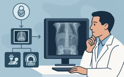
It’s 2 a.m. in the emergency department, and you’re evaluating a 2-year-old who just had a 20-minute seizure associated with a fever. The seizure started on the right side of his body before generalizing, and his parents are understandably terrified. He is now postictal but stable. While a simple febrile seizure often requires no imaging, this one had focal features and was prolonged, meeting the criteria for a complex febrile seizure. You are now faced with a critical decision: does this child need neuroimaging, and if so, which study is the right one to order? This article provides a detailed workflow for this specific scenario, guided by the American College of Radiology (ACR) Appropriateness Criteria, which rate MRI head without IV contrast as May be appropriate.
Who Fits This Clinical Scenario?
This guidance applies to a specific and well-defined patient population: children between 6 months and 5 years of age presenting with their initial complex febrile seizure.
To fit this scenario, the presentation must include a seizure in the context of a febrile illness (typically >38°C or 100.4°F) and at least one of the following “complex” features:
- Focal onset: The seizure begins in one part of the body (e.g., one arm or one side of the face).
- Prolonged duration: The seizure lasts longer than 15 minutes.
- Recurrence: More than one seizure occurs within a 24-hour period.
It is crucial to distinguish this presentation from similar but distinct clinical situations that follow different diagnostic pathways:
- Simple Febrile Seizures: This is the most common type of febrile seizure. It is generalized, lasts less than 15 minutes, and does not recur within 24 hours. For a child with a simple febrile seizure, the ACR rates all initial imaging as Usually not appropriate.
- Neonatal Seizures (0-29 days): Seizures in the first month of life have a different and more urgent differential diagnosis, often related to hypoxic-ischemic injury, inborn errors of metabolism, or congenital infections. The imaging workup is distinct.
- Afebrile Seizures: Any seizure occurring without a fever, regardless of age, falls outside this scenario and prompts a different workup for epilepsy or other non-infectious causes.
What Diagnoses Are You Working Up in This Scenario?
When a febrile seizure has complex features, the clinical concern shifts from a benign, self-limited event to the possibility of an underlying central nervous system (CNS) issue that requires urgent diagnosis and treatment. The decision to image is driven by the need to rule out these more serious conditions.
A primary concern is meningitis or encephalitis. While lumbar puncture is the gold standard for diagnosing meningitis, neuroimaging is critical for identifying complications like cerebral edema, hydrocephalus, empyema, or abscess. In encephalitis, imaging may be the first and only modality to reveal parenchymal inflammation.
A particularly feared diagnosis is Herpes Simplex Virus (HSV) encephalitis. This is a rare but treatable neurological emergency with high morbidity and mortality if diagnosis is delayed. MRI is highly sensitive for the characteristic asymmetric temporal lobe involvement, often showing cytotoxic edema on diffusion-weighted imaging (DWI) sequences long before other findings appear.
Less commonly, a complex febrile seizure can be the first manifestation of an underlying structural abnormality. A focal seizure, in particular, raises suspicion for a focal lesion like cortical dysplasia, a low-grade tumor, or a vascular malformation. The fever may have simply lowered the seizure threshold in a child with a pre-existing predisposition.
Finally, a prolonged seizure itself can cause brain injury. Imaging can help identify complications of status epilepticus, such as cortical T2 hyperintensity, cytotoxic edema, or thalamic and hippocampal changes that can have long-term prognostic implications.
Why Is MRI Head without IV Contrast Rated ‘May Be Appropriate’?
For a child with a complex febrile seizure, the ACR panel rates MRI head without IV contrast as May be appropriate. This rating reflects a nuanced clinical reality: not every child with a complex febrile seizure needs imaging, but for those who do, MRI is the superior modality. The decision to image should be based on clinical judgment, considering factors like prolonged postictal state, focal neurologic deficits, or high clinical suspicion for a CNS infection.
When imaging is indicated, MRI is the clear choice for several reasons:
- Superior Soft-Tissue Contrast: MRI is exquisitely sensitive for detecting the subtle parenchymal changes of early encephalitis, the cytotoxic edema of an ischemic insult or HSV infection, and the architectural abnormalities of cortical dysplasia. These findings are often invisible on other modalities.
- No Ionizing Radiation: This is a paramount consideration in young children. MRI uses magnetic fields and radio waves, carrying a radiation dose of 0 mSv. This avoids the cumulative risk associated with radiation exposure from CT scans.
The recommendation for a study without intravenous contrast is based on the high diagnostic yield of non-contrast sequences for the primary differential diagnoses. Diffusion-weighted imaging (DWI) and T2/FLAIR sequences are typically sufficient to identify encephalitis or seizure-related changes. Contrast is generally reserved for cases where there is a specific concern for an abscess, empyema, or enhancing tumor.
Why are alternative studies rated lower?
- CT head without IV contrast is rated Usually not appropriate. While faster and more accessible, non-contrast CT has poor sensitivity for the key diagnoses in this scenario. It can miss early encephalitis, subtle dysplasia, and the initial changes of HSV infection, providing false reassurance and delaying appropriate treatment. It also exposes the child to ionizing radiation (pediatric RRL ☢☢☢ 0.3-3 mSv).
- Ultrasound head is also rated Usually not appropriate. Its utility is severely limited in children over 6 months of age as the acoustic window provided by the anterior fontanelle begins to close. It cannot adequately visualize the brain parenchyma, especially the cortex and posterior fossa, where critical pathology may reside.
What’s Next After MRI Head without IV Contrast? Downstream Workflow
The results of the MRI will guide the subsequent clinical pathway. The workflow branches based on whether the findings are positive, negative, or indeterminate.
- If the MRI is positive: The next steps depend on the specific findings.
- Findings suggestive of HSV encephalitis (e.g., temporal or insular T2 hyperintensity/restricted diffusion): This is a neurologic emergency. Immediately initiate empiric intravenous acyclovir and perform a lumbar puncture for CSF HSV PCR analysis.
- Findings suggestive of a structural lesion (e.g., cortical thickening, abnormal gray-white differentiation): This warrants an urgent neurology consultation. The child will likely need an electroencephalogram (EEG) and may require further specialized imaging or long-term anti-epileptic drug management.
- Findings of meningitis complications (e.g., empyema, abscess): This requires immediate neurosurgical consultation in addition to aggressive antibiotic therapy.
- If the MRI is negative: A normal MRI is reassuring and significantly lowers the likelihood of a serious underlying structural or infectious cause. In a well-appearing child who has returned to their neurologic baseline, this result often supports a diagnosis of a complex febrile seizure without a sinister etiology. The next step is typically clinical observation and parental education on fever management and seizure first aid. A follow-up with their primary care physician or a pediatric neurologist may be arranged.
- If the MRI is indeterminate: Occasionally, findings may be subtle or non-specific. For example, mild diffuse edema could be from the seizure itself or early encephalitis. In these cases, the next step involves integrating the imaging findings with the full clinical picture, including CSF analysis from a lumbar puncture and potentially a follow-up MRI to assess for evolution of the findings.
Pitfalls to Avoid (and When to Get Help)
Navigating this scenario requires careful clinical judgment. Here are a few common pitfalls to avoid:
- Mistaking ‘May be appropriate’ for ‘Always order’: The ACR guidance does not recommend routine imaging for all complex febrile seizures. Reserve imaging for patients with concerning features like focal neurologic deficits, incomplete recovery, or signs of increased intracranial pressure.
- Defaulting to CT for convenience: While MRI may require sedation and more time, its superior diagnostic yield and lack of radiation make it the correct choice when imaging is necessary. Ordering a CT can lead to a missed diagnosis and unnecessary radiation exposure.
- Delaying empiric treatment: If you have a high clinical suspicion for HSV encephalitis, do not wait for the MRI result to start acyclovir. Treatment should be initiated as soon as the diagnosis is seriously considered.
If the patient has persistent, unexplained neurologic deficits, signs of elevated intracranial pressure, or is in status epilepticus, escalate care immediately by consulting with pediatric neurology and/or critical care specialists.
Related ACR Topics and Tools
For a comprehensive overview of all pediatric seizure scenarios, from simple febrile to post-traumatic, please consult our parent guide. For tools to assist in ordering the correct study and understanding the technical details, see the resources below.
- For breadth across all scenarios in Seizures-Child, see our parent guide: Seizures-Child: ACR Appropriateness Decoded.
- Imaging Appropriateness Selector — for adjacent scenarios
- Imaging Protocol Library — for technique on the recommended study
- Radiation Dose Calculator — for cumulative dose conversations
Frequently Asked Questions
Does every child with a complex febrile seizure need an MRI?
No. The American College of Radiology rates MRI as ‘May be appropriate,’ not ‘Usually appropriate.’ The decision to image should be based on clinical factors. Imaging is more strongly indicated if the child has focal neurologic deficits, has not returned to their baseline mental status, or if there is a high suspicion for a central nervous system infection.
Why is CT not recommended as a first-line study for complex febrile seizures?
CT is rated ‘Usually not appropriate’ because it has poor sensitivity for the most critical diagnoses in this scenario, such as early encephalitis (especially HSV), cortical dysplasia, or seizure-induced cytotoxic edema. A negative CT can be falsely reassuring. Furthermore, CT exposes the young child to ionizing radiation, whereas MRI does not.
What is the role of a lumbar puncture (LP) in this scenario?
A lumbar puncture is essential if there is any clinical suspicion of meningitis or encephalitis. In many cases, an LP is performed alongside imaging. The two tests are complementary: the LP diagnoses infection via CSF analysis, while the MRI identifies structural abnormalities and complications like abscess or edema. An LP should be considered in any child with a complex febrile seizure, especially if they are not fully immunized or appear ill.
If we proceed with MRI, will my young patient need sedation?
Most children between 6 months and 5 years of age cannot remain still enough to acquire high-quality MR images and will require sedation or general anesthesia. This requires coordination with the radiology department and anesthesia team and is a key logistical factor to consider when deciding to proceed with imaging.
Does one complex febrile seizure mean my child has epilepsy?
Not necessarily. While complex febrile seizures are associated with a slightly higher risk of developing epilepsy later in life compared to simple febrile seizures, the absolute risk is still low. A single complex febrile seizure does not equal a diagnosis of epilepsy. A negative MRI is reassuring, and follow-up with a pediatrician or pediatric neurologist will determine if further evaluation, like an EEG, is needed.
Reviewed by Pouyan Golshani, MD, Interventional Radiologist — May 26, 2026