What Is the Best Initial Imaging for a Child with Acute, Non-Traumatic Ataxia?
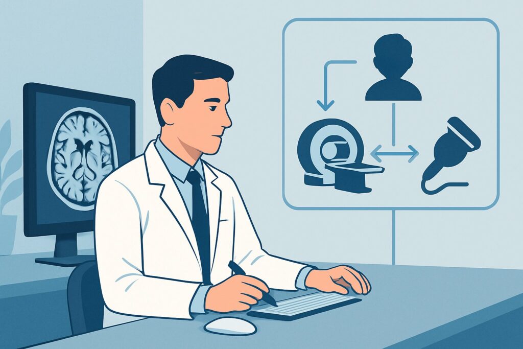
A 6-year-old is brought to the emergency department on a Tuesday evening. His parents report that he suddenly developed a “wobbly walk” and slurred speech over the past day. He hasn’t had any recent falls, injuries, or significant illness. On examination, he has a wide-based gait and clear dysmetria on finger-to-nose testing. The immediate clinical question is urgent: what is the best imaging study to evaluate a child with acute, non-traumatic ataxia? This is a high-stakes decision, as the differential includes both benign, self-limiting conditions and life-threatening emergencies. According to the American College of Radiology (ACR) Appropriateness Criteria, for this specific presentation, an MRI head without and with IV contrast is rated Usually Appropriate and is the recommended initial study.
Who Fits This Clinical Scenario for Acute Ataxia?
This clinical workflow is designed for a specific patient presentation: a child experiencing an acute onset of ataxia without any history of recent trauma. “Acute” typically means the symptoms have developed over hours to a few days. The core features are problems with coordination, such as an unsteady gait, difficulty with fine motor tasks (dysmetria), or changes in speech (dysarthria).
This guidance applies when you are ordering the initial imaging study. It is crucial to distinguish this scenario from similar but distinct clinical presentations that require different diagnostic approaches:
- Acute Ataxia with Recent Trauma: If the child has a history of a fall or head injury, the primary concern shifts towards traumatic injuries like intracranial hemorrhage, dissection, or posterior fossa contusion. This follows a different ACR variant.
- Recurrent or Episodic Ataxia: If the child has had similar episodes in the past with a return to a normal neurologic baseline in between, the differential diagnosis broadens to include metabolic disorders, channelopathies, and certain types of migraines.
- Chronic or Progressive Ataxia: If the ataxia has developed slowly over weeks, months, or years, the workup is less emergent and focuses on neurodegenerative diseases, chronic tumors, or genetic conditions.
This article focuses exclusively on the first, acute, non-traumatic presentation, where the goal of imaging is to rapidly identify or exclude urgent structural causes.
What Diagnoses Are You Working Up with Imaging in This Scenario?
The choice of imaging is driven by the need to evaluate a broad differential diagnosis, ranging from common and benign to rare and life-threatening. The primary goal is to assess the cerebellum and posterior fossa structures, the control center for balance and coordination.
A key concern driving the need for urgent imaging is a posterior fossa mass. This is the most feared diagnosis. Tumors such as medulloblastoma, ependymoma, and pilocytic astrocytoma can present with acute ataxia, sometimes complicated by obstructive hydrocephalus, which is a neurosurgical emergency. High-quality imaging is essential to rule this out promptly.
More commonly, acute ataxia in children is caused by acute post-infectious cerebellitis. This is an inflammatory condition of the cerebellum that often follows a recent viral illness. While it is typically self-limiting, imaging helps confirm the diagnosis by showing cerebellar swelling and enhancement, while also excluding a tumor that could mimic these symptoms.
Another important consideration is Acute Disseminated Encephalomyelitis (ADEM), an immune-mediated inflammatory and demyelinating condition that can affect the entire central nervous system, including the cerebellum. It is also frequently preceded by a viral infection or vaccination and presents with multifocal neurologic deficits, including ataxia.
Finally, while less common in children than adults, ischemic or hemorrhagic stroke must be considered. This can result from arterial dissection, venous sinus thrombosis, or underlying vasculopathy. Imaging is critical for detecting these vascular insults, which require specific and timely management.
Why Is MRI Head with and without IV Contrast Usually Appropriate for Acute Ataxia?
The ACR designates MRI head without and with IV contrast as Usually Appropriate because it provides the most comprehensive evaluation of the posterior fossa, directly addressing the critical differential diagnoses for this scenario.
The superior soft-tissue contrast of Magnetic Resonance Imaging (MRI) is its primary advantage over other modalities. It can clearly delineate the anatomy of the cerebellum, brainstem, and surrounding structures, making it highly sensitive for detecting tumors, inflammation, edema, and non-hemorrhagic stroke. This is a significant advantage in the posterior fossa, where bone artifact can severely limit the quality of Computed Tomography (CT) scans.
The addition of intravenous (IV) contrast is crucial. Many of the key pathologies have characteristic enhancement patterns that are essential for diagnosis. Tumors often show avid enhancement, inflammatory lesions in cerebellitis or ADEM demonstrate patchy or diffuse enhancement, and an abscess will typically rim-enhance. A non-contrast MRI could miss or mischaracterize these findings, delaying definitive diagnosis and treatment.
Comparing Alternative Studies:
- CT head without IV contrast: This study is also rated Usually Appropriate and is often used as a first-line test in emergency settings due to its speed and accessibility. It is excellent for identifying acute hemorrhage and hydrocephalus. However, its utility is limited by poor visualization of the posterior fossa (due to beam-hardening artifact) and low sensitivity for non-hemorrhagic causes like cerebellitis, ADEM, small tumors, or early ischemia. A “normal” CT can be falsely reassuring. It also involves ionizing radiation (Pediatric RRL: ☢☢☢ 0.3-3 mSv).
- CT head with IV contrast: This study is rated Usually not appropriate. The diagnostic information gained from adding IV contrast to a CT is substantially less than that provided by a contrast-enhanced MRI, while still carrying the limitations of CT in the posterior fossa and the burden of ionizing radiation. If a contrast-enhancing lesion is suspected, MRI is the definitive test.
Ultimately, MRI provides the highest diagnostic yield without using ionizing radiation (Pediatric RRL: O 0 mSv), a critical consideration in the pediatric population. While it may require sedation for younger children and takes longer to acquire, its ability to definitively evaluate for tumor, inflammation, and stroke makes it the preferred initial study in a stable patient.
What Is the Downstream Workflow After the Initial Head MRI?
The results of the head MRI will guide the subsequent clinical pathway. The downstream workflow depends on whether the findings are positive, negative, or indeterminate.
- If the MRI shows a mass: This constitutes a medical emergency. The immediate next step is an urgent consultation with pediatric neurosurgery and pediatric neuro-oncology. Further imaging, such as an MRI of the complete spine to evaluate for leptomeningeal spread (“drop metastases”), is often required for staging before any surgical intervention.
- If the MRI is negative or normal: This is a frequent and often reassuring outcome. In the absence of a structural lesion, the diagnosis frequently becomes one of exclusion, pointing towards a benign post-infectious cerebellitis (which can sometimes present with a normal MRI), a toxic ingestion, or a metabolic disturbance. The next step is close clinical observation. If the ataxia resolves over the following days or weeks, no further imaging is typically needed. If symptoms persist or worsen, the workup may transition towards that of chronic ataxia, involving a neurology consultation for potential metabolic and genetic testing.
- If the MRI suggests inflammation (cerebellitis or ADEM): A pediatric neurology consultation is warranted. Management often involves supportive care, and in more severe cases, treatments like corticosteroids or intravenous immunoglobulin (IVIG) may be considered. A lumbar puncture may be performed to analyze the cerebrospinal fluid for signs of inflammation.
- If the MRI is indeterminate or suggests a vascular cause: If a stroke, dissection, or venous thrombosis is suspected, additional vascular imaging may be necessary. An MRA head and neck or an MRV (Magnetic Resonance Venography), both rated May be appropriate, can be added to the examination to clarify vascular pathology and guide specific treatments like anticoagulation.
What Are Common Pitfalls to Avoid in This Acute Ataxia Workup?
Navigating the workup for acute pediatric ataxia requires avoiding several common pitfalls that can delay diagnosis or lead to unnecessary testing.
1. Over-reliance on a Normal CT Scan: Do not be falsely reassured by a normal non-contrast CT of the head. This study has significant limitations in visualizing the posterior fossa and can easily miss small tumors, inflammation, or non-hemorrhagic strokes. If clinical suspicion for a central cause remains high despite a normal CT, proceeding to an MRI is essential.
2. Unnecessary Delay in Imaging: While many causes of acute ataxia are benign, the possibility of a posterior fossa tumor causing obstructive hydrocephalus makes this a time-sensitive presentation. Unwarranted delays in obtaining definitive imaging can have serious consequences.
3. Forgetting the Spine in the Context of a Tumor: If the head MRI does reveal a tumor like a medulloblastoma or ependymoma, the diagnostic workup is incomplete without imaging the entire spine. These tumors have a high propensity for spreading through the cerebrospinal fluid. While not always part of the initial scan, it is a critical next step.
4. Not Preparing for Sedation: Young children often cannot remain still for the duration of an MRI scan. Failing to anticipate the need for sedation or anesthesia can lead to motion-degraded, non-diagnostic studies and further delays. Coordinate with the radiology department and anesthesia team in advance.
If a child with acute ataxia presents with altered mental status, signs of increased intracranial pressure (e.g., papilledema, vomiting, headache), or cranial nerve palsies, escalate immediately for emergent imaging and consultation with pediatric neurology and neurosurgery.
Related ACR Topics and Tools
For a comprehensive overview of all clinical scenarios related to pediatric ataxia, further reading and specialized tools can provide additional context and support for clinical decision-making.
- For breadth across all scenarios in Ataxia-Child, see our parent guide: Ataxia-Child: ACR Appropriateness Decoded.
- To explore adjacent clinical scenarios and their corresponding ACR ratings, use the ACR Appropriateness Criteria Lookup.
- For detailed technical parameters on how to perform the recommended study, consult the Imaging Protocol Library.
- To discuss radiation exposure from alternative studies like CT with families, the Radiation Dose Calculator can help frame the conversation.
Frequently Asked Questions
Why not just get a CT scan first since it’s faster?
While a CT is faster, it is significantly less sensitive for the key diagnoses in the posterior fossa, such as small tumors, inflammation from cerebellitis, or non-hemorrhagic stroke. Beam-hardening artifact often obscures detail in this area. A normal CT can be falsely reassuring, and MRI is considered the definitive initial study for a stable patient.
Is contrast always necessary for the initial MRI in this scenario?
While an ‘MRI head without IV contrast’ is also rated ‘Usually Appropriate,’ the addition of contrast is highly recommended. Contrast is essential for characterizing many of the most serious causes, including tumors, abscesses, and inflammatory lesions like ADEM. Omitting contrast can lead to a missed or delayed diagnosis, so it is best to order the study ‘without and with’ contrast unless a specific contraindication exists.
What should I do if the MRI is normal but the child is still ataxic?
A normal MRI is a common outcome and often points toward a benign, self-limiting cause like post-infectious cerebellitis (which can have a normal MRI) or a toxic ingestion. The next step is close clinical follow-up. If the ataxia persists, worsens, or recurs, the workup may shift to a different clinical scenario, prompting a neurology consultation for metabolic or genetic evaluation.
Does this imaging guidance apply if the child also has a fever?
Yes, this guidance is still appropriate. Fever can accompany infectious or post-infectious causes like cerebellitis or ADEM, which MRI is excellent at detecting. However, the presence of fever and nuchal rigidity should also raise strong suspicion for meningitis, making a lumbar puncture a critical part of the workup, typically performed after imaging has ruled out a mass effect or hydrocephalus.
When should I add spine imaging to the initial workup for acute ataxia?
An ‘MRI complete spine’ is rated ‘May be appropriate’ but is not a routine part of the initial workup for isolated ataxia. Spine imaging becomes essential as a next step if the head MRI reveals a tumor known to metastasize to the spine (like medulloblastoma or ependymoma). It should also be considered if there are clinical signs of spinal cord involvement, such as bowel/bladder dysfunction or a distinct sensory level.
Reviewed by Pouyan Golshani, MD, Interventional Radiologist — May 30, 2026