What Imaging Should You Order for a Generalized Seizure in a Neurologically Abnormal Child?
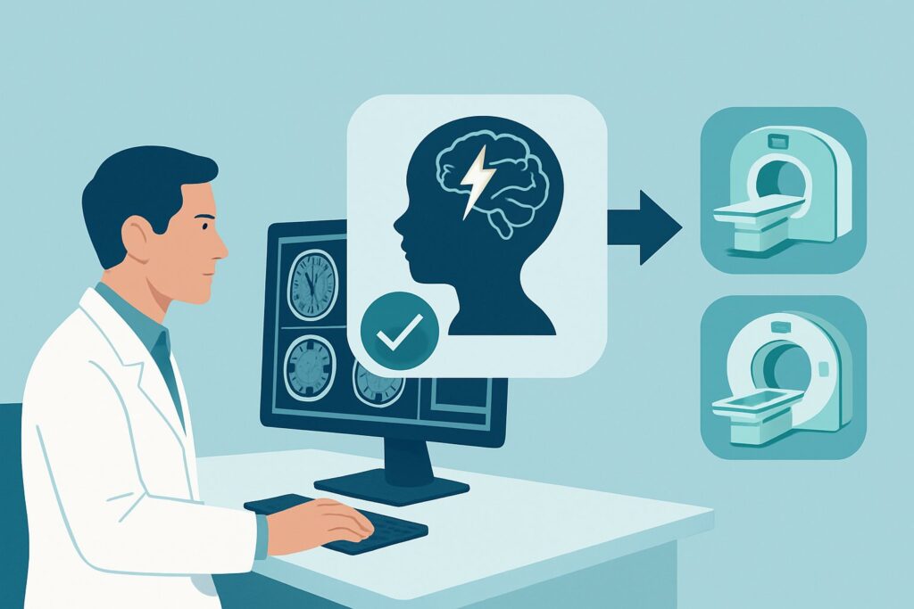
A 9-year-old with a known history of developmental delay presents to the emergency department after his first generalized tonic-clonic seizure, which lasted approximately two minutes. His postictal period is resolving, but his neurologic exam remains consistent with his abnormal baseline—no new focal deficits are present. You have stabilized the patient, and initial labs are pending. The immediate question is what, if any, neuroimaging is required for this initial workup. This scenario, a first-time generalized seizure in a child with a pre-existing neurologic abnormality, requires a specific diagnostic approach to identify potential underlying structural causes. According to the American College of Radiology (ACR) Appropriateness Criteria, the definitive initial study for this presentation is MRI head without IV contrast, which is rated as Usually Appropriate.
Who Fits This Clinical Scenario?
This guidance applies to a specific pediatric population: children aged 1 month to 17 years who present with their first generalized seizure and have a known, pre-existing abnormal neurologic examination or history. This includes patients with conditions like cerebral palsy, developmental delay, intellectual disability, or known focal deficits. The key distinction is the abnormal neurologic baseline, which raises the pre-test probability of an underlying structural brain abnormality as the cause of the seizure.
This workflow should be distinguished from several similar, but distinct, clinical situations:
- Primary Generalized Seizure (Neurologically Normal): If the same-aged child has a completely normal neurologic exam and developmental history, the diagnostic yield of imaging is lower, and the imaging recommendations differ.
- Neonatal Seizures (0 to 29 days): Seizures in the first month of life have a unique differential diagnosis (e.g., hypoxic-ischemic injury, inborn errors of metabolism), prompting a different imaging pathway.
- Febrile Seizures: Both simple and complex febrile seizures in children aged 6 months to 5 years are evaluated under separate criteria and often do not require neuroimaging.
- Post-Traumatic Seizures: Seizures occurring after a known head injury follow a trauma-focused imaging protocol, where the primary goal is to identify acute injury like hemorrhage or contusion.
Applying this article’s guidance to those other scenarios would be inappropriate.
What Diagnoses Are You Working Up in This Scenario?
In a child with a known neurologic abnormality, a new seizure prompts a search for an underlying structural epileptogenic focus. The differential diagnosis is broad, but imaging is primarily aimed at identifying conditions that require specific management or inform the patient’s long-term prognosis.
A primary consideration is a congenital malformation of cortical development. These are structural brain abnormalities that occurred during fetal development, such as focal cortical dysplasia, lissencephaly (smooth brain), polymicrogyria (too many small folds), or heterotopia (collections of gray matter in the wrong location). These conditions are highly associated with epilepsy and are often the underlying cause of the patient’s baseline neurologic deficits. MRI is exceptionally sensitive for detecting these subtle but significant findings.
Another important category includes the phakomatoses, or neurocutaneous syndromes. Conditions like tuberous sclerosis complex can present with cortical tubers and subependymal nodules, while Sturge-Weber syndrome is associated with leptomeningeal angiomatosis. These syndromes have systemic implications beyond seizures, and identifying their characteristic brain findings on MRI is crucial for diagnosis and comprehensive care.
Finally, imaging helps identify acquired structural lesions. These can include the sequelae of a remote injury, such as gliosis or encephalomalacia from a prior stroke, infection (e.g., meningitis or encephalitis), or hypoxic-ischemic event. While less common in this context, it is also critical to exclude an underlying brain tumor or a vascular malformation (like a cavernoma or arteriovenous malformation) that could present with a seizure.
Why Is MRI Head without IV Contrast the Recommended Study for This Presentation?
The ACR designates MRI head without IV contrast as Usually Appropriate because it provides the highest diagnostic yield for the most likely underlying causes in this specific clinical scenario, without exposing the child to ionizing radiation.
The superior soft-tissue contrast of Magnetic Resonance Imaging (MRI) is essential for visualizing the brain’s gray-white matter differentiation. This makes it the unparalleled modality for detecting subtle malformations of cortical development, such as focal cortical dysplasia or gray matter heterotopia, which are often invisible on Computed Tomography (CT). For identifying the tubers of tuberous sclerosis or the gliotic scars of a prior injury, MRI is substantially more sensitive than any other imaging test.
For an initial evaluation, intravenous contrast is typically not necessary. The primary differential includes non-enhancing structural abnormalities. Adding contrast does not usually increase the detection of cortical dysplasia and is reserved for cases where there is a specific concern for a tumor, active inflammation/infection, or a vascular lesion. This is reflected in the ACR rating for MRI head without and with IV contrast as May be appropriate—a reasonable alternative but not the required first step.
Let’s consider the alternatives and why they are rated lower:
- CT head without IV contrast: This study is rated May be appropriate. While fast and widely available, making it useful in an unstable patient or to rule out an acute bleed, its utility here is limited. The radiation dose (pediatric RRL ☢☢☢, 0.3-3 mSv) is a significant consideration in children. More importantly, a normal CT scan does not adequately rule out the key epileptogenic lesions this workup is designed to find. It can easily miss cortical dysplasia or small, low-grade tumors.
- US head: Rated Usually not appropriate, head ultrasound is only useful in infants with an open anterior fontanelle. For the majority of the age range covered in this scenario (1 month to 17 years), the fontanelle is closed, rendering ultrasound diagnostically inadequate for evaluating the brain parenchyma.
The choice of non-contrast MRI prioritizes diagnostic accuracy for the most probable causes while adhering to the principle of using non-radiation-emitting modalities in children whenever possible (MRI RRL O, 0 mSv).
What’s Next After MRI Head without IV Contrast? Downstream Workflow
The results of the MRI will significantly guide the subsequent clinical pathway, in close collaboration with a pediatric neurologist.
- If the MRI is positive for a structural lesion (e.g., cortical dysplasia, tumor, or vascular malformation): This finding provides a definitive etiology for the seizures. It allows for more targeted anti-epileptic drug therapy and informs the prognosis. For certain lesions, particularly focal cortical dysplasia or a low-grade tumor, the patient may be referred for an epilepsy surgery evaluation to determine if resecting the lesion could lead to seizure freedom.
- If the MRI is negative: A normal MRI is a very common outcome and does not mean the patient does not have epilepsy. Epilepsy is a clinical diagnosis based on the occurrence of seizures, often supported by electroencephalogram (EEG) findings. A negative MRI simply rules out a major underlying structural cause. Management will then be guided by the clinical seizure type and EEG results. In cases of medically refractory epilepsy despite a normal initial MRI, a follow-up study using a high-field-strength magnet (e.g., 3T) with a dedicated epilepsy protocol may be considered.
- If the MRI is indeterminate: Sometimes, a finding may be non-specific, such as a small area of abnormal signal in the white matter. In these cases, discussion with a neuroradiologist is critical. The next step might be to perform a follow-up MRI with contrast or to obtain advanced imaging sequences. The decision will be based on integrating the imaging finding with the patient’s specific clinical and EEG data.
Pitfalls to Avoid (and When to Get Help)
Navigating this workup requires careful attention to the specific clinical context to avoid common errors.
- Misclassifying the patient: Do not apply this guidance to a child with a normal neurologic exam or a simple febrile seizure, where the risk-benefit balance of imaging is different.
- Accepting CT as definitive: While a CT may be obtained in the emergency setting for expediency, do not consider a negative CT sufficient to rule out an underlying structural cause. The patient still requires the definitive MRI.
- Failing to plan for sedation: A high-quality MRI requires a motionless patient. Most young children and many older children with developmental delays will require sedation or general anesthesia. Plan for this in advance to avoid a non-diagnostic, motion-degraded study.
- Ignoring urgent red flags: If the child presents in status epilepticus, has a new and persistent focal neurologic deficit, or shows signs of increased intracranial pressure, the workup must be expedited. In these emergent situations, a non-contrast head CT is often the appropriate first step to rapidly assess for hemorrhage, hydrocephalus, or a large mass while arranging for the more definitive MRI.
Related ACR Topics and Tools
For a comprehensive overview of all pediatric seizure scenarios, further reading on imaging techniques, or to discuss radiation dose with families, the following resources are valuable.
- For breadth across all scenarios in Seizures-Child, see our parent guide: Seizures-Child: ACR Appropriateness Decoded.
- Imaging Appropriateness Selector — for adjacent scenarios
- Imaging Protocol Library — for technique on the recommended study
- Radiation Dose Calculator — for cumulative dose conversations
Frequently Asked Questions
Why not just get a CT scan in the emergency department since it’s faster?
A CT scan is appropriate in an emergency to quickly rule out life-threatening conditions like a large bleed, hydrocephalus, or a significant mass. However, for a stable child with a first-time seizure and a known neurologic abnormality, the primary goal is to find a subtle underlying structural cause like cortical dysplasia. CT has very low sensitivity for these findings. An MRI is the definitive diagnostic study, and a normal CT should not prevent a patient from getting a subsequent MRI.
What if my patient cannot hold still for an MRI scan?
This is a critical practical consideration. A motion-degraded MRI is often non-diagnostic and a waste of resources. For young children or any child who cannot cooperate, the MRI should be scheduled with sedation or general anesthesia. This requires coordination with the radiology department and anesthesia team but is essential for obtaining high-quality, interpretable images.
Does a normal MRI mean my patient doesn’t have epilepsy?
No. Epilepsy is a clinical diagnosis defined by the predisposition to have unprovoked seizures. An MRI is performed to look for a structural cause or ‘lesion’ that is triggering the seizures. Many forms of epilepsy, including genetic and metabolic types, are not associated with any visible abnormality on an MRI. A normal MRI is reassuring but does not change the diagnosis of epilepsy.
Is intravenous contrast ever needed for this type of seizure workup?
Yes, but not always as the first step. The ACR rates MRI with and without contrast as ‘May be appropriate.’ Contrast is typically added if the non-contrast images show a finding suspicious for a tumor, an active infection/inflammatory process, or a certain type of vascular malformation. The decision to add contrast is often made by the radiologist at the time of the scan or based on specific clinical red flags.
How does this workup differ from imaging a child with a focal seizure?
The choice of imaging modality—MRI head without contrast—is the same for both focal and generalized seizures in a neurologically abnormal child. However, the pre-test probability of finding a focal structural lesion is even higher in a patient presenting with a focal seizure. This makes imaging arguably more urgent and essential in the workup of focal seizures, as the findings are very likely to directly guide management and potential surgical planning.
Reviewed by Pouyan Golshani, MD, Interventional Radiologist — May 26, 2026