What Imaging Should You Order for Significant Knee Trauma Like an MVA or Dislocation?
A 34-year-old is brought into the emergency department after a high-speed motor vehicle accident. Their left knee is visibly deformed and exquisitely tender. As the primary clinician, you need to rapidly assess for fracture, dislocation, and the dreaded complication of vascular injury. The immediate decision is which imaging study to order first to guide management without delay. This article provides a step-by-step workflow for this specific high-acuity scenario, based on the American College of Radiology (ACR) Appropriateness Criteria. For this presentation, the ACR rates both Radiography of the knee and Computed Tomography Angiography (CTA) of the lower extremity as Usually appropriate, reflecting the dual priorities of assessing bony and vascular structures.
Who Fits This Clinical Scenario for Significant Knee Trauma?
This guidance applies to a specific, high-energy patient presentation: an adult or child aged 5 years or older who has sustained significant trauma to the knee. The mechanism of injury is a key differentiator. This includes events like a motor vehicle accident (MVA), a fall from a significant height, a direct high-impact blow to the knee, or a knee dislocation, which may have been reduced in the field or may still be present on arrival.
This workflow is distinct from those for lower-energy injuries. It is crucial not to apply this guidance to patients with different presentations, such as:
- Fall or twisting trauma with no focal tenderness or effusion: These patients often do not require imaging and can be managed based on clinical decision rules like the Ottawa Knee Rules.
- Fall or twisting trauma with focal tenderness: This common presentation follows a different diagnostic algorithm, typically starting with radiographs to rule out a simple fracture.
- Suspected occult fracture after normal radiographs: In cases where initial X-rays are negative but clinical suspicion for a fracture remains high, the next step is often Magnetic Resonance Imaging (MRI), particularly in skeletally immature patients.
The defining feature of the current scenario is the high-energy mechanism, which raises the clinical suspicion for complex fractures, dislocation, and limb-threatening vascular compromise, demanding a more urgent and potentially more comprehensive imaging strategy.
What Diagnoses Are You Working Up in This High-Energy Scenario?
In the setting of significant knee trauma, the differential diagnosis extends beyond a simple fracture to include severe, time-sensitive injuries. The initial imaging choice is driven by the need to rapidly identify or exclude these conditions.
Knee Dislocation: This is a true orthopedic emergency. Even if the knee has spontaneously reduced and appears aligned, the injury to the supporting ligaments is severe. The primary concern with dislocation is not the bony alignment itself, which can be corrected, but the high incidence of associated popliteal artery injury.
Complex Fractures: High-energy mechanisms can cause severe fracture patterns that are not always seen with simple falls. These include tibial plateau fractures, distal femur fractures (especially supracondylar or intercondylar), and patellar fractures. These often require surgical fixation, and detailed imaging is necessary for preoperative planning.
Popliteal Artery Injury: This is the most feared complication of a knee dislocation, occurring in a substantial number of cases. The popliteal artery is tethered both proximally and distally, making it susceptible to traction injury and transection during a dislocation. A missed vascular injury can lead to limb ischemia and amputation.
Compartment Syndrome: While a clinical diagnosis, compartment syndrome is often precipitated by a high-energy fracture (like a tibial plateau fracture) causing bleeding and swelling within the fascial compartments of the lower leg. Imaging helps identify the inciting injury that places the patient at high risk.
Why Are Both Radiography and CTA Considered the Recommended Studies?
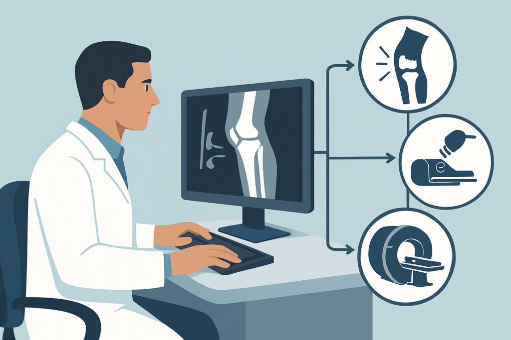
For significant knee trauma, the ACR designates two studies as Usually appropriate: Radiography of the knee and CTA of the lower extremity with IV contrast. This dual recommendation reflects the need to evaluate two distinct but equally critical organ systems: the bones and the blood vessels. The optimal workflow often involves a rapid radiograph followed by a CTA if indicated by the mechanism or physical exam.
Radiography knee is the essential first step. It is fast, widely available, and uses a very low radiation dose (☢ <0.1 mSv for adults). Radiographs are excellent for identifying fractures and confirming the presence and direction of a dislocation. Anteroposterior (AP) and lateral views are standard. These initial images guide immediate management, such as the need for urgent reduction of a dislocation.
CTA lower extremity with IV contrast is also rated Usually appropriate because of the high risk of popliteal artery injury, especially with a knee dislocation. CTA provides a detailed, non-invasive assessment of the arterial system from the aorta to the ankle, identifying intimal tears, thrombosis, or transection. While it involves a higher radiation dose (☢☢☢ 1-10 mSv for adults) and IV contrast, its ability to definitively rule out a limb-threatening vascular injury is paramount in this clinical context.
Alternative studies are rated lower for specific reasons in this initial, acute setting:
- MRI knee without IV contrast is rated May be appropriate. While it is the best test for evaluating ligaments, menisci, and cartilage, it is not the ideal first-line study in a high-energy trauma setting. It is slower to acquire, less sensitive for certain fracture patterns than CT, and does not assess for acute vascular injury. It is often used as a second-line study after acute bony and vascular injuries have been addressed.
- US knee is rated Usually not appropriate. Ultrasound is limited in its ability to visualize bone and deep structures of the knee. While it can be used for a focused vascular assessment, it is operator-dependent and not as comprehensive or reliable as CTA for ruling out popliteal artery injury in the setting of high-energy trauma.
What’s Next After Initial Imaging? Downstream Workflow
The results of the initial radiographs and potential CTA will dictate the subsequent clinical pathway. The workflow is a decision tree based on identifying and managing the most severe injuries first.
If radiographs show a knee dislocation: The immediate next step is urgent reduction, followed by post-reduction radiographs to confirm alignment. After reduction, the limb should be splinted and a thorough neurovascular exam performed. Given the high association with vascular injury, most protocols mandate a CTA of the lower extremity to definitively rule out a popliteal artery injury, even if distal pulses are palpable. An Ankle-Brachial Index (ABI) may be performed, with a value less than 0.9 also triggering a CTA.
If radiographs show a complex fracture (e.g., tibial plateau): A CT knee without IV contrast (rated May be appropriate) is typically the next step. This provides the detailed bony anatomy, including the degree of articular depression and comminution, that orthopedic surgeons need for preoperative planning.
If radiographs are negative but clinical suspicion is high: If the initial X-rays show no fracture or dislocation but the patient has hard or soft signs of a vascular injury (diminished pulses, expanding hematoma, nerve deficit), proceed directly to CTA. If vascular injury is ruled out and suspicion for a significant soft tissue (ligamentous) injury remains, an MRI knee without IV contrast (May be appropriate) can be considered, often on a less urgent basis or as an outpatient.
Pitfalls to Avoid (and When to Get Help)
In this high-stakes clinical scenario, several common pitfalls can lead to poor outcomes. Awareness of these issues is key to effective management.
- Assuming a normal pulse rules out vascular injury: A palpable distal pulse or a “warm foot” does not exclude a significant popliteal artery intimal tear that can later thrombose. Maintain a very high index of suspicion for vascular injury in all knee dislocations.
- Delaying reduction to obtain imaging: A persistent dislocation is a medical emergency. If imaging is not immediately available, reduction should not be delayed. Obtain pre-reduction films if possible without causing delay, but always obtain post-reduction films.
- Failing to assess for compartment syndrome: After a severe fracture, especially of the tibial plateau, frequent re-assessment for the “5 Ps” of compartment syndrome is critical. A rising compartment pressure is a surgical emergency.
- Underestimating pediatric injuries: In children, be vigilant for physeal (growth plate) fractures, such as a Salter-Harris fracture, which can have long-term growth implications if missed or improperly managed.
If there are any hard signs of arterial injury (e.g., absent pulses, rapidly expanding hematoma, pulsatile bleeding), an immediate consultation with vascular surgery is warranted, sometimes even before advanced imaging is completed.
Related ACR Topics and Tools
For a comprehensive overview of all clinical variants related to acute knee trauma, from low-energy twists to post-operative complications, please see our parent guide. It provides a hub-and-spoke model to help you navigate to the specific scenario that matches your patient.
- For breadth across all scenarios in Acute Trauma to the Knee, see our parent guide: Acute Trauma to the Knee: ACR Appropriateness Decoded.
- To explore adjacent clinical scenarios, use the Imaging Appropriateness Selector.
- For details on imaging techniques, consult the Imaging Protocol Library.
- To discuss radiation exposure with patients, the Radiation Dose Calculator can help frame the conversation.
Frequently Asked Questions
Why are both X-ray and CTA listed as ‘Usually Appropriate’ for significant knee trauma?
They evaluate for two different, equally critical emergencies. Radiography (X-ray) is the fastest and best initial test for bony injuries like fractures and dislocations. CTA is the definitive test for the associated high-risk vascular injury, particularly popliteal artery damage after a knee dislocation. The typical workflow is X-ray first, followed by CTA if a dislocation is found or if there are clinical signs of vascular compromise.
If a knee dislocation was reduced in the field, do I still need an X-ray first?
Yes. An X-ray is still essential to rule out associated fractures that may have occurred during the injury or even during the reduction. Management of a fracture-dislocation is different from that of a pure ligamentous dislocation. After the X-ray, a CTA is still strongly recommended to rule out vascular injury.
Is an Ankle-Brachial Index (ABI) a reliable substitute for CTA after knee dislocation?
While an ABI is a useful tool, its role as a standalone screening test is debated. An ABI of less than 0.9 is a strong indicator for further vascular imaging. However, some significant arterial injuries (like an intimal flap) can be present even with a normal ABI. For this reason, many trauma centers and society guidelines recommend CTA for all knee dislocations, regardless of the initial ABI value.
When should I order an MRI in this high-trauma setting?
MRI is typically a second-line or delayed imaging modality in this scenario. Its primary role is to evaluate for soft tissue injuries like ligament tears (ACL, PCL, collateral), meniscal tears, and cartilage damage. These are rarely surgical emergencies. The priority is to first rule out limb-threatening fracture-dislocations and vascular injuries with X-ray and CTA. An MRI is often ordered days later, once the patient is stable, to plan for future ligamentous reconstruction.
How does this scenario differ from the one for a known tibial plateau fracture on radiographs?
This scenario covers the initial imaging for a patient presenting with high-energy trauma where the specific diagnosis is not yet known. The other scenario, ‘Tibial plateau fracture on radiographs,’ addresses the next step in imaging once that specific fracture has already been identified on an X-ray. In that case, the primary question is about surgical planning, making a non-contrast CT the most appropriate next study.
Reviewed by Pouyan Golshani, MD, Interventional Radiologist — May 26, 2026