What Imaging Should You Order for Unilateral Vocal Cord or Palatal Paralysis (CN X Palsy)?
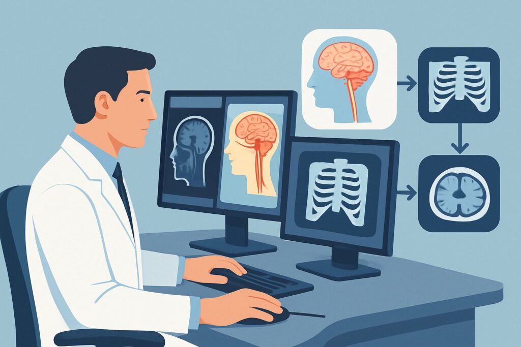
A 58-year-old patient presents to your clinic with a two-month history of progressive hoarseness and occasional choking on liquids. A flexible laryngoscopy performed by an otolaryngologist confirms left vocal cord paralysis. There is no history of recent surgery, trauma, or infection. You are now faced with the critical task of identifying the underlying cause of this isolated vagal nerve (Cranial Nerve X) palsy. The nerve’s long, tortuous path from the brainstem to the larynx means the lesion could be anywhere from the skull base to the chest. This article provides a clinical workflow for this specific scenario, explaining why the American College of Radiology (ACR) finds MRI head without and with IV contrast to be a Usually Appropriate initial imaging study.
Who Fits This Clinical Scenario for Vagal Nerve Palsy?
This guidance is for patients presenting with a new, unilateral, and isolated paralysis of the palate or a vocal cord (or both), where the clinical suspicion is a non-traumatic, non-iatrogenic lesion of the vagus nerve (CN X). The key term is “isolated.” The workup described here assumes the patient’s neurologic examination is otherwise normal, with no other cranial nerve deficits, long tract signs, or cerebellar findings.
This workflow is NOT intended for:
- Patients with a clear iatrogenic cause: For example, vocal cord paralysis that develops immediately following thyroid, carotid, or cardiothoracic surgery.
- Patients with multiple cranial nerve palsies: If the patient also presents with weakness of the sternocleidomastoid muscle (CN XI) or tongue deviation (CN XII), this suggests a more complex skull base lesion (e.g., jugular foramen syndrome) and may require a different imaging strategy.
- Patients with oropharyngeal dysphagia as the primary symptom: While CN X is involved, a primary complaint of dysphagia without confirmed vocal cord paralysis may point toward a glossopharyngeal nerve (CN IX) issue, which is a distinct ACR scenario.
The focus here is on identifying an unknown primary cause for an isolated CN X deficit.
What Diagnoses Are You Working Up in This Scenario?
The vagus nerve has the longest course of any cranial nerve, originating in the medulla oblongata and descending through the neck into the thorax. A lesion anywhere along this path can cause vocal cord paralysis. The imaging workup is designed to interrogate this entire length to identify the point of compression or injury. The differential diagnosis is broad and categorized by location.
Intracranial and Skull Base Lesions: Pathologies at the nerve’s origin or as it exits the skull are a primary concern. This includes brainstem strokes, demyelinating plaques (e.g., multiple sclerosis), or tumors like schwannomas, meningiomas, or paragangliomas (e.g., glomus jugulare tumors) located at the jugular foramen.
Head and Neck Lesions: As the nerve descends through the carotid space in the neck, it is vulnerable to compression from primary neck cancers (e.g., squamous cell carcinoma), metastatic lymph nodes, thyroid malignancies, or carotid artery aneurysms or dissections. Benign nerve sheath tumors like vagal schwannomas can also arise here.
Chest and Mediastinal Lesions: This is a critical and often-overlooked area, particularly for left-sided vocal cord paralysis. The left recurrent laryngeal nerve, a branch of the vagus, loops under the aortic arch, making it susceptible to compression from lung apex tumors (Pancoast tumors), mediastinal lymphadenopathy (from lymphoma or metastatic disease), or aortic arch aneurysms. This is the basis for the classic “hoarseness as a sign of lung cancer” teaching.
Why Is MRI of the Head and Neck the Recommended Approach?
For an initial workup of isolated, unilateral vagal nerve palsy, the ACR designates both MRI head without and with IV contrast and MRI orbits face neck without and with IV contrast as Usually Appropriate. The choice between them depends on the highest clinical suspicion, but both offer superior soft-tissue contrast for visualizing the nerve and surrounding structures compared to other modalities.
The rationale for MRI is its exceptional ability to evaluate the entire potential course of the nerve from the brainstem to the thoracic inlet. Intravenous contrast is essential for highlighting abnormal enhancement in tumors, inflammatory processes, or areas of nerve enhancement (neuritis). MRI can clearly delineate skull base foramina, identify subtle brainstem lesions, and characterize masses within the soft tissues of the neck.
While MRI is the preferred starting point, other studies are also rated for this scenario:
- CT neck with IV contrast is also rated Usually Appropriate. It is an excellent alternative, especially if MRI is contraindicated or unavailable. CT provides superior bone detail of the skull base and is faster to acquire. It is particularly effective for identifying pathology in the neck and upper mediastinum. However, it involves ionizing radiation (1-10 mSv) and is less sensitive for subtle intracranial pathology like small brainstem strokes or demyelination.
- CT chest with IV contrast is rated May be appropriate. This study becomes essential if the initial head and neck imaging is negative, as the cause may lie in the mediastinum, especially for left-sided palsies. Some institutions proceed directly to a combined CT of the neck and chest to cover the entire course in one study.
- MRA head and neck and US neck are both rated Usually not appropriate for the initial workup. While MRA is useful for vascular pathology like aneurysms, it is not the optimal screening tool for the broad differential. Ultrasound has a limited role as it cannot visualize the intracranial, skull base, or mediastinal segments of the nerve.
What’s Next After Imaging? Downstream Workflow
The results of the initial imaging study will guide the subsequent clinical pathway. The goal is to find a treatable cause and provide a prognosis.
If the study is positive for a mass: A definitive finding, such as a skull base tumor, neck mass, or apical lung lesion, dictates the next steps. This typically involves referral to the appropriate subspecialist—neurosurgery for intracranial lesions, otolaryngology–head and neck surgery for neck masses, or thoracic surgery/oncology for chest pathology. A biopsy is often required to establish a histologic diagnosis and guide treatment planning.
If the study is negative: A negative high-quality MRI or CT from the skull base through the upper chest is reassuring but does not end the workup. The next step is often to obtain a dedicated CT chest with IV contrast (if not already performed) to fully evaluate the mediastinum and aortopulmonary window, which is a common site for pathology affecting the left recurrent laryngeal nerve. If the entire course of the nerve is imaged and found to be normal, the paralysis is deemed idiopathic. In these cases, management focuses on voice therapy and potential surgical interventions (e.g., vocal cord injection or medialization) to improve voice quality and prevent aspiration.
If the study is indeterminate: Ambiguous findings, such as subtle nerve enhancement or non-specific soft tissue thickening, may require further investigation. This could involve a follow-up scan to assess for change over time, a different imaging modality (e.g., PET/CT if malignancy is suspected), or direct laryngoscopy with palpation of the post-cricoid region under anesthesia.
Pitfalls to Avoid (and When to Get Help)
In the workup of vocal cord paralysis, several common pitfalls can delay diagnosis. First, failing to image the entire course of the vagus nerve from the brainstem to the aortic arch is a frequent error. A negative MRI of the head or neck does not rule out a thoracic cause. Second, omitting intravenous contrast significantly reduces the sensitivity of both MRI and CT for detecting neoplastic and inflammatory causes. Finally, attributing new-onset hoarseness in a high-risk patient (e.g., a smoker) to laryngitis without performing laryngoscopy can miss an underlying paralysis and the serious pathology it may represent. If the clinical picture is complex or imaging findings are unclear, consultation with neuroradiology, otolaryngology, or neurology is essential to ensure a comprehensive and accurate diagnostic plan.
Related ACR Topics and Tools
The ACR Appropriateness Criteria are extensive, covering thousands of clinical scenarios. For breadth across all variants in Cranial Neuropathy, see our parent guide: Cranial Neuropathy: ACR Appropriateness Decoded. For additional decision support, the following tools can help refine your imaging orders and patient conversations.
- ACR Appropriateness Criteria Lookup — for adjacent scenarios
- Imaging Protocol Library — for technique on the recommended study
- Radiation Dose Calculator — for cumulative dose conversations
Frequently Asked Questions
Why is a chest X-ray rated ‘Usually not appropriate’ if a lung tumor is on the differential?
While a chest radiograph can sometimes detect a large apical lung mass or significant mediastinal widening, it has low sensitivity for the subtle pathology that can cause vagal nerve palsy. A normal chest X-ray does not rule out a small tumor, adenopathy, or an aortic aneurysm compressing the recurrent laryngeal nerve. A contrast-enhanced CT of the chest is far more sensitive and is the appropriate study to evaluate the thoracic course of the nerve if initial head and neck imaging is negative.
My patient has a left vocal cord paralysis. Should I just order a CT of the chest?
While the cause is often in the chest for left-sided palsies, starting with only a chest CT would miss important pathology in the brainstem, skull base, and neck. The most life-threatening diagnoses (e.g., brainstem stroke, skull base malignancy) are located intracranially or in the head/neck. Therefore, the standard of care is to image the entire nerve pathway. A common approach is to start with an MRI or CT of the head and neck, and if negative, proceed to a CT of the chest.
Is there a difference in the workup for right-sided versus left-sided vocal cord paralysis?
Yes, slightly. The right recurrent laryngeal nerve has a shorter course, looping under the right subclavian artery in the root of the neck. It does not enter the mediastinum in the same way the left nerve does. Therefore, a right-sided paralysis is less likely to be caused by deep mediastinal pathology. However, the initial workup is the same, as intracranial, skull base, and neck lesions can affect either side. If initial imaging of the head and neck is negative, the focus for a right-sided palsy remains on the thoracic inlet and apex of the right lung.
If my patient has a pacemaker, can they still get an MRI for this workup?
Many modern pacemakers and implantable cardiac devices are MRI-conditional, meaning they are safe for MRI under specific protocols. However, this requires careful coordination with the radiology department and cardiology. The device must be checked and put into a safe ‘MRI mode’ before the scan and reprogrammed afterward. If the patient’s device is not MRI-safe or if these resources are not available, a contrast-enhanced CT of the neck and chest is an excellent alternative and is also rated ‘Usually Appropriate’ by the ACR for the neck portion.
What if the patient’s only symptom is palatal paralysis without vocal cord involvement?
Isolated paralysis of the soft palate is also a sign of a vagus nerve (CN X) lesion, as the levator veli palatini muscle is innervated by the pharyngeal branch of the vagus. The workup is identical to that for vocal cord paralysis because the lesion can still be located anywhere from the brainstem to the high cervical region, before the recurrent laryngeal nerve branches off. The same differential diagnoses apply, and the imaging approach should cover the intracranial, skull base, and cervical portions of the nerve.
Reviewed by Pouyan Golshani, MD, Interventional Radiologist — June 27, 2026