Which Imaging Is Best for Congenital Hearing Loss or Cochlear Implant Planning?
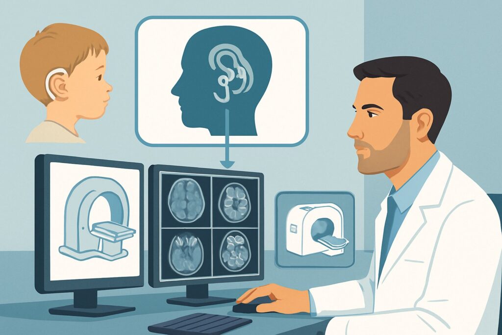
A 15-month-old presents to your otolaryngology clinic with profound bilateral sensorineural hearing loss, confirmed on auditory brainstem response testing. The family is motivated, and the child is an excellent candidate for cochlear implantation. Before scheduling surgery, you need to assess the underlying anatomy of the inner ear and the status of the vestibulocochlear nerves. The critical question is which imaging study provides the necessary detail to ensure a successful outcome and identify any potential contraindications or surgical challenges. This article details the American College of Radiology (ACR) recommended imaging workflow for this specific scenario: congenital hearing loss or evaluation for cochlear implantation. For this presentation, the ACR rates `MRI head and internal auditory canal without and with IV contrast` as Usually Appropriate.
Who Fits This Clinical Scenario?
This guidance applies to a specific patient population being evaluated for auditory rehabilitation with a cochlear implant. The key factor is the need for detailed anatomical information for surgical planning.
Inclusion criteria for this workflow:
- Infants and children with congenital sensorineural hearing loss (SNHL) who are candidates for cochlear implantation.
- Adults with acquired profound SNHL or total deafness who are being evaluated for a cochlear implant.
- Patients with known inner ear malformations where implantation is being considered, requiring a detailed preoperative map.
It is crucial to distinguish this scenario from other presentations of hearing loss or vertigo, which follow different diagnostic pathways.
Exclusion criteria (patients who fit a different ACR variant):
- Acquired, non-profound SNHL: A patient with new-onset, asymmetric, or progressive hearing loss who is not yet a candidate for implantation fits the Acquired sensorineural hearing loss variant, which focuses on identifying a cause like a vestibular schwannoma.
- Conductive Hearing Loss: A patient with hearing loss due to problems in the outer or middle ear (e.g., otosclerosis, ossicular chain disruption) follows the Acquired conductive hearing loss pathway.
- Primary Symptom is Vertigo: If the dominant clinical feature is dizziness or imbalance, the workup aligns with the Episodic vertigo or Persistent vertigo scenarios.
What Diagnoses Are You Working Up in This Scenario?
For cochlear implant candidates, imaging is not primarily for diagnosing the cause of hearing loss but for assessing anatomical feasibility and guiding the surgical approach. The key questions are structural.
Cochlear Nerve Status: The most critical finding is the presence and caliber of the cochlear nerve. A cochlear implant stimulates this nerve directly. If the nerve is absent (aplasia) or severely underdeveloped (hypoplasia), the implant will fail. MRI is the only modality that can reliably visualize the nerve within the internal auditory canal.
Inner Ear Malformations: A wide spectrum of congenital anomalies can affect the cochlea and vestibular system. These include Mondini dysplasia (an incomplete cochlea), common cavity deformity (undifferentiated cochlea and vestibule), and enlarged vestibular aqueduct. Identifying these malformations is essential as they can alter the surgical technique and increase the risk of complications, such as a “gusher” of cerebrospinal fluid during surgery.
Cochlear Patency: The cochlea must be a fluid-filled space to allow for the insertion of the implant’s electrode array. In some cases, particularly following bacterial meningitis, the cochlea can become filled with fibrous tissue or new bone (labyrinthitis ossificans). This obliteration can make electrode insertion difficult or impossible and must be identified preoperatively.
Anatomic Variants: Imaging also maps the precise location of critical structures the surgeon must avoid, such as an aberrant course of the facial nerve or a high-riding jugular bulb. This information is vital for preventing iatrogenic injury.
Why MRI Head and Internal Auditory Canal Is the Recommended Study for This Presentation
The ACR designates three studies as Usually Appropriate for this scenario, with MRI being central to answering the most critical questions about the cochlear nerve and fluid spaces.
MRI head and internal auditory canal without and with IV contrast is rated Usually Appropriate. Its strength is unparalleled soft-tissue and fluid visualization. High-resolution T2-weighted sequences (e.g., CISS, FIESTA) are the workhorse of the exam, providing clear depiction of the four distinct nerves within the internal auditory canal and the fluid signal within the cochlea and semicircular canals. This directly assesses cochlear nerve presence and cochlear patency. The addition of IV contrast can help identify active inflammation or enhancement of the vestibulocochlear nerve, though it is not always necessary for purely anatomical planning.
MRI head and internal auditory canal without IV contrast is also rated Usually Appropriate. For many congenital cases, a non-contrast study is sufficient to answer the key surgical questions. Omitting contrast is often preferred in the pediatric population to avoid the risks associated with IV access and contrast administration.
CT temporal bone without IV contrast is the third Usually Appropriate study. While MRI excels at soft tissue, CT is superior for delineating fine bony anatomy. It provides a precise map of the otic capsule, mastoid air cell development, the round window, and the bony course of the facial nerve canal. For this reason, many implant centers obtain both an MRI and a non-contrast CT to have a complete picture. CT does involve ionizing radiation (ACR Relative Radiation Level ☢☢☢ 1-10 mSv), a key consideration in pediatric patients.
Why Alternative Studies Are Rated Lower
- CT temporal bone with IV contrast is rated Usually not appropriate. The addition of IV contrast provides no significant benefit for assessing the bony structures relevant to implant surgery and adds unnecessary risk and radiation dose.
- MRA head or MRV head are rated Usually not appropriate. These are vascular studies designed to evaluate arteries and veins, respectively. Their protocols are not optimized for the high-resolution anatomical detail required to assess the inner ear structures and cochlear nerve.
What’s Next After MRI? Downstream Workflow
The imaging results directly shape the subsequent clinical and surgical pathway. The radiologist’s report should be reviewed in detail with the surgical team.
- Favorable Findings: If MRI confirms the presence of a normal-caliber cochlear nerve and a patent, fluid-filled cochlea, and CT (if performed) shows no prohibitive bony anomalies, the patient is an excellent anatomical candidate. The next step is to proceed with surgical scheduling and device selection.
- Cochlear Nerve Aplasia: If the cochlear nerve is definitively absent on high-resolution MRI, a cochlear implant is contraindicated as there is no neural structure to stimulate. The conversation then shifts to alternative technologies, such as an auditory brainstem implant (ABI), which requires a different surgical approach (typically via neurosurgery) and has different outcome expectations.
- Cochlear Obliteration: If imaging reveals labyrinthitis ossificans, the surgeon is forewarned. They may plan for a more complex procedure involving drilling out the obliterated portion of the cochlea or select a specialized electrode array designed for these cases. In severe cases, implantation in that ear may be deemed impossible.
- Anatomic Malformations: The specific type of malformation dictates the plan. For example, an enlarged vestibular aqueduct alerts the surgeon to a higher risk of a perilymph gusher, prompting specific intraoperative maneuvers to mitigate it. The choice of electrode may also be tailored to the malformed cochlea’s dimensions.
Pitfalls to Avoid (and When to Get Help)
Navigating the imaging workup for cochlear implant candidates requires careful attention to detail to avoid common errors.
- Pitfall: Ordering a routine “MRI Brain.” A standard brain MRI protocol lacks the thin, high-resolution sequences focused on the internal auditory canals and will fail to adequately visualize the cochlear nerve. The order must specifically request evaluation of the internal auditory canals/cranial nerve VIII.
- Pitfall: Relying solely on CT. While excellent for bone, a non-contrast CT of the temporal bones cannot confirm the presence or absence of the cochlear nerve. A decision to implant based on a normal CT alone risks surgical failure if the nerve is aplastic.
- Pitfall: Underestimating sedation needs. In infants and young children, MRI and CT require the patient to be perfectly still. This almost always necessitates coordination with pediatric anesthesiology for deep sedation or general anesthesia, which should be planned well in advance.
If imaging reveals an unexpected mass, such as a vestibular schwannoma or other cerebellopontine angle tumor, an immediate escalation to a neuro-otologist or neurosurgeon is warranted for further management before any cochlear implant surgery is considered.
Related ACR Topics and Tools
This article covers one specific scenario within the broader topic of hearing loss and vertigo. For a comprehensive overview of all related clinical variants and their appropriate imaging workups, please consult our parent guide. Additional GigHz tools can help you navigate other aspects of imaging decisions.
- For breadth across all scenarios in Hearing Loss and/or Vertigo, see our parent guide: Hearing Loss and/or Vertigo: ACR Appropriateness Decoded.
- To look up other clinical scenarios, use the Imaging Appropriateness Selector.
- For technical details on performing the recommended studies, see the Imaging Protocol Library.
- To discuss radiation exposure with patients, especially for pediatric CT, use the Radiation Dose Calculator.
Frequently Asked Questions
Do all cochlear implant candidates need both an MRI and a CT scan?
Not necessarily. While many centers prefer both to get a complete picture (MRI for the nerve/fluid, CT for the bone), some may proceed with only a high-resolution MRI if it clearly shows a patent cochlea and a present cochlear nerve. The decision often depends on institutional protocol and surgeon preference. CT becomes more critical if there is a history of meningitis or suspicion of a bony anomaly.
Is an MRI with contrast always required for this workup?
No. The ACR rates both MRI with and without contrast as ‘Usually Appropriate.’ A non-contrast MRI is often sufficient for answering the primary surgical questions about anatomy, especially in children with congenital hearing loss. Contrast is more useful if there is a clinical suspicion of an active inflammatory process, infection, or tumor.
What is the most important sequence on the MRI for evaluating the cochlear nerve?
Thin-section, heavily T2-weighted, high-resolution sequences are the most critical. These go by various names depending on the MRI manufacturer, such as CISS (Constructive Interference in Steady State) or FIESTA (Fast Imaging Employing Steady-state Acquisition). These sequences provide excellent contrast between the dark bone, bright cerebrospinal fluid, and the intermediate signal of the nerves, allowing for clear visualization of the cochlear nerve.
Can imaging predict how well a patient will do with a cochlear implant?
Imaging can predict failure but not guarantee success. If imaging shows an absent cochlear nerve, the implant will not work. If it shows favorable anatomy (nerve present, cochlea patent), it confirms the patient is a good anatomical candidate. However, the ultimate level of success also depends on many other factors, including the duration of deafness, device programming, and intensive post-operative auditory therapy.
What if the patient has a contraindication to MRI, like an older incompatible metallic implant?
In the rare case that a high-quality MRI is not possible, a high-resolution CT of the temporal bone is the next best study. While it cannot visualize the cochlear nerve directly, it can assess for cochlear patency and identify bony malformations. The inability to confirm nerve presence preoperatively increases the surgical risk, and this limitation must be discussed thoroughly with the patient or family.
Reviewed by Pouyan Golshani, MD, Interventional Radiologist — May 26, 2026