Which Imaging Study Should You Order for a Seizure Surgery Candidate?
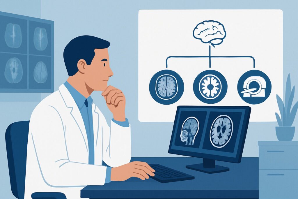
A 34-year-old patient with a long-standing diagnosis of focal epilepsy returns to your neurology clinic. Despite trials of three different anti-epileptic drugs, they continue to have disabling seizures several times a month. You and the patient agree it’s time to consider surgical options, and you refer them to the comprehensive epilepsy center for evaluation. The first step in their pre-surgical workup is advanced imaging to identify a potential structural cause for their seizures. This article details the imaging workflow for a patient with a known seizure disorder who is a candidate for surgical planning. For this specific clinical scenario, the American College of Radiology (ACR) rates MRI head without and with IV contrast as Usually Appropriate.
Who Fits This Clinical Scenario?
This guidance applies to patients with an established diagnosis of a seizure disorder, specifically those with medically refractory epilepsy who are being evaluated for potential surgical intervention. This includes procedures like resective surgery, laser interstitial thermal therapy (LITT), or implantation of neurostimulation devices. The primary goal of imaging in this context is to identify a discrete, surgically-treatable epileptogenic lesion that corresponds to the patient’s clinical and electroencephalographic (EEG) findings.
This workflow is distinct from other seizure-related scenarios. This article does not apply to:
- Patients with a new-onset seizure: These patients require an initial diagnostic workup to determine the cause of the seizure, which follows a different imaging pathway.
- Patients with a known seizure disorder whose seizures are well-controlled on medication: If there is no change in seizure type or frequency and surgery is not being considered, routine surveillance imaging is often not indicated.
- Patients with a known seizure disorder and a history of a brain tumor: While there is overlap, imaging for these patients is primarily focused on tumor surveillance and assessing for recurrence or treatment-related changes.
What Diagnoses Are You Working Up in This Scenario?
In the pre-surgical evaluation of drug-resistant epilepsy, imaging aims to uncover a structural abnormality responsible for generating seizures. The differential diagnosis is focused on identifying a specific, often subtle, epileptogenic lesion.
Mesial Temporal Sclerosis (MTS): This is the most common structural abnormality found in adults with focal epilepsy undergoing surgical evaluation. Also known as hippocampal sclerosis, it involves neuronal loss and gliosis in the hippocampus. High-resolution MRI is highly sensitive for detecting the characteristic findings of hippocampal atrophy and increased T2 signal.
Focal Cortical Dysplasia (FCD): FCDs are malformations of cortical development and are a leading cause of intractable epilepsy, particularly in children and young adults. These lesions can be extremely subtle, appearing as areas of abnormal cortical thickening, blurring of the gray-white matter junction, or abnormal signal. Identifying an FCD is critical as its complete resection is associated with excellent seizure-free outcomes.
Low-Grade Neoplasms: Slow-growing tumors, such as gangliogliomas or dysembryoplastic neuroepithelial tumors (DNETs), are a well-known cause of chronic epilepsy. They are often discovered during the pre-surgical workup. Contrast enhancement can be a key feature in identifying and characterizing these lesions.
Vascular Malformations: Cavernous malformations (cavernomas) are clusters of abnormal, thin-walled blood vessels that can cause seizures due to chronic microhemorrhages and surrounding hemosiderin deposition. MRI is the modality of choice for identifying these lesions.
Why Is MRI Head Without and With IV Contrast the Recommended Study?
For a patient with a known seizure disorder being considered for surgery, the ACR Appropriateness Criteria rate MRI head without and with IV contrast as Usually Appropriate. The goal is not just to find a lesion, but to characterize it with the highest possible anatomical detail to guide surgical planning.
The superior soft-tissue contrast of Magnetic Resonance Imaging (MRI) is essential for detecting the subtle structural abnormalities that cause epilepsy, such as focal cortical dysplasia or mesial temporal sclerosis. A standard brain MRI protocol may not be sufficient; a dedicated high-resolution epilepsy protocol MRI is crucial. This typically includes thin-section T1-weighted, T2-weighted, and FLAIR sequences oriented perpendicular to the long axis of the hippocampus to maximize detection of MTS.
The addition of intravenous contrast is important for identifying underlying low-grade tumors or vascular malformations that might not be apparent on non-contrast imaging. While many epileptogenic lesions do not enhance, the potential to uncover a treatable neoplasm makes contrast administration a key part of the comprehensive evaluation.
Let’s consider the alternatives:
- CT head without or with IV contrast is rated as May be appropriate. While useful for detecting large tumors, acute hemorrhage, or calcification, CT lacks the sensitivity to detect the subtle, non-calcified lesions like FCD or MTS that are the most common targets of epilepsy surgery. Its use of ionizing radiation (☢☢☢ 1-10 mSv) is another disadvantage compared to MRI (O 0 mSv).
- FDG-PET/CT brain is rated Usually appropriate. This is a functional study, not a structural one. It identifies areas of decreased glucose metabolism (interictal hypometabolism) that often correspond to the seizure focus. It is an extremely valuable tool, but it complements structural MRI rather than replacing it. It is most often used when the high-resolution MRI is negative or “non-lesional.”
What’s Next After MRI Head Without and With IV Contrast? Downstream Workflow
The results of the epilepsy protocol MRI are a critical branch point in the patient’s surgical evaluation. The findings must be interpreted in the context of the patient’s seizure semiology and EEG data at a multidisciplinary epilepsy conference.
- If the MRI is positive (lesional): When a clear structural lesion corresponding to the EEG findings is identified (e.g., MTS, FCD, or a small tumor), the patient may proceed toward surgical planning. The next step is often functional mapping to ensure the planned resection will not harm eloquent cortex. This may involve MRI functional (fMRI) head without IV contrast (May be appropriate) to map motor and language areas, or Magnetoencephalography (MEG) (May be appropriate) to precisely localize the seizure focus in relation to the lesion and eloquent cortex.
- If the MRI is negative (non-lesional): A negative or normal MRI does not mean the patient is not a surgical candidate. This is a common outcome. The workup then shifts to functional imaging to localize the seizure onset zone. The most common next step is an FDG-PET/CT brain (Usually appropriate) to search for a zone of interictal hypometabolism. Another option is SPECT or SPECT/CT brain perfusion (May be appropriate), which can be performed during a seizure (ictal) and between seizures (interictal) to identify areas of hyperperfusion and hypoperfusion, respectively.
- If the findings are indeterminate: Ambiguous findings require careful review and correlation. This may lead to repeat imaging, additional advanced MRI sequences, or proceeding to invasive EEG monitoring with stereotactically placed electrodes (SEEG) to confirm the seizure onset zone before any surgical decision is made.
Pitfalls to Avoid (and When to Get Help)
Navigating the pre-surgical epilepsy workup requires precision. Here are common pitfalls to avoid:
- Ordering a “routine” brain MRI: A standard MRI protocol lacks the thin, high-resolution sequences needed to detect subtle epileptogenic lesions. Always specify “epilepsy protocol” when ordering.
- Accepting a negative CT scan as definitive: CT has a very low yield for the most common causes of medically refractory epilepsy and should not be used to rule out a surgical substrate.
- Ignoring the EEG data: Imaging findings are only meaningful when they correlate with the clinical and electrophysiologic picture. A lesion on MRI that is distant from the EEG-defined seizure focus may be an incidental finding.
If a high-quality epilepsy protocol MRI is negative but the clinical suspicion for a focal lesion remains high, it is time to escalate. This patient should be discussed at a multidisciplinary epilepsy surgery conference at a comprehensive epilepsy center.
Related ACR Topics and Tools
For a comprehensive overview of all clinical scenarios related to seizures, please consult our parent guide. For tools to help you implement the recommendations in this article, see the resources below.
- For breadth across all scenarios in Seizures and Epilepsy, see our parent guide: Seizures and Epilepsy: ACR Appropriateness Decoded.
- Imaging Appropriateness Selector — for adjacent scenarios
- Imaging Protocol Library — for technique on the recommended study
- Radiation Dose Calculator — for cumulative dose conversations
Frequently Asked Questions
Why is intravenous contrast necessary if we are primarily looking for mesial temporal sclerosis or cortical dysplasia?
While mesial temporal sclerosis and most focal cortical dysplasias do not enhance, a significant minority of patients with drug-resistant epilepsy have an underlying low-grade neoplasm (like a ganglioglioma) or a vascular malformation as the cause. These lesions often enhance with contrast. Administering contrast is part of a comprehensive evaluation to ensure these potentially treatable causes are not missed.
What is an ‘epilepsy protocol’ MRI and how is it different from a standard brain MRI?
An epilepsy protocol MRI is a specialized study designed to maximize the detection of subtle epileptogenic lesions. Compared to a routine brain MRI, it uses higher magnetic field strength (3T is preferred), thinner image slices (1-3 mm), and specific imaging sequences (like high-resolution T2 and FLAIR) angled perpendicular to the hippocampus to best visualize its internal architecture and detect atrophy or signal change characteristic of mesial temporal sclerosis.
If the MRI is negative, does that mean the patient is not a candidate for epilepsy surgery?
No, a negative or ‘non-lesional’ MRI does not rule out a patient as a surgical candidate. Many patients with focal epilepsy have no visible abnormality on structural MRI. In these cases, the workup proceeds to advanced functional imaging tests like FDG-PET, SPECT, or MEG to localize the seizure focus. If these are suggestive, the patient may proceed to invasive EEG monitoring to confirm the location before surgery.
What is the role of functional MRI (fMRI) in this scenario?
Functional MRI (fMRI) is rated as ‘May be appropriate’ and is not used to find the seizure focus itself. Instead, it is a mapping tool used after a potential surgical target has been identified. fMRI helps locate critical brain areas, such as those responsible for language and motor function (‘eloquent cortex’), in relation to the lesion. This information is vital for surgical planning to maximize seizure control while minimizing the risk of postoperative neurologic deficits.
If the MRI is negative, is FDG-PET or SPECT better for finding the seizure focus?
Both FDG-PET and SPECT are valuable functional imaging tools for non-lesional epilepsy. FDG-PET, rated ‘Usually appropriate,’ identifies the seizure focus by detecting areas of reduced glucose metabolism between seizures (interictal hypometabolism). SPECT, rated ‘May be appropriate,’ can be performed both during a seizure (ictal) to show hyperperfusion and between seizures (interictal) to show hypoperfusion. The choice often depends on institutional expertise and the ability to inject the SPECT tracer during a typical seizure. Many centers start with FDG-PET due to its logistical simplicity.
Reviewed by Pouyan Golshani, MD, Interventional Radiologist — May 26, 2026