Which Imaging Is Best for Cognitive Deficits After Subacute Head Trauma?
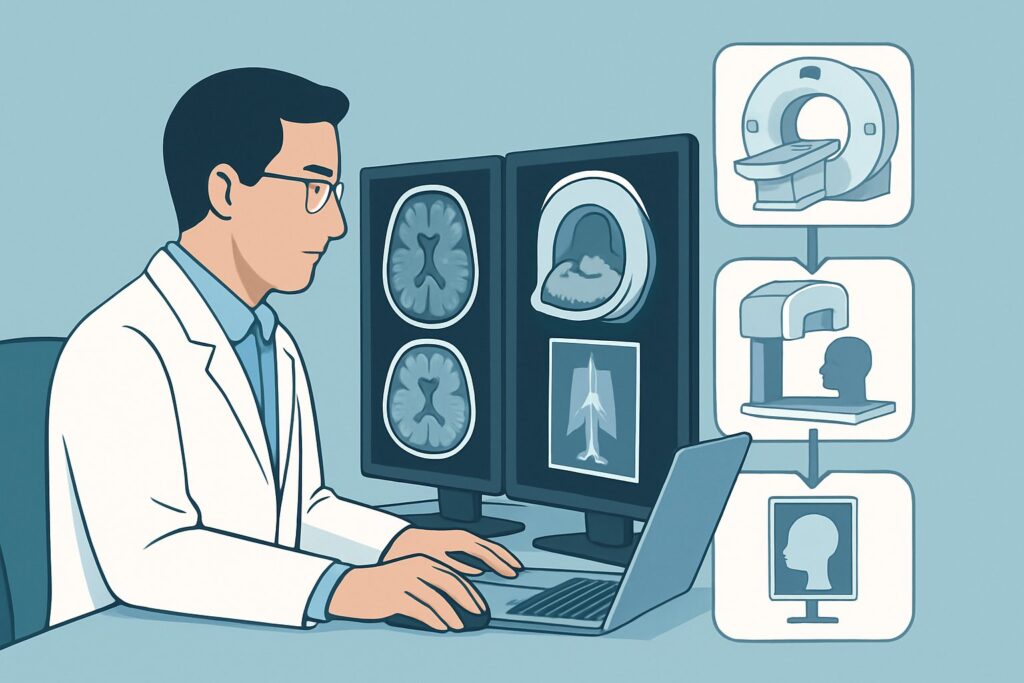
A 68-year-old male is brought to your clinic by his family. They report he had a ground-level fall about a month ago where he hit his head but “seemed fine” and refused to be seen. Over the last two weeks, however, he has become progressively more forgetful, confused, and his gait seems unsteady. He has no acute symptoms, but the change is undeniable. You suspect a delayed consequence of his injury, but the differential is broad. What is the right initial imaging study to order to evaluate these insidious, progressive neurologic deficits in the setting of subacute or chronic head trauma? This article provides a clinical workflow based on the American College of Radiology (ACR) Appropriateness Criteria, which rates MRI head without IV contrast as Usually Appropriate for this specific presentation.
Who Fits This Clinical Scenario?
This guidance applies to patients presenting with new or worsening cognitive or neurologic deficits that have developed in a subacute (days to weeks) or chronic (weeks to months) timeframe following a known or suspected head injury. The key features are the delayed onset and progressive nature of the symptoms. Common presentations include:
- Insidious onset of confusion or memory impairment
- Personality changes or abulia (lack of initiative)
- New or worsening gait instability or frequent falls
- Headaches that are persistent or worsening
- Subtle focal neurologic signs, such as mild hemiparesis or aphasia
It is crucial to distinguish this scenario from others. This workflow does not apply to:
- Acute Head Trauma: Patients presenting to the emergency department immediately after an injury are evaluated under different criteria, often guided by clinical decision rules like the Canadian CT Head Rule. Their workup is detailed in the ACR variant for acute, moderate, or severe head trauma.
- New Deficits in the Hospital: A patient with a known acute traumatic brain injury who develops new symptoms during their initial hospitalization follows a separate short-term follow-up imaging pathway.
- Cognitive Decline Without Trauma: An elderly patient with cognitive decline but no clear history of trauma would typically be evaluated under a dementia or encephalopathy pathway, which has a different differential and imaging approach.
What Diagnoses Are You Working Up in This Scenario?
When a patient presents with delayed neurologic changes after head trauma, imaging is primarily used to identify treatable structural causes. The differential diagnosis guides the choice of modality.
Chronic Subdural Hematoma (cSDH): This is the most common and critical diagnosis to exclude. Minor trauma, especially in older adults or those on anticoagulants, can tear bridging veins. The resulting slow venous bleed accumulates in the subdural space. Over weeks, membranes form around the hematoma, which can re-bleed, causing the collection to expand and exert mass effect on the brain, leading to the insidious onset of symptoms.
Post-traumatic Hydrocephalus: Trauma can disrupt the normal circulation and absorption of cerebrospinal fluid (CSF), leading to communicating hydrocephalus. This can present with the classic triad of cognitive impairment, gait disturbance, and urinary incontinence, often mimicking other neurologic conditions. Imaging is essential to identify ventricular enlargement out of proportion to sulcal widening.
Diffuse Axonal Injury (DAI): While DAI is an injury that occurs at the moment of impact, its long-term consequences manifest as cognitive and functional deficits. Shearing forces cause widespread microscopic damage to white matter tracts. While severe DAI is apparent acutely, the chronic sequelae and associated microhemorrhages are best visualized on specific MRI sequences weeks to months later.
Encephalomalacia and Gliosis: These terms refer to brain volume loss and scarring, respectively, that occur at the site of a previous contusion or hemorrhage. This area of damaged brain tissue can become a focus for post-traumatic seizures or contribute to focal neurologic deficits that emerge or worsen over time.
Why Is MRI Head without IV Contrast the Recommended Initial Study?
The ACR panel rates MRI head without IV contrast as Usually Appropriate because it provides the highest diagnostic yield for the key pathologies in this clinical context. The rationale is rooted in its superior soft-tissue contrast and sensitivity for blood products of varying ages.
The primary advantage of MRI is its ability to definitively characterize extra-axial fluid collections. A subacute subdural hematoma can become isodense to brain parenchyma on a CT scan (appearing the same shade of gray), making it notoriously difficult to detect. On MRI, however, different sequences (T1, T2, FLAIR, and susceptibility-weighted imaging) can clearly distinguish the hematoma from CSF and brain tissue, regardless of its age. MRI is also far more sensitive for detecting the subtle petechial hemorrhages of chronic diffuse axonal injury and for characterizing parenchymal scarring from old contusions.
While MRI is the preferred study, it’s important to understand why other modalities are rated differently for this specific scenario:
- CT head without IV contrast is also rated Usually Appropriate. It is faster, more accessible, and excellent for ruling out acute hemorrhage or significant mass effect. However, its primary limitation is the potential to miss an isodense subacute subdural hematoma. It remains a strong first choice if MRI is contraindicated (e.g., incompatible pacemaker) or not readily available.
- Radiography skull is rated Usually not appropriate. A skull fracture does not correlate well with the presence or absence of underlying intracranial injury in the subacute setting. The critical pathology is within the brain and its coverings, which are not visualized on a plain radiograph.
- MRI head with IV contrast is rated Usually not appropriate for initial evaluation. Contrast is typically unnecessary to diagnose a cSDH or hydrocephalus. It is reserved for cases where there is a concern for an alternative diagnosis, such as an underlying tumor, infection, or to better visualize the membranes of a cSDH if surgical planning is contemplated.
A key advantage of MRI is the absence of ionizing radiation (0 mSv), which is a consideration for all patients. In contrast, a non-contrast head CT carries a dose of 1-10 mSv.
What’s Next After MRI? Downstream Workflow
The results of the non-contrast head MRI will guide your next steps, creating a clear decision tree for patient management.
If the MRI is positive for a chronic subdural hematoma (cSDH): The immediate next step is a neurosurgical consultation. The decision for intervention (e.g., burr hole drainage, craniotomy) versus conservative management will depend on the size of the hematoma, the degree of mass effect, and the severity of the patient’s symptoms. The neurosurgeon may order a follow-up CT or MRI with contrast to aid in surgical planning.
If the MRI is positive for post-traumatic hydrocephalus: A neurology or neurosurgery consultation is warranted. Further evaluation may include a lumbar puncture with a high-volume tap (CSF removal) to see if symptoms transiently improve. If so, the patient may be a candidate for a ventriculoperitoneal (VP) shunt.
If the MRI is negative for a structural cause: A negative scan is a crucial finding. It effectively rules out cSDH and hydrocephalus, shifting the focus to non-structural causes. The diagnosis may be post-concussive syndrome, a medication side effect, or an unrelated underlying neurodegenerative condition. The next steps involve a comprehensive neurologic evaluation, neuropsychological testing, and management of symptoms like headache or sleep disturbance.
If the MRI is indeterminate or shows unexpected findings: If the findings are unclear (e.g., an unusual extra-axial collection) or suggest an alternative diagnosis like a tumor or abscess, ordering a follow-up MRI head with IV contrast may be the appropriate next step to better characterize the lesion.
Pitfalls to Avoid (and When to Get Help)
Navigating this scenario requires avoiding several common pitfalls to ensure timely and accurate diagnosis.
- Attribution Error: Do not automatically attribute all new neurologic symptoms in an older adult to “dementia” without considering a remote trauma history. Always ask about falls.
- Over-relying on CT: While CT is a valuable tool, remember its limitations in the subacute phase. If a non-contrast head CT is negative but your clinical suspicion for cSDH remains high, proceeding to MRI is the correct step.
- Ignoring Anticoagulation: The risk of cSDH is significantly higher in patients on anticoagulants or antiplatelet agents. Maintain a lower threshold for imaging in this population.
- Dismissing “Minor” Trauma: A simple ground-level fall can be sufficient to cause a cSDH, especially in patients with cerebral atrophy, which puts tension on bridging veins.
If the initial non-contrast MRI is negative but the patient’s neurologic decline continues, it is time to escalate. A consultation with a neurologist is essential to broaden the differential diagnosis and consider less common etiologies.
Related ACR Topics and Tools
This article focuses on a single clinical variant. For a comprehensive overview of imaging for all types of traumatic brain injury, from acute to chronic, please consult our parent guide. You can also use the tools below to explore adjacent scenarios, review imaging techniques, or discuss radiation dose with your patients.
- For breadth across all scenarios in Head Trauma, see our parent guide: Head Trauma: ACR Appropriateness Decoded.
- To explore other clinical presentations, use the Imaging Appropriateness Selector.
- For details on imaging techniques, visit the Imaging Protocol Library.
- To help in patient conversations about medical imaging, see the Radiation Dose Calculator.
Frequently Asked Questions
Why is MRI without contrast preferred over MRI with contrast for this initial workup?
For the primary differential diagnoses in this scenario—chronic subdural hematoma (cSDH) and hydrocephalus—intravenous contrast is not necessary for diagnosis. The inherent contrast between brain, CSF, and blood products of different ages on standard non-contrast MRI sequences (like T1, T2, and FLAIR) is sufficient. Contrast is reserved for cases where the initial study is indeterminate or if there’s a suspicion of an alternative diagnosis like a tumor or infection.
If my hospital has long wait times for MRI, is it acceptable to start with a non-contrast head CT?
Yes. The ACR rates both non-contrast head MRI and non-contrast head CT as ‘Usually Appropriate.’ While MRI is preferred for its higher sensitivity, CT is a very reasonable and practical first step, especially if it can be obtained much faster. A CT can quickly identify a large hematoma, significant mass effect, or hydrocephalus. However, if the CT is negative and your clinical suspicion remains high, you should still proceed with an MRI to rule out a CT-isodense subdural hematoma.
What if the patient has a pacemaker or other contraindication to MRI?
In cases where MRI is contraindicated, a non-contrast head CT is the best alternative and the appropriate initial study. If the non-contrast CT is equivocal for a suspected isodense subdural hematoma, a CT with intravenous contrast may be helpful, as the enhancing membranes or displaced cortical veins can sometimes reveal the collection.
Does this guidance apply to children with delayed symptoms after head trauma?
While the principles are similar, pediatric head trauma has unique considerations, including different injury patterns and a stronger emphasis on minimizing radiation exposure. The ACR criteria provide separate radiation relative level (RRL) estimates for pediatric patients. For a child with this presentation, consultation with a pediatric neurologist or radiologist is strongly recommended to ensure the most appropriate imaging plan.
The patient’s family is convinced he has Alzheimer’s. How does a negative MRI help?
A negative MRI is a highly valuable result. It effectively rules out major structural and surgically correctable causes of cognitive decline, such as a chronic subdural hematoma or normal pressure hydrocephalus. This allows you to confidently pivot the workup toward other causes, including neurodegenerative diseases like Alzheimer’s, metabolic encephalopathy, or post-concussive syndrome, and to counsel the family on the appropriate next steps for that diagnostic pathway.
Reviewed by Pouyan Golshani, MD, Interventional Radiologist — May 26, 2026