What Is the Right Imaging for Surveillance of Cerebral Venous Sinus Thrombosis in Adults?
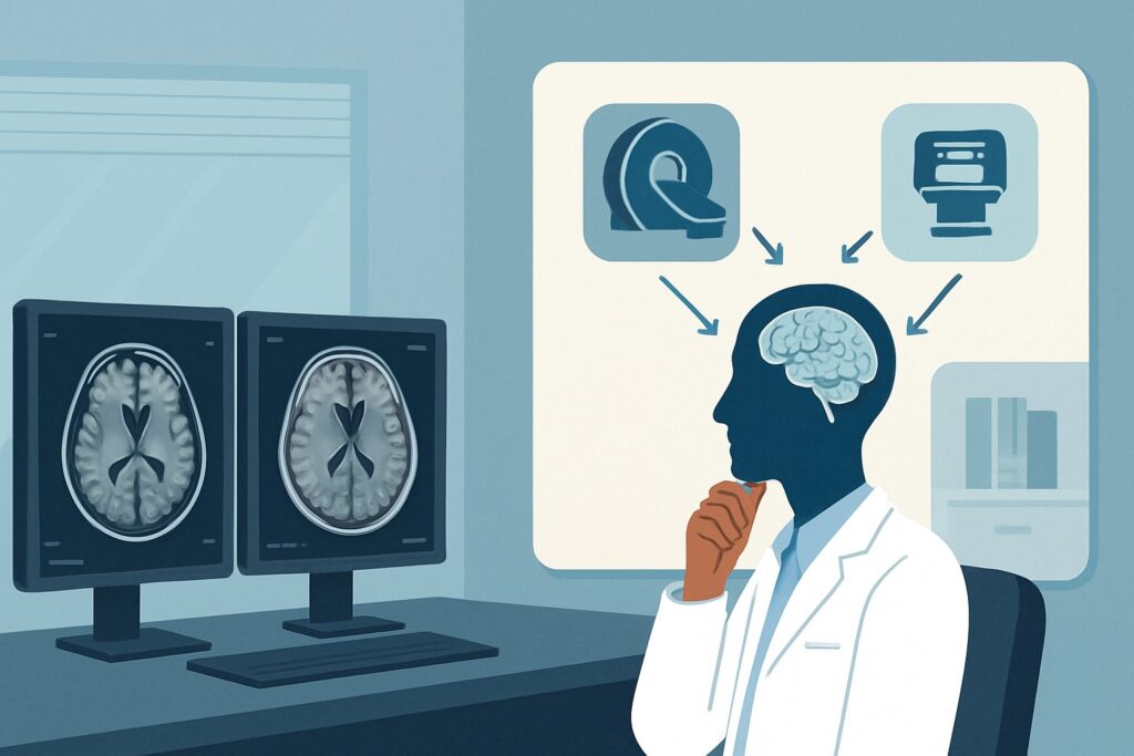
A 45-year-old female with a history of cerebral venous sinus thrombosis (CVST), diagnosed three months prior and attributed to oral contraceptive use, presents for her scheduled follow-up. She has been compliant with therapeutic anticoagulation, and her initial severe headaches have fully resolved. Now, you must decide on the appropriate surveillance imaging to assess for sinus recanalization, which will guide the duration of her treatment. This decision requires a study that can evaluate both the venous structures and the brain parenchyma for any secondary changes. According to the American College of Radiology (ACR) Appropriateness Criteria, the recommended study for this specific clinical scenario is `MRI head without and with IV contrast`, which is rated Usually appropriate.
Who Fits This Clinical Scenario?
This guidance applies specifically to adult patients with a previously confirmed diagnosis of cerebral venous sinus thrombosis (CVST) who are now undergoing follow-up or surveillance imaging. The typical patient is clinically stable or improving on medical therapy (usually anticoagulation) and the purpose of the imaging is to monitor the status of the thrombus and assess for complications or resolution.
This workflow is distinct from the initial diagnostic workup. It is crucial not to apply this surveillance protocol to patients presenting with new, acute symptoms. The following scenarios require different imaging pathways:
- Acute Focal Neurologic Deficit: A patient presenting with new-onset weakness, aphasia, or sensory loss requires an emergent workup for acute ischemic stroke, which prioritizes different imaging sequences and goals.
- Transient Ischemic Attack (TIA): A patient whose symptoms have completely resolved after a brief, stroke-like episode falls under the TIA imaging guidelines, which focus on identifying an embolic source.
- Known Intraparenchymal Hemorrhage: A patient being followed for a brain bleed has a separate follow-up protocol focused on hematoma evolution and stability, which may not require the same venous-specific imaging.
This article is exclusively for the non-acute, follow-up setting of a known CVST diagnosis.
What Diagnoses Are You Working Up in This Scenario?
In surveillance imaging for known CVST, the goal is not to establish a new diagnosis but to assess the evolution of the known condition and guide management. The key clinical questions you are trying to answer with the study are:
Degree of Venous Recanalization: This is the primary objective. The imaging aims to determine if the previously thrombosed dural venous sinus or cerebral vein has reopened. Complete, partial, or non-recanalization of the sinus is a critical factor in the clinical decision to continue, modify, or discontinue anticoagulation therapy.
Evolution of Parenchymal Injury: CVST can cause secondary injury to the brain tissue, such as venous infarction or hemorrhage. Surveillance imaging assesses the chronic effects of these injuries, looking for changes like encephalomalacia (brain tissue loss) or hemosiderin deposition from a prior bleed. It also ensures no new parenchymal abnormalities have developed.
Thrombus Stability and Extension: The study evaluates whether the existing thrombus is stable, regressing, or, in concerning cases, extending into adjacent sinuses or veins. New or extending thrombus despite anticoagulation would signal a need for a significant change in management and a workup for underlying hypercoagulable states.
Development of Late Complications: Although less common, chronic venous hypertension from a persistently occluded sinus can lead to the formation of a dural arteriovenous fistula (dAVF). While not the primary purpose of routine surveillance, imaging may reveal indirect signs like venous congestion or abnormal flow patterns that could prompt further investigation.
Why Is MRI Head Without and With IV Contrast the Recommended Study?
For surveillance of known cerebral venous sinus thrombosis, `MRI head without and with IV contrast` is rated Usually appropriate because it provides a comprehensive assessment of both the venous anatomy and the brain parenchyma without using ionizing radiation.
The power of this study lies in its combination of sequences. Non-contrast sequences (like T1, T2, FLAIR, and DWI) are highly sensitive for evaluating the brain tissue for the sequelae of CVST, including chronic venous infarcts, hemorrhage, and edema. These sequences can characterize the age of a thrombus, which may appear different on T1 and T2 sequences depending on the stage of hemoglobin breakdown.
The addition of intravenous contrast is critical for definitive assessment of the venous sinuses. A contrast-enhanced MR Venogram (MRV) is typically performed as part of the protocol. This technique directly visualizes blood flow within the dural sinuses and deep cerebral veins. A persistent thrombus will appear as a “filling defect”—an area within the sinus that does not enhance with contrast—clearly delineating the extent of any remaining occlusion. This is superior to non-contrast MRV techniques, where slow flow can sometimes mimic thrombosis.
Several other imaging modalities are also rated for this scenario, but are often less ideal:
- CTV head with IV contrast: This study is also rated Usually appropriate and is an excellent alternative, particularly for patients with contraindications to MRI (e.g., an incompatible implanted device). It offers high spatial resolution and clearly depicts the venous sinuses. However, it requires IV iodinated contrast and involves ionizing radiation (☢☢☢ 1-10 mSv).
- MRA head without IV contrast: This study is rated Usually not appropriate. Magnetic Resonance Angiography (MRA) sequences are optimized to visualize high-flow arterial structures. They are not designed to evaluate the slower-flow venous system and can produce artifacts that either obscure the sinuses or mimic thrombosis, making it an unreliable tool for this specific clinical question.
Given its lack of radiation (O 0 mSv) and its dual capability to assess parenchyma and venous patency with high fidelity, MRI with and without contrast remains the primary recommendation for CVST surveillance.
What’s Next After MRI? Downstream Workflow
The results of the surveillance MRI will directly influence your patient’s management plan, primarily concerning anticoagulation. The workflow typically branches based on the degree of sinus recanalization and the presence of any new findings.
If the study shows complete or near-complete recanalization: This is the desired outcome. In conjunction with the patient’s clinical stability and the initial suspected cause of the CVST (e.g., a transient risk factor like oral contraceptive use that has been stopped), this finding may support a decision to discontinue anticoagulation after a standard course (typically 3-12 months).
If the study shows partial recanalization or persistent occlusion: If a significant filling defect remains, particularly if the patient has an ongoing prothrombotic condition or experienced a severe initial presentation, extending the duration of anticoagulation is often warranted. The decision becomes more nuanced and depends on balancing the risk of recurrent thrombosis against the risk of bleeding from long-term anticoagulation.
If the study shows new or extended thrombus: This is a significant finding that indicates treatment failure or a potent underlying thrombophilia. This result should prompt an immediate re-evaluation of the anticoagulation regimen (e.g., dose, agent) and a comprehensive workup for hypercoagulable disorders if not already performed. Escalation to a hematologist or stroke neurologist is appropriate.
If the study is negative for thrombus but shows new parenchymal changes: New areas of venous infarction or hemorrhage despite treatment are concerning. This requires urgent clinical correlation to see if the patient has had new, subtle symptoms and may necessitate a change in management similar to that for thrombus extension.
Pitfalls to Avoid (and When to Get Help)
Navigating surveillance for CVST requires careful ordering and interpretation. Here are a few common pitfalls to avoid:
- Ordering an Arterial Study: A frequent error is ordering an MRA or CTA instead of an MRV or CTV. Ensure the order explicitly requests venography to evaluate the dural sinuses.
- Omitting Contrast Unnecessarily: While non-contrast MRI can suggest thrombosis, IV contrast is essential for a definitive assessment of recanalization. Omitting it can lead to an indeterminate or misleading result.
- Misinterpreting Chronic Findings: A chronically occluded sinus may become small or “atretic.” Differentiating this from a subacute or acute-on-chronic thrombus requires careful review of prior imaging and the MR signal characteristics.
- Ignoring Clinical Context: Do not treat the image in isolation. A patient with persistent, severe headaches despite imaging that shows recanalization may have elevated intracranial pressure requiring further management, such as ophthalmologic evaluation for papilledema.
If new, severe, or progressive neurologic symptoms develop at any point, do not wait for routine surveillance. Escalate care immediately for an urgent re-evaluation, as this may represent a new thrombotic event.
Related ACR Topics and Tools
For a comprehensive overview of imaging for all stroke and stroke-related conditions, this deep-dive article is best used alongside its parent topic guide. The following resources provide additional context for evidence-based imaging decisions.
- For breadth across all scenarios in Cerebrovascular Diseases-Stroke and Stroke-Related Conditions, see our parent guide: Cerebrovascular Diseases-Stroke and Stroke-Related Conditions: ACR Appropriateness Decoded.
- To explore adjacent clinical scenarios and their corresponding ACR ratings, use the ACR Appropriateness Criteria Lookup.
- For detailed technical parameters of the recommended study, consult the Imaging Protocol Library.
- To discuss radiation exposure from alternative studies like CTV, the Radiation Dose Calculator can help frame conversations with patients.
Frequently Asked Questions
How often should surveillance imaging for cerebral venous sinus thrombosis be performed?
The optimal timing and frequency are not rigidly defined and depend on clinical factors. A common approach is to perform the first follow-up scan at 3 to 6 months after diagnosis to assess for recanalization and guide the initial decision on anticoagulation duration. Further imaging is typically reserved for patients with persistent symptoms or those with continued occlusion on the first follow-up scan.
Is intravenous contrast always necessary for follow-up MRI scans?
Yes, for the definitive assessment of venous sinus patency, IV contrast is highly recommended. Contrast-enhanced MR Venography (MRV) is the most reliable non-invasive method to distinguish between slow-flowing blood and an occlusive thrombus. A non-contrast study is often insufficient and can lead to diagnostic uncertainty.
What is the best alternative if my patient has a contraindication to MRI?
If a patient cannot undergo an MRI (e.g., due to an incompatible pacemaker or severe claustrophobia), a CT Venogram (CTV) with IV contrast is the best alternative. The ACR rates this study as ‘Usually appropriate’ for this scenario. It provides excellent visualization of the dural sinuses, though it involves ionizing radiation and iodinated contrast.
Can I just order an MRV instead of a full brain MRI with and without contrast?
While the MRV sequence is the key part for assessing the sinuses, ordering a full diagnostic brain MRI is crucial. The standard MRI sequences (T1, T2, FLAIR, DWI) are necessary to evaluate the brain parenchyma for secondary complications like venous infarction, hemorrhage, or edema, which are essential for comprehensive management.
What is the main difference between MRV and CTV for CVST surveillance?
Both are excellent for visualizing the venous sinuses. The main difference is the technology used. MRV uses strong magnetic fields and radio waves, avoiding ionizing radiation. CTV uses X-rays (CT) and requires iodinated contrast. MRI also provides superior soft tissue contrast for evaluating the brain parenchyma itself. CTV is generally faster and is the preferred alternative when MRI is contraindicated.
Reviewed by Pouyan Golshani, MD, Interventional Radiologist — May 30, 2026