What Is the Best Imaging for Local T-Staging in Pretreatment Cervical Cancer?
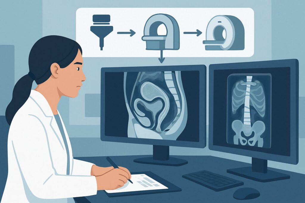
A 48-year-old woman presents to your gynecologic oncology clinic after a Pap test and subsequent biopsy confirmed invasive squamous cell carcinoma of the cervix. On speculum exam, you visualize a 3 cm exophytic lesion. Before you can formulate a treatment plan—deciding between primary surgery or chemoradiation—you need to precisely define the local extent of the tumor. Has it invaded the parametrial tissues, the vaginal walls, or adjacent pelvic organs? This critical question of local tumor extension, or T staging, is the primary determinant of the initial therapeutic approach. For this specific clinical scenario, the American College of Radiology (ACR) Appropriateness Criteria rate MRI pelvis without and with IV contrast as Usually Appropriate, providing the detailed soft-tissue anatomy required for this crucial decision.
Who Fits This Clinical Scenario for Cervical Cancer Staging?
This guidance is specifically for patients with a new, biopsy-proven diagnosis of invasive cervical cancer where a lesion is clinically visible on physical examination. The primary clinical question is the initial local staging (T staging) to determine the tumor’s size and direct extension into adjacent structures before any treatment has been initiated. This includes assessing for parametrial, vaginal, bladder, or rectal involvement.
This workflow is distinct from other common scenarios in cervical cancer care. It does not apply to patients who:
- Require systemic staging: If the primary question is the assessment of lymph node involvement or distant metastases (N and M staging), the imaging strategy is different. That evaluation represents a separate, though often concurrent, clinical question.
- Are undergoing treatment response assessment: Imaging performed during or after a course of chemoradiation to evaluate tumor response follows a different set of recommendations.
- Are being monitored for recurrence: Surveillance imaging for asymptomatic patients or workup for a suspected recurrence in a previously treated patient are distinct clinical scenarios with their own appropriate imaging pathways.
This article focuses exclusively on the initial local staging of a known, visible primary tumor.
What Diagnoses Are You Working Up in This Scenario?
In this context, the “differential diagnosis” is less about identifying the disease—which is already known from biopsy—and more about differentiating the stage of the disease. The imaging study is tasked with answering specific anatomical questions that directly correspond to the FIGO (International Federation of Gynecology and Obstetrics) staging system. The key distinctions you are trying to make will determine whether the patient is a candidate for surgery or requires primary chemoradiation.
The most critical question is the presence or absence of parametrial invasion. A tumor confined to the cervix is Stage I. Once it invades the parametrium (the connective tissue and fat adjacent to the uterus), it becomes at least Stage IIB. This is a pivotal distinction, as Stage IIB and higher are typically treated with primary chemoradiation, whereas earlier stages may be amenable to radical hysterectomy.
Another key determination is the extent of vaginal involvement. A tumor extending to the upper two-thirds of the vagina is Stage IIA, but extension to the lower third of the vagina immediately upstages the disease to Stage IIIA. This finding also significantly alters the treatment approach, particularly the radiation field design.
Finally, imaging must assess for invasion into adjacent pelvic organs. Evidence of tumor invading the bladder or rectal mucosa constitutes Stage IVA disease. This is a less common finding at initial presentation but is a critical one to identify, as it necessitates a more complex and aggressive treatment strategy, often involving pelvic exenteration in select surgical candidates.
Why Is MRI Pelvis without and with IV contrast the Recommended Study for This Presentation?
For assessing local tumor extension in cervical cancer, MRI provides unparalleled soft-tissue resolution, making it the superior modality for answering the critical T-staging questions. The ACR rates MRI pelvis without and with IV contrast as Usually Appropriate for this indication.
The strength of MRI lies in its ability to distinguish the tumor from the normal cervical stroma. On T2-weighted images, the cervical tumor typically appears as a high-signal-intensity mass, contrasting sharply with the low-signal-intensity of the normal fibrous cervical stroma. This clear demarcation allows for precise measurement of tumor size and assessment of deep stromal invasion. Most importantly, it is highly sensitive and specific for detecting the subtle signs of parametrial invasion, such as disruption of the low-signal-intensity stromal ring and direct tumor extension into the adjacent T2-hyperintense parametrial fat.
The addition of intravenous contrast enhances the delineation of the tumor, which typically shows early arterial enhancement. This can be particularly helpful in distinguishing viable tumor from post-biopsy changes or necrosis.
Alternative studies are rated lower for specific reasons:
- CT pelvis with IV contrast is rated May be appropriate. While excellent for detecting bulky lymphadenopathy and distant metastases, CT has inferior soft-tissue contrast compared to MRI. It is significantly less sensitive for detecting subtle parametrial invasion, which is the key determination in this scenario. CT carries a radiation dose of 1-10 mSv (☢☢☢).
- US pelvis transvaginal is also rated May be appropriate. Transvaginal ultrasound can be very effective for measuring the primary tumor’s size and assessing its relationship to the internal os. However, its field of view is limited, and its ability to reliably assess deep parametrial extension to the pelvic sidewall is operator-dependent and often inferior to MRI.
Given that MRI provides the most detailed anatomical information for local staging without using ionizing radiation (0 mSv), it is the cornerstone of modern pretreatment T-staging for clinically visible cervical cancer.
What’s Next After MRI Pelvis without and with IV contrast? Downstream Workflow
The results of the pelvic MRI are a critical input for the multidisciplinary tumor board and directly guide the next steps in management. The downstream workflow branches based on the T-stage determined by the imaging findings, correlated with the clinical exam.
If the MRI confirms the tumor is confined to the cervix (Stage IB or IIA1), with no evidence of parametrial invasion, the patient is often a candidate for primary surgical management, such as a radical hysterectomy with pelvic lymph node dissection. The next step in this pathway is typically systemic staging to evaluate for nodal and distant metastases, which may involve PET/CT. This addresses the “Initial systemic staging” sibling scenario.
If the MRI demonstrates definite parametrial invasion (Stage IIB or greater), invasion to the lower third of the vagina (Stage IIIA), or extension to the pelvic sidewall (Stage IIIB), the patient is generally not a candidate for primary surgery. The standard of care becomes definitive chemoradiation. The MRI findings are then used by the radiation oncologist to plan the radiation treatment fields with high precision.
If the MRI findings are indeterminate or equivocal, for example, showing subtle parametrial stranding that could be either inflammation or early tumor infiltration, the case requires careful multidisciplinary discussion. An examination under anesthesia may be performed to clinically assess the parametria. In some cases, a PET/CT or PET/MRI, which is also rated Usually Appropriate, may be considered to help differentiate metabolically active tumor from benign changes.
Pitfalls to Avoid (and When to Get Help)
Accurate local staging is paramount, and several pitfalls can compromise the utility of the imaging workup.
- Timing After Biopsy: Performing the MRI too soon after a large cervical biopsy can lead to post-procedural inflammation and hemorrhage, which can mimic or obscure parametrial tumor extension. Allowing a week or two between biopsy and MRI can improve diagnostic accuracy.
- Suboptimal Technique: For optimal delineation, the MRI protocol should include high-resolution, small field-of-view T2-weighted images in the sagittal, axial, and oblique axial planes (perpendicular to the long axis of the endocervical canal). Failure to use an optimized protocol can limit the study’s value.
- Ignoring the Clinical Exam: FIGO staging for cervical cancer is unique in that it is a clinical staging system. Imaging findings are used to supplement, not replace, the physical examination. Discrepancies between the exam and MRI findings should be carefully reviewed.
If imaging findings are equivocal or conflict with the clinical assessment, the case should be escalated for review at a gynecologic oncology multidisciplinary tumor board, where radiologists, gynecologic oncologists, radiation oncologists, and pathologists can reach a consensus on the patient’s definitive stage and treatment plan.
Related ACR Topics and Tools
This article covers a single, specific scenario. For a comprehensive overview of all related clinical variants, from systemic staging to post-treatment surveillance, please consult the parent topic hub.
- For breadth across all scenarios in Pretreatment Evaluation and Follow-up of Invasive Cancer of the Cervix, see our parent guide: Pretreatment Evaluation and Follow-up of Invasive Cancer of the Cervix: ACR Appropriateness Decoded.
- To explore adjacent clinical questions and their recommended imaging studies, use the ACR Appropriateness Criteria Lookup.
- For detailed technical specifications on performing the recommended study, browse the Imaging Protocol Library.
- To discuss cumulative radiation exposure with patients when considering CT or PET/CT for other staging needs, the Radiation Dose Calculator can be a helpful tool.
Frequently Asked Questions
Why is MRI so much better than CT for local cervical cancer staging?
MRI’s superiority lies in its excellent soft-tissue contrast resolution. It can clearly distinguish the tumor from the normal fibrous cervical stroma and detect subtle infiltration into the adjacent parametrial tissues. CT, while useful for detecting lymph nodes and distant disease, cannot provide this level of detail for the primary tumor, making it less reliable for local T-staging.
If the cervical lesion looks very small on my clinical exam, is an MRI still necessary?
Yes. Clinical examination can underestimate tumor size and cannot reliably assess the depth of stromal invasion or detect microscopic parametrial extension. An MRI is essential to rule out occult local spread that would upstage the disease and change the treatment from surgery to chemoradiation. It provides critical information that the physical exam alone cannot.
What about PET/CT? I thought that was the standard for cancer staging.
PET/CT is excellent for systemic staging—detecting involved lymph nodes (N-staging) and distant metastases (M-staging). However, for local T-staging of the primary tumor, its spatial resolution is inferior to MRI. While FDG-PET/MRI is also rated ‘Usually Appropriate’ and combines the strengths of both, a dedicated pelvic MRI is the primary modality for answering the specific question of local tumor extension.
Does the patient need vaginal gel for this MRI study?
Using intravaginal ultrasound gel is a common and recommended technique for pelvic MRI in cervical cancer staging. The gel distends the vaginal fornices, allowing for better visualization of the vaginal walls and clear assessment of any tumor extension into these areas. It is an important part of an optimized imaging protocol.
Can I order a non-contrast MRI if my patient has a severe contrast allergy or renal failure?
A non-contrast MRI of the pelvis is rated as ‘May be appropriate’ by the ACR. The high-resolution T2-weighted sequences provide the most critical information for T-staging, particularly parametrial invasion. While contrast enhancement improves tumor conspicuity and delineation, a high-quality non-contrast study is still far superior to CT and is a reasonable alternative when gadolinium-based contrast is contraindicated.
Reviewed by Pouyan Golshani, MD, Interventional Radiologist — May 30, 2026