What Is the Best Initial Imaging for Abnormal Uterine Bleeding? An ACR-Guided Workflow
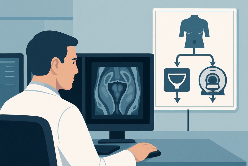
It’s 3 PM in the outpatient clinic. A 42-year-old patient presents with several months of progressively heavier and more prolonged menstrual periods. Her history is otherwise unremarkable, a pregnancy test is negative, and the bimanual exam reveals a slightly bulky, non-tender uterus. You suspect a structural cause for her abnormal uterine bleeding (AUB), but the physical exam is non-specific. The immediate clinical question is which imaging study to order first to evaluate the uterine anatomy and guide subsequent management. For this common presentation—the initial imaging workup of abnormal uterine bleeding—the American College of Radiology (ACR) Appropriateness Criteria rate US duplex Doppler pelvis as Usually Appropriate. This article provides a detailed workflow for this specific scenario.
Who Fits This Clinical Scenario for Initial AUB Imaging?
This guidance applies to patients presenting with abnormal uterine bleeding where a structural etiology is suspected after an initial clinical evaluation, including a history, physical examination, and a negative pregnancy test. The patient is typically hemodynamically stable and the evaluation is occurring in an outpatient or non-emergent setting. This workflow is designed for the first imaging study ordered for this problem.
This article is NOT for patients who:
- Have had a prior inconclusive ultrasound. These patients fall into a different clinical scenario requiring problem-solving with more advanced imaging. Their workflow is covered in the ACR variant for follow-up imaging when ultrasound is inconclusive.
- Are undergoing surveillance for a known condition. A patient with known uterine fibroids who requires periodic re-evaluation follows a separate surveillance pathway.
- Present with acute, life-threatening hemorrhage. In cases of hemodynamic instability, the priority is resuscitation and potential immediate surgical or interventional radiology consultation, which may supersede or alter the standard diagnostic imaging sequence.
Correctly identifying the patient’s clinical context is crucial for applying the right imaging criteria and avoiding unnecessary or low-yield studies.
What Diagnoses Are You Working Up With Initial Imaging for AUB?
The differential diagnosis for abnormal uterine bleeding is broad, often categorized by the PALM-COEIN acronym (Polyp, Adenomyosis, Leiomyoma, Malignancy/hyperplasia; Coagulopathy, Ovulatory dysfunction, Endometrial, Iatrogenic, Not yet classified). Initial imaging is primarily focused on identifying the structural causes (PALM).
Leiomyomas (Fibroids): These are the most common benign uterine tumors and a frequent cause of AUB, particularly heavy menstrual bleeding. Ultrasound is excellent for identifying their presence, number, size, and location (submucosal, intramural, subserosal). Submucosal fibroids, which impinge on the endometrial cavity, are most strongly associated with AUB, and their precise localization is critical for planning hysteroscopic resection.
Adenomyosis: This condition, where endometrial glands and stroma are present within the myometrium, is a common but often under-recognized cause of heavy, painful periods. On ultrasound, it can present with characteristic findings like a bulky, globular uterus, myometrial cysts, or indistinctness of the endometrial-myometrial junction.
Endometrial Polyps: These are focal benign overgrowths of the endometrium that can cause intermenstrual or heavy menstrual bleeding. While sometimes visible on standard 2D ultrasound as focal areas of endometrial thickening, they are best characterized with sonohysterography. However, the initial ultrasound is key to identifying the endometrial thickening that prompts further evaluation.
Endometrial Hyperplasia and Malignancy: While less common in premenopausal women without specific risk factors, excluding endometrial pathology is a primary goal of the AUB workup, especially in postmenopausal patients or those with risk factors like obesity or polycystic ovary syndrome. Ultrasound assesses endometrial thickness, which is a key triage tool. A thickened or heterogeneous endometrium often requires subsequent endometrial sampling.
Why Is Pelvic Ultrasound the Recommended First Step for Abnormal Uterine Bleeding?
The ACR designates US pelvis transabdominal, US pelvis transvaginal, and US duplex Doppler pelvis as Usually Appropriate for the initial evaluation of AUB. A comprehensive pelvic ultrasound typically incorporates all three components to provide a complete, high-yield diagnostic assessment.
The rationale for this recommendation is multifactorial. First, ultrasound offers excellent spatial resolution of the female pelvis, providing detailed views of the myometrium, endometrium, and adnexa. The transabdominal approach gives a broad overview of the entire pelvis, while the transvaginal approach provides higher-resolution detail of the uterus and ovaries. This combination is highly sensitive and specific for detecting the most common structural causes of AUB, including fibroids and adenomyosis, and for measuring endometrial thickness.
The addition of Duplex Doppler is crucial for assessing vascularity. It can help characterize an endometrial or myometrial mass, such as identifying the feeding vessel of an endometrial polyp or evaluating the vascular pattern of a fibroid. This information can help differentiate between pathologies and guide further management.
Most importantly, ultrasound involves no ionizing radiation (adult_rrl=O 0 mSv), a critical safety consideration, particularly in premenopausal and reproductive-age patients who may require future imaging. It is also widely accessible, relatively inexpensive, and well-tolerated.
Why Lower-Rated Alternatives Are Not First-Line
- MRI Pelvis without and with IV contrast: Rated Usually not appropriate for initial imaging. While MRI provides superior soft-tissue contrast and is the gold standard for characterizing and mapping fibroids or complex adenomyosis, it is reserved as a problem-solving tool. It is used when ultrasound findings are inconclusive or when precise pre-procedural mapping is required before uterine artery embolization or myomectomy. Using it as a first-line test is not cost-effective and represents a misallocation of resources.
- CT Pelvis with IV contrast: Rated Usually not appropriate. CT has poor intrinsic soft-tissue contrast for evaluating the endometrial-myometrial interface, making it significantly inferior to ultrasound for assessing for polyps, adenomyosis, or subtle endometrial abnormalities. Furthermore, it exposes the patient to ionizing radiation (adult_rrl=☢☢☢ 1-10 mSv) without providing the necessary diagnostic information for this specific clinical question.
What’s Next After Pelvic Ultrasound? Downstream Workflow
The results of the initial pelvic ultrasound will direct the subsequent clinical pathway. The goal is to move from a general diagnosis of AUB to a specific underlying cause that can be targeted with treatment.
- If the ultrasound is positive for a clear etiology (e.g., large fibroids): The next step is typically a discussion with the patient about management options, which may range from medical therapy (hormonal IUD, oral medications) to procedural interventions (myomectomy, uterine artery embolization, hysterectomy), guided by the patient’s symptoms and desire for future fertility.
- If the ultrasound is negative or non-specific: If no clear structural cause is identified and the endometrium is thin and regular, the workup shifts toward non-structural causes (the “COEIN” categories). This may involve laboratory testing for coagulopathies or hormonal evaluation for ovulatory dysfunction.
- If the ultrasound is indeterminate or suspicious for an intracavitary lesion: If the endometrium is globally thickened or a focal lesion like a suspected polyp or submucosal fibroid is seen, the next step is often direct visualization or sampling. US sonohysterography (rated May be appropriate (Disagreement)) is an excellent next test to better delineate intracavitary pathology. Alternatively, the clinician may proceed directly to hysteroscopy with or without endometrial biopsy. This decision point aligns with the ACR scenario for follow-up imaging when original ultrasound is inconclusive.
Pitfalls to Avoid (and When to Get Help)
Several common pitfalls can occur in the initial workup of abnormal uterine bleeding. First, failing to perform a transvaginal ultrasound when feasible can lead to missed pathology; the transabdominal view alone is often insufficient for detailed endometrial and myometrial evaluation. Second, misinterpreting a normal post-menstrual thin endometrium as atrophy in a patient with cyclical bleeding can delay diagnosis. Third, not correlating the imaging findings with the patient’s menstrual cycle phase can lead to confusion, as endometrial thickness varies significantly.
Escalate care if you encounter red flags such as suspected malignancy on imaging, particularly in a postmenopausal patient, or if the patient is hemodynamically unstable from acute blood loss. In these cases, an urgent referral to a gynecologist or the emergency department is warranted.
Related ACR Topics and Tools
This article covers one specific scenario within the broader topic of Abnormal Uterine Bleeding. For a comprehensive overview of all related clinical variants, from follow-up imaging to surveillance, please see our parent guide. The following GigHz tools can also support your clinical workflow.
- For breadth across all scenarios in Abnormal Uterine Bleeding, see our parent guide: Abnormal Uterine Bleeding: ACR Appropriateness Decoded.
- ACR Appropriateness Criteria Lookup — for adjacent scenarios
- Imaging Protocol Library — for technique on the recommended study
- Radiation Dose Calculator — for cumulative dose conversations
Frequently Asked Questions
Why is a transvaginal ultrasound necessary if a transabdominal one was already done?
The transabdominal ultrasound provides a wide field of view, which is excellent for assessing large fibroids and the overall pelvic anatomy. However, the transvaginal probe uses a higher frequency, allowing for much more detailed, high-resolution images of the endometrium, myometrium, and ovaries. It is superior for detecting subtle abnormalities like small polyps, adenomyosis, or early endometrial changes that can be missed on the transabdominal view alone. A complete pelvic ultrasound includes both for this reason.
Is an ultrasound necessary for all patients with abnormal uterine bleeding?
Not always, but it is the recommended initial imaging test when a structural cause is suspected. In very young patients or those where anovulatory bleeding is the clear cause, clinicians may opt for a trial of medical management first. However, for persistent AUB, new-onset AUB in patients over 40, or when the physical exam is abnormal, imaging is crucial to rule out structural pathology like fibroids, polyps, or endometrial hyperplasia.
When should I consider ordering a sonohysterogram instead of a standard pelvic ultrasound?
A sonohysterogram is typically a second-line, problem-solving test, not an initial one. The ACR rates it ‘May be appropriate (Disagreement)’ for initial imaging. It is most valuable when a standard ultrasound shows a thickened endometrium or a suspected intracavitary lesion (like a polyp or submucosal fibroid). By instilling saline into the endometrial cavity, it distends the walls and provides clear delineation of these lesions, which is superior to standard 2D ultrasound.
Can pelvic ultrasound reliably diagnose uterine cancer?
Ultrasound cannot definitively diagnose endometrial cancer, but it is a critical triage tool. Its primary role is to measure endometrial thickness. In a postmenopausal woman with bleeding, a very thin endometrium (<4-5 mm) has a very high negative predictive value for cancer, potentially avoiding the need for a biopsy. Conversely, a thickened, irregular, or heterogeneous endometrium is a significant red flag that mandates an endometrial biopsy for a definitive histologic diagnosis.
Does the timing of the ultrasound within the menstrual cycle matter?
Yes, timing can be very important. The best time to evaluate the endometrium for focal lesions like polyps is in the early proliferative phase (days 4-10 of the cycle), when the endometrium is at its thinnest. Performing the scan during the secretory phase, when the endometrium is naturally thick and echogenic, can obscure small polyps or submucosal fibroids. While an ultrasound can be performed at any time to look for larger structural issues, scheduling it for the early part of the cycle is ideal if an endometrial lesion is the primary concern.
Reviewed by Pouyan Golshani, MD, Interventional Radiologist — June 27, 2026