What Is the Best Imaging for New Cervical Pain in a Patient with Cancer?
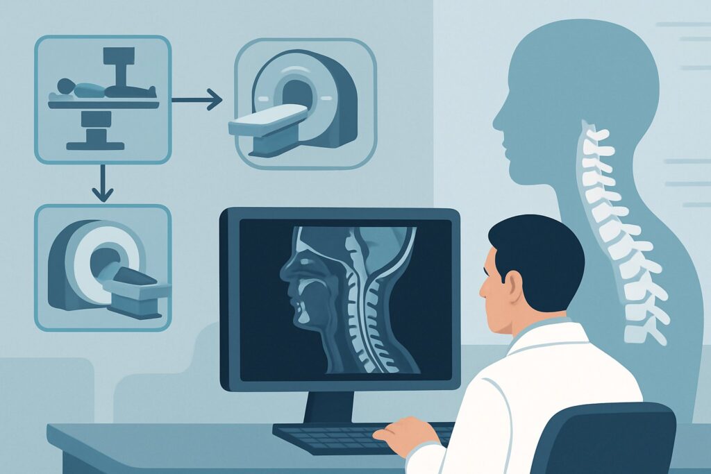
A 62-year-old woman with a history of breast cancer, currently on hormonal therapy, presents to her oncologist’s office with three weeks of worsening neck pain and new, tingling paresthesias radiating into her right thumb and index finger. She denies any recent falls or trauma. The primary clinical question is urgent: is this a new metastatic lesion causing nerve root compression, a complication of therapy, or simply unrelated degenerative disc disease? Choosing the right initial imaging study is critical for timely diagnosis and management. For this specific scenario, the American College of Radiology (ACR) designates one imaging modality as the most effective first step. According to the ACR Appropriateness Criteria, MRI of the cervical spine without and with IV contrast is Usually appropriate.
## Who Fits This Clinical Scenario?
This imaging workflow is designed for a specific patient population: an adult with a known diagnosis of malignancy who presents with new, acute, or progressively worsening cervical pain or radiculopathy. The absence of recent, significant trauma is a key qualifier, as that would trigger a different diagnostic algorithm focused on fracture and ligamentous injury.
This guidance applies regardless of the primary cancer type, as many solid tumors (notably breast, lung, prostate, kidney, and thyroid) and hematologic malignancies can metastasize to the spine.
It is crucial to distinguish this presentation from similar, yet distinct, clinical situations that require different approaches:
- Suspected Spinal Infection: If the patient presents with concurrent fever, elevated inflammatory markers, a history of intravenous drug use, or other signs of infection, the workup should follow the ACR variant for suspected spinal infection, where the differential includes discitis-osteomyelitis and epidural abscess.
- No Malignancy or Red Flags: An adult with acute neck pain or radiculopathy but no history of cancer and no constitutional “red flag” symptoms (e.g., unexplained weight loss, fever, night sweats) would follow a more conservative initial pathway, often starting with non-imaging management.
- Post-Surgical Anatomy: A patient with a history of prior cervical spine surgery (e.g., fusion, laminectomy) presenting with new pain follows a separate variant, as hardware can create imaging artifacts and the differential includes post-surgical complications.
## What Diagnoses Are You Working Up in This Scenario?
In a patient with known cancer, new spinal symptoms demand a high index of suspicion for oncologic causes. The imaging choice is tailored to differentiate between several critical possibilities.
The foremost concern is vertebral metastatic disease. Cancer can spread to the vertebral bodies, leading to pain from bone destruction, pathologic compression fractures, and instability. More critically, tumor can extend from the bone into the epidural space, directly compressing the spinal cord or nerve roots and causing myelopathy or radiculopathy. Contrast-enhanced MRI is exceptionally sensitive for detecting marrow replacement by tumor and delineating the extent of any epidural involvement.
A related but distinct diagnosis is leptomeningeal disease, where cancer cells spread to the cerebrospinal fluid and coat the surfaces of the spinal cord and nerve roots. This can cause diffuse radicular symptoms and is often invisible on non-contrast imaging. The administration of intravenous contrast is essential, as it will highlight the inflamed, tumor-coated meninges.
Less commonly, the symptoms may stem from complications of cancer treatment. Radiation therapy to the neck or chest can, over time, cause radiation-induced myelopathy or brachial plexopathy, which can mimic tumor recurrence. Imaging helps exclude a structural lesion, although specific imaging findings of radiation injury can also be identified.
Finally, it is essential to remember that patients with cancer are just as susceptible to unrelated degenerative spine disease as the general population. A herniated disc, foraminal stenosis from osteoarthritis, or spondylosis are common causes of neck pain and radiculopathy. A high-quality MRI can confidently diagnose these benign conditions, preventing unnecessary changes to the patient’s cancer therapy and guiding appropriate conservative management.
## Why Is MRI of the Cervical Spine Without and With IV Contrast the Recommended Study?
The ACR rates MRI of the cervical spine without and with IV contrast as Usually appropriate because it provides the most comprehensive evaluation for the specific differential diagnoses in this high-stakes scenario. This single examination directly assesses bone marrow, the spinal cord, nerve roots, intervertebral discs, and the surrounding soft tissues with unparalleled detail.
The rationale for this specific protocol includes:
- Superior Soft Tissue and Marrow Evaluation: The non-contrast sequences are highly sensitive for detecting the replacement of normal fatty bone marrow with tumor. T1-weighted images are particularly effective at showing abnormal marrow signal. T2-weighted sequences are excellent for identifying spinal cord edema, a critical sign of compression (myelomalacia).
- Essential Role of IV Contrast: The addition of a gadolinium-based contrast agent is what makes this study definitive. Malignant tumors, particularly metastases and leptomeningeal disease, are typically vascular and will demonstrate enhancement. Contrast administration is crucial for identifying and delineating epidural tumor extension, which can be subtle on non-contrast images, and is the only reliable way to visualize leptomeningeal spread.
- Safety Profile: MRI involves no ionizing radiation (Relative Radiation Level: O), a significant benefit in patients who may have already undergone multiple radiation-based imaging studies or therapies.
Why are alternative studies rated lower for this initial workup?
- Radiography (X-Rays): Rated Usually not appropriate. Plain films are notoriously insensitive for detecting early metastatic disease. A significant amount of bone (30-50%) must be destroyed before a lytic lesion becomes visible. Furthermore, radiographs provide zero information about the spinal cord, nerves, or epidural space—the most critical structures to evaluate.
- CT of the Cervical Spine: Rated May be appropriate (Disagreement). CT is superior to radiography for assessing bone detail and can detect lytic and blastic lesions earlier. However, its ability to evaluate the spinal cord, nerve roots, and early marrow infiltration is significantly inferior to MRI. It also exposes the patient to ionizing radiation (RRL: ☢☢☢). CT is primarily reserved for patients with a contraindication to MRI.
Once you’ve decided on the recommended study, understanding the technical aspects is key. For a detailed look at the foundational sequences, contrast considerations, and interpretation principles, see our protocol guide: MRI Cervical Spine Without Contrast.
## What’s Next After MRI? Downstream Workflow
The results of the contrast-enhanced cervical MRI will dictate the subsequent clinical pathway. The workflow branches based on whether the findings are positive, negative, or indeterminate for malignancy.
If the MRI is positive for metastatic disease:
- Spinal Cord or Cauda Equina Compression: This is a neurologic emergency. An immediate consultation with neurosurgery and radiation oncology is required for consideration of surgical decompression and/or emergent radiation therapy to preserve neurologic function.
- Leptomeningeal Disease: This finding warrants a consultation with neuro-oncology. The next step often involves a lumbar puncture to obtain cerebrospinal fluid for cytologic analysis to confirm the diagnosis, which guides further treatment like intrathecal chemotherapy or radiation.
- Osseous Metastasis without Neural Compression: The patient’s primary oncologist should be engaged to consider adjustments to systemic therapy. A consultation with radiation oncology is also standard for consideration of palliative radiation to control pain and prevent future fracture or compression.
If the MRI is negative for malignancy:
A negative study is highly reassuring and effectively rules out metastatic disease as the cause of the patient’s symptoms. The focus can then shift to managing the identified benign cause, such as degenerative disc disease or foraminal stenosis. This typically involves a referral to physical therapy, pain management, or physiatry.
If the MRI is indeterminate:
Occasionally, a finding may be equivocal (e.g., a small, vaguely enhancing lesion). In this situation, a follow-up MRI in several weeks may show stability or progression. Alternatively, a more metabolically sensitive study like an FDG-PET/CT of the whole body (rated May be appropriate) can be considered. If the cervical spine lesion is metabolically active (FDG-avid) and other sites of metastatic disease are found, it strongly suggests the lesion is malignant.
## Pitfalls to Avoid (and When to Get Help)
Navigating this clinical scenario requires avoiding several common diagnostic traps that can delay care or lead to misinterpretation.
- Pitfall 1: Omitting IV Contrast. Ordering a non-contrast MRI is a frequent error. While it can detect significant marrow-replacing lesions and disc herniations, it can easily miss epidural and leptomeningeal disease, which are critical diagnoses in this patient population. Always specify “without and with IV contrast.”
- Pitfall 2: Accepting Negative Radiographs. Do not be falsely reassured by normal cervical spine X-rays. Their low sensitivity for early metastases makes them an inappropriate screening tool in this high-risk context. Proceed directly to MRI.
- Pitfall 3: Attribution Error. While malignancy is the primary concern, do not automatically assume it is the cause. If the high-quality MRI is negative for cancer, trust the result and pivot the workup to address the more common degenerative causes that were likely identified.
- Pitfall 4: Forgetting the Rest of the Patient. A new cervical spine metastasis is rarely an isolated event. The finding should prompt a review or initiation of systemic staging to assess the overall disease burden, which is critical for oncologic treatment planning.
If the MRI demonstrates impending or actual spinal cord compression, escalate immediately to the on-call neurosurgery and radiation oncology services.
## Related ACR Topics and Tools
This article focuses on a single, specific clinical variant. For a comprehensive overview of imaging for all types of neck pain, from acute trauma to chronic degenerative conditions, please see our parent guide.
- For breadth across all scenarios in Cervical Pain or Cervical Radiculopathy, see our parent guide: Cervical Pain or Cervical Radiculopathy: ACR Appropriateness Decoded
To explore other scenarios or understand the technical details of various imaging studies, these GigHz resources can help:
- ACR Appropriateness Criteria Lookup — for adjacent scenarios
- Imaging Protocol Library — for technique on the recommended study
- Radiation Dose Calculator — for cumulative dose conversations
Frequently Asked Questions
Why is MRI with and without contrast preferred over a whole-body PET/CT for initial evaluation?
While FDG-PET/CT is excellent for systemic staging and assessing metabolic activity, MRI provides far superior anatomical detail of the spinal cord, nerve roots, and epidural space. An MRI can precisely define the extent of neural compression, which is the most urgent clinical question. A PET/CT is often a downstream study used for systemic staging or to clarify an indeterminate finding on MRI.
What if my patient has a contraindication to MRI, like a non-compatible pacemaker?
In cases where MRI is contraindicated, the next best test is a CT of the cervical spine with IV contrast, which the ACR rates as ‘May be appropriate (Disagreement)’. If IV contrast is also contraindicated, a CT myelogram may be necessary to evaluate the spinal canal and nerve roots, though this is a more invasive procedure.
Does the type of primary cancer change the recommendation for MRI?
No, the initial imaging recommendation for MRI of the cervical spine with and without contrast remains the same regardless of the primary tumor type. While some cancers (like prostate) tend to cause blastic (bone-forming) metastases and others (like kidney) cause lytic (bone-destroying) metastases, MRI is sensitive to the marrow replacement process common to both.
If the MRI shows only degenerative changes, can I be confident there is no malignancy?
Yes, a high-quality contrast-enhanced MRI of the cervical spine has a very high negative predictive value for clinically significant metastatic disease. If the study clearly demonstrates a degenerative cause (e.g., a large disc herniation compressing a nerve root) that correlates with the patient’s symptoms and shows no evidence of marrow lesions or abnormal enhancement, you can confidently proceed with treatment for the degenerative condition.
Should I order imaging of the entire spine (cervical, thoracic, and lumbar) at the same time?
This depends on the clinical presentation. If the symptoms are localized purely to the neck and a specific cervical dermatome, imaging the cervical spine alone is appropriate. However, if the patient has back pain, bilateral leg symptoms, or signs of myelopathy without a clear cervical cause, imaging of the thoracic and/or lumbar spine may be warranted. Oncologists may also request whole-spine screening MRI in certain high-risk scenarios.
Reviewed by Pouyan Golshani, MD, Interventional Radiologist — May 29, 2026