What Is the Best Imaging for Renal Cell Carcinoma Follow-up After Nephrectomy?

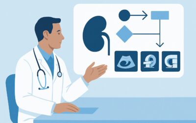
A 58-year-old patient is in your urology clinic for their one-year follow-up after a robotic partial nephrectomy for a pT1b clear cell renal cell carcinoma. They feel well and have no new complaints. Your surveillance protocol calls for cross-sectional imaging, but the choice of modality—and the rationale behind it—is critical for long-term management. You need to select the study that best balances diagnostic accuracy for detecting recurrence against the cumulative risks of repeated scans over many years. This article details the clinical workflow for this exact scenario, explaining why the American College of Radiology (ACR) gives its highest rating to a specific imaging test. For this patient, ‘MRI abdomen without and with IV contrast’ is rated Usually Appropriate.
Who Fits This Clinical Scenario for Renal Cell Carcinoma Follow-up?
This guidance applies specifically to patients who have undergone curative-intent surgery—either a radical or partial nephrectomy—for clinically localized renal cell carcinoma (RCC). The primary goal of imaging in this context is surveillance: the scheduled, periodic search for disease recurrence in an asymptomatic patient.
This workflow is distinct from other, similar-sounding clinical situations. It is crucial to differentiate this scenario from:
- Follow-up after ablation: Patients treated with thermal ablation (e.g., cryoablation, radiofrequency ablation) have different imaging needs. The post-ablation zone can be complex, and the patterns of local recurrence differ from those seen after surgical excision. This requires a separate imaging strategy, detailed in a different ACR variant.
- Active surveillance of a small renal mass: This applies to patients who have not undergone treatment and are being monitored to assess a tumor’s growth rate and characteristics. The imaging goal here is to track the primary tumor, not to search for post-treatment recurrence.
- Staging or restaging of known metastatic disease: This workflow is not intended for patients with known metastatic RCC or those with new symptoms concerning for metastasis. Symptomatic patients require a diagnostic workup, which may involve different imaging protocols or modalities.
This article focuses exclusively on the asymptomatic, post-nephrectomy patient undergoing routine surveillance.
What Diagnoses Are You Working Up in Post-Nephrectomy Surveillance?
Surveillance imaging after nephrectomy is not a general screen; it is a focused search for specific patterns of disease recurrence. The differential diagnosis, while narrow, is critical, as is the ability to distinguish recurrence from expected post-surgical changes.
The most important consideration is local recurrence. This refers to the regrowth of cancer in the surgical bed—the nephrectomy fossa after a radical nephrectomy, or at the resection margin after a partial nephrectomy. Differentiating enhancing tumor nodularity from non-enhancing scar tissue or benign post-operative fluid collections (seromas) is the central challenge for the radiologist and the primary task of the imaging study.
Next is the detection of regional recurrence. This typically involves metastatic spread to the retroperitoneal or hilar lymph nodes. Enlarged or newly appeared nodes are highly suspicious and must be carefully evaluated.
The imaging study must also assess for new primary tumors in the contralateral kidney. While less common than recurrence of the original cancer, a second, independent RCC can develop, particularly in patients with underlying genetic syndromes. Surveillance provides an opportunity to detect these new tumors at an early, treatable stage.
Finally, abdominal imaging provides a screen for distant metastases to common sites in the upper abdomen, such as the liver, adrenal glands, and pancreas. While the lungs are the most frequent site of RCC metastasis, these abdominal organs are also at risk and are well-covered by the recommended studies.
Why Is MRI Abdomen Without and With IV Contrast Usually Appropriate for This Follow-up?
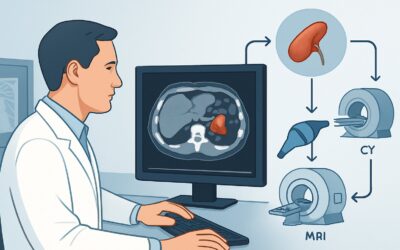
The ACR designates ‘MRI abdomen without and with IV contrast’ as Usually Appropriate for post-nephrectomy RCC surveillance, highlighting its unique advantages in this clinical setting. The rationale is rooted in its superior diagnostic capabilities and safety profile for long-term follow-up.
The primary strength of MRI is its exceptional soft-tissue contrast resolution. This allows for a more confident differentiation between post-surgical scar tissue, which may show some mild, persistent enhancement, and a true recurrent tumor, which typically demonstrates more avid, nodular enhancement. The use of subtraction imaging, where the pre-contrast images are digitally subtracted from the post-contrast images, is particularly powerful for isolating even small areas of true enhancement that might be obscured on other modalities.
A crucial advantage of MRI is the complete absence of ionizing radiation (Adult RRL: O 0 mSv). Patients with RCC often require many years of surveillance with repeated scans. Choosing a radiation-free modality from the outset helps minimize the cumulative radiation dose over the patient’s lifetime, a significant consideration, especially for younger patients.
How do alternative studies compare?
- CT abdomen with IV contrast: This study is also rated Usually Appropriate and is an excellent alternative, particularly if MRI is contraindicated (e.g., incompatible hardware, severe claustrophobia) or less available. However, it involves a moderate dose of ionizing radiation (Adult RRL: ☢☢☢ 1-10 mSv). While CT provides superb anatomic detail, it can sometimes be less definitive than MRI in characterizing subtle enhancement in the surgical bed.
- US kidneys retroperitoneal: Rated May be appropriate, ultrasound is not a primary tool for comprehensive surveillance. While it can effectively evaluate the contralateral kidney for new masses, its sensitivity for detecting small recurrences in the surgical bed or for evaluating retroperitoneal lymph nodes is limited by patient body habitus and overlying bowel gas.
For optimal results when ordering the recommended study, be sure to specify “without and with IV contrast” and provide a clear clinical history on the requisition, such as “Follow-up imaging for status post right partial nephrectomy for clear cell RCC.” This context is vital for the radiologist to tailor the protocol and interpret the findings accurately.
What’s the Next Step After the Surveillance MRI?
The results of the surveillance MRI guide the subsequent clinical workflow, which branches into distinct pathways based on the findings.
- If the study is negative: A negative or benign-appearing scan is the most common outcome. The patient continues with their established, risk-stratified surveillance schedule. This typically involves annual or biennial imaging for a period of years, as determined by the tumor’s original stage and pathology. No immediate action is needed beyond scheduling the next follow-up appointment.
- If the study is positive for recurrence: A definitive finding of local recurrence, regional nodal disease, or new distant metastasis is a significant event. The next step is typically a discussion at a multidisciplinary tumor board involving urology, medical oncology, and radiology. This may lead to a biopsy to confirm the pathology, followed by treatment options such as metastasectomy, systemic therapy (e.g., immunotherapy or targeted therapy), or stereotactic radiation.
- If the study is indeterminate: Indeterminate findings, such as subtle soft-tissue thickening or equivocal enhancement in the surgical bed, present a clinical dilemma. The downstream workflow may involve several options. One common approach is short-interval follow-up imaging, often with the same modality (e.g., repeat MRI in 3-6 months), to assess for stability or change. Alternatively, a different modality, such as an FDG-PET/CT (rated May be appropriate), may be considered to evaluate for metabolic activity if clinical suspicion remains high. In select cases, a percutaneous biopsy may be pursued if the finding is accessible and suspicious enough to alter management.
Pitfalls to Avoid (and When to Get Help)
Navigating post-nephrectomy surveillance requires attention to several common pitfalls to ensure comprehensive and accurate follow-up.
- Forgetting Chest Imaging: The lungs are the most common site of RCC metastasis. Abdominal imaging alone is incomplete surveillance. Protocols must include dedicated chest imaging, typically with a non-contrast CT chest or chest radiography (both rated May be appropriate), at specified intervals.
- Ordering Without IV Contrast: Non-contrast scans are insufficient for recurrence detection. The key feature of a viable tumor is enhancement. Omitting intravenous contrast severely limits the diagnostic utility of either CT or MRI for this indication.
- Misinterpreting Early Post-Surgical Changes: In the first few months after surgery, normal healing tissue and inflammation can mimic recurrence. It is crucial to have a baseline post-operative scan (often done 3-6 months after surgery) to which all future surveillance scans can be compared.
- Ignoring Renal Function: Gadolinium-based contrast agents for MRI and iodinated contrast for CT both carry risks in patients with significant renal impairment. Always check the patient’s estimated Glomerular Filtration Rate (eGFR) before ordering a contrasted study.
If you encounter a complex or indeterminate finding on surveillance imaging, escalation to a multidisciplinary tumor board or a direct consultation with the interpreting radiologist is the most appropriate next step.
Related ACR Topics and Tools
For further reading and to explore adjacent clinical scenarios, the following resources provide authoritative guidance and practical tools for imaging decisions.
- For breadth across all scenarios in Post Treatment Follow-up and Active Surveillance of Renal Cell Carcinoma, see our parent guide: Post Treatment Follow-up and Active Surveillance of Renal Cell Carcinoma: ACR Appropriateness Decoded.
- ACR Appropriateness Criteria Lookup: https://gighz.com/acr-appropriateness-criteria/
- Imaging Protocol Library: https://gighz.com/imaging-protocol-library/
- Radiation Dose Calculator: https://gighz.com/radiation-dose-calculator/
Frequently Asked Questions
Is CT an acceptable alternative to MRI for post-nephrectomy surveillance?
Yes. CT of the abdomen with IV contrast is also rated ‘Usually Appropriate’ by the ACR for this scenario. It is a strong alternative, especially if a patient has contraindications to MRI. The main advantage of MRI is the lack of ionizing radiation, which is preferable for patients requiring long-term, repeated surveillance.
How often should surveillance imaging be performed after nephrectomy?
The frequency and duration of surveillance imaging depend on the pathologic stage and risk features of the original tumor. Guidelines from organizations like the American Urological Association (AUA) provide risk-stratified schedules, but a typical regimen for an intermediate-risk tumor might involve annual imaging for 3-5 years, followed by less frequent or no further imaging.
Do I need to order separate imaging for the chest?
Yes. Standard abdominal CT or MRI protocols do not adequately visualize the entire lung fields, which are the most common site for renal cell carcinoma metastasis. Surveillance plans should include dedicated chest imaging, such as a non-contrast CT chest or a chest radiograph, at intervals determined by the patient’s risk category.
What if my patient has impaired renal function (low eGFR)?
Significant renal impairment requires careful consideration of contrast agents. For patients with very low eGFR, gadolinium-based contrast for MRI may be contraindicated due to the risk of nephrogenic systemic fibrosis (NSF). Similarly, iodinated contrast for CT can cause contrast-induced nephropathy. In these cases, a non-contrast MRI or ultrasound may be considered, or a consultation with radiology is recommended to determine the safest and most effective imaging plan.
Can ultrasound be used for primary surveillance after a nephrectomy?
Ultrasound is rated ‘May be appropriate’ but is not recommended as the primary or sole modality for comprehensive surveillance. While it is useful for evaluating the contralateral kidney, it has significant limitations in visualizing the surgical bed and retroperitoneum, making it less sensitive for detecting local or regional recurrence compared to CT or MRI.
Reviewed by Pouyan Golshani, MD, Interventional Radiologist — May 29, 2026