Why Is Duplex Ultrasound the First Imaging Step for Adult Renal Transplant Dysfunction?
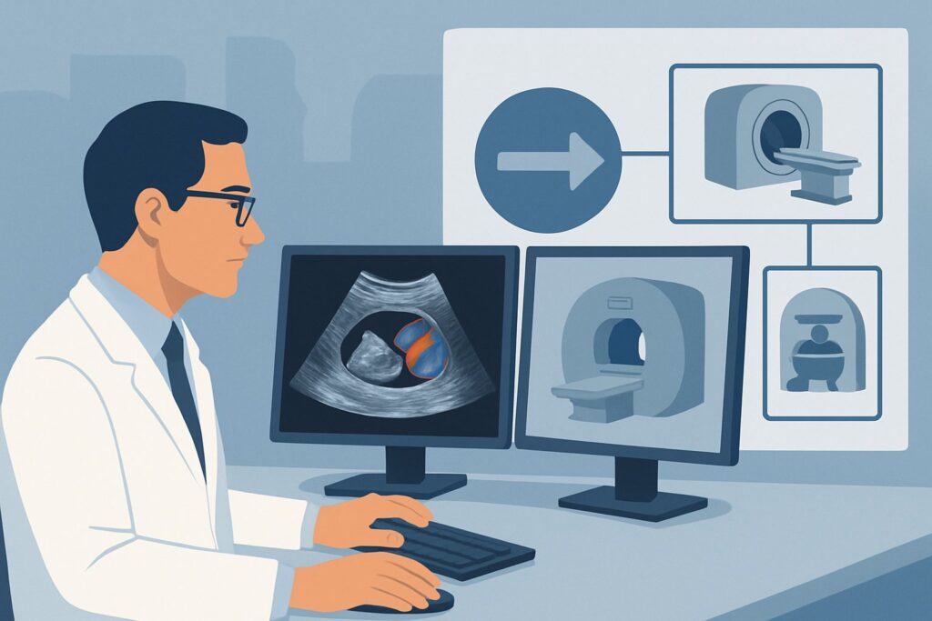
It’s 8 PM on a Tuesday, and you’re covering the nephrology service. The lab calls a critical value: the creatinine on your patient, a 54-year-old who received a kidney transplant six months ago, has jumped from a baseline of 1.4 mg/dL to 2.8 mg/dL over 48 hours. The patient is asymptomatic, and the physical exam is unrevealing. You need to quickly and safely evaluate the allograft for the cause of this acute dysfunction. The immediate question is which imaging study to order first to assess for vascular, urologic, or parenchymal causes without causing further harm. This article details the clinical workflow for this exact scenario, explaining why the American College of Radiology (ACR) designates US duplex Doppler kidney transplant as Usually Appropriate for the initial imaging of suspected renal transplant dysfunction in an adult.
Who Fits This Clinical Scenario for Renal Transplant Dysfunction?
This guidance applies to a specific, common clinical situation: an adult patient with a transplanted kidney who presents with new or worsening dysfunction, defined by rising serum creatinine, decreased urine output, or other clinical signs. This workflow is intended for the initial imaging workup, where no prior imaging for the current episode of dysfunction has been performed.
The key inclusion criteria are:
- Patient: Adult with a history of renal transplantation.
- Presentation: Clinical or laboratory evidence of allograft dysfunction (e.g., elevated creatinine, oliguria).
- Timing: This is the first imaging study being ordered for this specific episode of dysfunction.
It is crucial to distinguish this scenario from related but distinct clinical questions. This guidance does not apply if an initial ultrasound has already been performed and was inconclusive or suggested a specific problem. For those situations, the imaging choice is different. For example, this article is not for a patient whose initial ultrasound was:
- Suspicious for an arterial etiology: Such as focal high velocities suggesting renal artery stenosis.
- Suspicious for a venous etiology: Such as poor or reversed diastolic flow suggesting renal vein thrombosis.
- Unremarkable or indeterminate: Where the initial ultrasound showed no clear cause for the dysfunction.
These represent separate downstream decision points, each with its own recommended imaging pathway. This article focuses exclusively on getting the first step right.
What Diagnoses Are You Working Up in This Scenario?
When a renal allograft fails, the differential diagnosis is broad, spanning vascular, urologic, and parenchymal causes. The goal of initial imaging is to rapidly identify or exclude surgically correctable conditions. The primary diagnostic considerations in this setting fall into three categories.
Vascular Complications: These are among the most urgent diagnoses to consider. Renal artery stenosis (RAS) can occur at the anastomosis site, leading to hypertension and ischemic graft dysfunction. Conversely, renal artery or vein thrombosis is a catastrophic event that can cause irreversible graft loss if not identified immediately. Pseudoaneurysms or arteriovenous fistulas, often related to prior biopsies, are less common but can also compromise graft function.
Urologic and Perinephric Complications: Obstruction is a frequent and often reversible cause of graft dysfunction. Hydronephrosis, caused by a ureteral stricture, stone, or external compression, is a key finding. Perinephric fluid collections are also common and include urinomas (from a urine leak at the ureteral anastomosis), hematomas (post-operative or post-biopsy), and lymphoceles (from disrupted lymphatic channels during surgery). These collections can compress the ureter, leading to secondary obstruction.
Parenchymal Abnormalities: While biopsy is the gold standard for diagnosing parenchymal disease, imaging provides important clues. Acute rejection may cause graft swelling, altered cortical echogenicity, and changes in arterial resistance on Doppler. Acute tubular necrosis (ATN), common in the early post-operative period, can have similar, often nonspecific, imaging findings. Imaging helps exclude other causes and can guide the decision and timing for a biopsy.
Why Is US Duplex Doppler the Recommended Initial Study for This Presentation?
For the initial evaluation of a dysfunctional renal transplant, US duplex Doppler kidney transplant is rated Usually Appropriate by the ACR. This recommendation is based on the modality’s high diagnostic utility, safety profile, and ability to assess the key components of graft health in a single, non-invasive examination.
The strength of duplex ultrasound lies in its comprehensive, multi-faceted evaluation:
- Grayscale Imaging: Provides detailed anatomical information, assessing graft size, cortical thickness, and echotexture. It is highly sensitive for detecting hydronephrosis, which indicates a collecting system obstruction, and for identifying and characterizing perinephric fluid collections like hematomas, urinomas, or lymphoceles.
- Color Doppler: Maps blood flow throughout the graft, allowing for rapid assessment of global and segmental perfusion. It can immediately identify a lack of flow, a critical finding concerning for vascular thrombosis.
- Pulsed-Wave Doppler: Quantifies blood flow dynamics. By sampling velocities in the main renal artery, segmental arteries, and interlobar arteries, sonographers can calculate the Resistive Index (RI) and Pulsatility Index (PI). Elevated RIs can be a nonspecific sign of dysfunction from rejection, ATN, or obstruction, while abnormally high velocities in the main renal artery are highly suggestive of stenosis.
Critically, ultrasound involves no ionizing radiation (0 mSv) and does not require iodinated contrast, avoiding the risk of contrast-induced nephropathy in a patient with already compromised renal function.
Alternative cross-sectional imaging modalities are rated Usually not appropriate as a first-line test. For example:
- CT abdomen and pelvis with IV contrast: While providing excellent anatomic detail, it exposes the patient to significant ionizing radiation (☢☢☢ 1-10 mSv) and requires nephrotoxic contrast media, making it a poor initial choice.
- MRA abdomen and pelvis without and with IV contrast: This avoids radiation but is more expensive, less readily available, and the use of gadolinium-based contrast agents is a concern in patients with significantly reduced renal function due to the risk of nephrogenic systemic fibrosis.
These advanced modalities are reserved for problem-solving when ultrasound is inconclusive or points toward a specific abnormality that requires more precise characterization.
Once you’ve decided on duplex ultrasound, our protocol guide covers the technique, contrast, and reading principles: US Carotid Doppler.
What’s Next After US Duplex Doppler? Downstream Workflow
The results of the initial duplex ultrasound will guide your next steps, often routing you to a different, more specific clinical scenario. The workflow branches based on the findings.
- If the study is positive for a specific finding:
- High-grade hydronephrosis: This suggests obstruction. The next step is often a percutaneous nephrostomy tube placement for decompression and an antegrade nephrostogram to define the level and cause of obstruction. This falls under the workup for a suspected extrinsic or urologic etiology.
- Elevated peak systolic velocities in the renal artery (>200-250 cm/s): This is highly suspicious for renal artery stenosis. The patient now fits the scenario “US suspicious for, but not conclusive for arterial etiology,” and the next logical step is often CTA, MRA, or conventional angiography.
- Absent flow in the renal vein: This indicates renal vein thrombosis, a surgical emergency. The patient now fits the “US suspicious for, but not conclusive for venous etiology” scenario, and a confirmatory study like CT venography may be performed while mobilizing the surgical team.
- A large or complex perinephric fluid collection: This may require image-guided drainage for both diagnostic and therapeutic purposes.
- If the study is negative or indeterminate:
- When the ultrasound is completely normal or shows only nonspecific findings like mildly elevated resistive indices, it effectively rules out urgent surgical causes like high-grade obstruction or vascular thrombosis. The patient now fits the “US unremarkable or indeterminate” scenario. In this case, the underlying cause is more likely to be parenchymal (e.g., acute rejection, ATN, or drug toxicity), and the definitive next step is almost always a renal allograft biopsy.
Pitfalls to Avoid (and When to Get Help)
Navigating the workup of renal transplant dysfunction requires careful attention to detail. Here are a few common pitfalls to avoid:
- Not providing clinical context: The quality of a duplex ultrasound report depends heavily on the information provided. Always include the time since transplant, baseline creatinine, current creatinine, and specific clinical concern (e.g., rule out RAS, rule out obstruction) on the order.
- Ignoring prior studies: Comparison to previous ultrasounds is essential. A subtle change in graft size, RI values, or the size of a fluid collection can be the only clue to an evolving problem.
- Accepting a limited study: If the patient has a body habitus that limits visualization of the vascular anastomosis, do not accept an indeterminate report. Escalate to the radiologist to discuss whether a different modality like CTA or MRA is warranted.
- Delaying the scan: In cases of acute anuria or rapid creatinine rise, time is kidney. Ultrasound should be obtained emergently to rule out a catastrophic vascular event or high-grade obstruction. If you suspect graft torsion or thrombosis, escalate immediately to your transplant surgery colleagues.
Related ACR Topics and Tools
For a comprehensive overview of all clinical scenarios related to imaging renal transplants, and for tools to help you order the right study every time, please see the following resources.
- For breadth across all scenarios in Renal Transplant Dysfunction, see our parent guide: Renal Transplant Dysfunction: ACR Appropriateness Decoded.
- To look up other clinical presentations, use the Imaging Appropriateness Selector tool.
- To review the technical details of imaging studies, explore the Imaging Protocol Library.
- To discuss radiation exposure with patients, consult the Radiation Dose Calculator.
Frequently Asked Questions
Why not order a CT or MRI first to get a more detailed look at the transplant?
While CT and MRI provide excellent anatomical detail, they are considered ‘Usually not appropriate’ for the initial workup. The primary reasons are safety and utility. CT involves ionizing radiation and often requires iodinated contrast, which can be harmful to a struggling kidney. MRI may require gadolinium, which carries risks in patients with poor renal function. Duplex ultrasound is non-invasive, uses no radiation or nephrotoxic contrast, and can effectively evaluate the three most critical areas: blood flow, collecting system, and parenchyma, making it the ideal first-line test.
What specific information should I include when ordering the duplex ultrasound?
To maximize the diagnostic yield, provide key clinical context. Include the date of transplant, the patient’s baseline and current serum creatinine levels, blood pressure, any relevant medications (especially calcineurin inhibitors), and the specific clinical question (e.g., ‘evaluate for renal artery stenosis,’ ‘rule out hydronephrosis’). This information helps the sonographer and radiologist tailor the exam and interpret the findings accurately.
Does a normal duplex ultrasound rule out acute rejection?
No, a normal ultrasound does not rule out acute rejection. While severe rejection can cause findings like graft swelling, increased cortical echogenicity, and elevated resistive indices (RIs), early or mild rejection may have no sonographic signs. The primary role of the initial ultrasound is to exclude surgical or vascular causes of dysfunction. If the ultrasound is normal, the next step is typically a graft biopsy to diagnose parenchymal processes like rejection or acute tubular necrosis (ATN).
What is the role of a nuclear medicine scan (like a MAG3 or DTPA scan) in this initial workup?
According to the ACR, nuclear medicine renal scans are ‘Usually not appropriate’ for the initial imaging of transplant dysfunction. While these scans are excellent for quantifying graft function, perfusion, and excretion, they provide limited anatomical information. Duplex ultrasound is superior for the initial step because it can simultaneously assess anatomy, vascularity, and potential urologic complications. A nuclear scan may be used later as a problem-solving tool, for example, to differentiate ATN from rejection or to confirm a urine leak suspected on other imaging.
How soon after transplant can these complications occur?
Complications can occur at any time, but the timing can offer clues. Hyperacute rejection occurs within minutes to hours. Vascular thrombosis is most common in the first few days to weeks. Acute tubular necrosis (ATN) typically presents in the first week. Urine leaks and urinomas often appear in the first few weeks. Acute cellular rejection is most common in the first few months. Chronic issues like renal artery stenosis or ureteral strictures can develop months to years after transplantation.
Reviewed by Pouyan Golshani, MD, Interventional Radiologist — June 27, 2026