What Is the Best Imaging for Staging Vulvar Cancer (≤4 cm, >1 mm Invasion)?
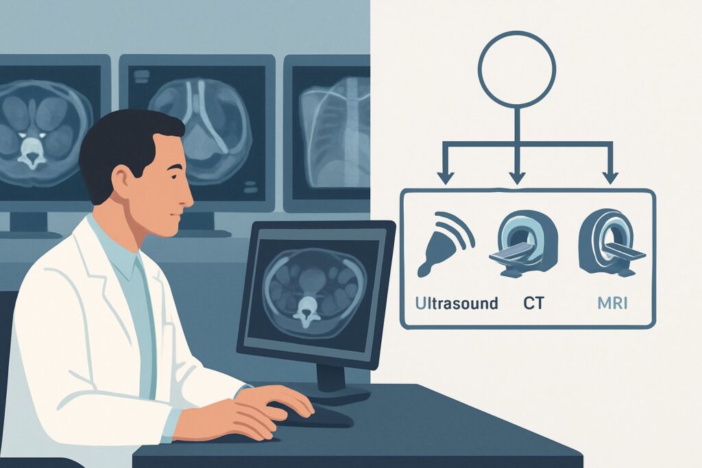
A 68-year-old woman presents to your gynecologic oncology clinic with a 3 cm lesion on her left labia majora. A recent biopsy confirmed squamous cell carcinoma with 3 mm of stromal invasion. On physical examination, the tumor appears confined to the vulva with no palpable groin lymphadenopathy. To plan for either wide local excision with sentinel lymph node biopsy or a more extensive resection, you need to accurately assess the local extent of the tumor and evaluate the regional lymph nodes. This article details the American College of Radiology (ACR) guided workflow for this specific clinical scenario. For initial staging of a vulvar tumor ≤ 4 cm with >1 mm invasion, MRI pelvis without and with IV contrast is rated Usually appropriate.
Who Fits This Clinical Scenario for Staging Vulvar Cancer?
This imaging workflow is specifically for patients with newly diagnosed, untreated vulvar cancer who meet a precise set of clinical and pathologic criteria. The recommendations apply when the primary tumor has been biopsied and confirmed as malignant, with findings that place it beyond the earliest stage but not yet in the advanced category.
Inclusion Criteria for This Workflow:
- Tumor Size: The primary tumor measures 4 cm or less in its greatest dimension.
- Stromal Invasion: The biopsy confirms stromal invasion of greater than 1 mm. This is a critical threshold, as it significantly increases the risk of lymph node metastasis compared to microinvasive disease.
- Tumor Location: The cancer is confined to the vulva or perineum, or has only minimal, early involvement of adjacent structures like the distal urethra, distal vagina, or anus.
This guidance should not be applied to patients with different presentations. For instance, a patient with a smaller tumor (≤ 2 cm) and minimal stromal invasion (≤ 1 mm) falls into a different ACR variant where advanced imaging may not be necessary. Conversely, a patient with a tumor larger than 4 cm or one that grossly invades the bladder, rectum, or pelvic bone requires a more extensive workup, often involving different imaging modalities to assess for distant disease.
What Are You Evaluating During the Staging Workup?
While the diagnosis of vulvar cancer is already established by biopsy, the purpose of staging imaging is to answer critical questions about the extent of the disease, which directly dictates the surgical and adjuvant therapy plan. The imaging workup is focused on assessing three key areas.
Local Tumor Extent: The primary goal is to precisely delineate the boundaries of the tumor. Imaging must determine if the cancer extends to or invades adjacent critical structures, such as the clitoris, urethra, vagina, or anus. The degree of involvement in these areas can change the surgical approach from a wide local excision to a more radical vulvectomy or necessitate neoadjuvant therapy.
Inguinal and Femoral Lymph Node Status: This is the most important prognostic factor in early-stage vulvar cancer. The lymphatic drainage of the vulva primarily flows to the superficial and deep inguinal lymph nodes. Imaging aims to identify nodes that are suspicious for metastasis based on criteria like size, loss of the fatty hilum, irregular borders, and central necrosis. This information is crucial for planning either a sentinel lymph node biopsy (SLNB) or a full inguinofemoral lymphadenectomy.
Pelvic Lymph Node Status: While less common in this specific scenario, metastasis to the pelvic lymph nodes (external iliac, internal iliac, obturator) can occur, typically after inguinal node involvement. Identifying pelvic node disease upstages the cancer to FIGO Stage IVB and radically alters treatment, often shifting the plan from surgery-centric to primary chemoradiation. Imaging provides a non-invasive survey of these deeper nodal basins.
Why Is MRI of the Pelvis the Recommended Study for Staging This Tumor?
The ACR designates MRI pelvis without and with IV contrast as Usually appropriate for this scenario because of its superior ability to evaluate both the primary tumor and regional lymph nodes without using ionizing radiation.
The excellent soft-tissue contrast of Magnetic Resonance Imaging (MRI) is unmatched for delineating the primary vulvar tumor from surrounding normal tissues. It can accurately depict the tumor’s size, depth of invasion, and relationship to the urethra, vagina, and anal sphincter complex. The addition of IV gadolinium-based contrast enhances the tumor, making its margins more conspicuous and helping to identify subtle areas of invasion that might be missed on non-contrast sequences.
For nodal assessment, MRI evaluates lymph nodes based on both size and morphologic features. While size is an imperfect criterion, features like central necrosis, spiculated margins, and rounded shape are highly suggestive of metastatic involvement. High-resolution, small field-of-view T2-weighted and diffusion-weighted imaging (DWI) sequences can further increase the sensitivity for detecting abnormal nodes in the inguinal and pelvic regions.
Why Other Studies Are Rated Lower:
- CT abdomen and pelvis with IV contrast is rated Usually not appropriate. While CT is excellent for evaluating pelvic nodes and distant disease in many other cancers, its soft-tissue resolution is significantly lower than MRI’s for characterizing the primary vulvar tumor. It also exposes the patient to a moderate dose of ionizing radiation (ACR Relative Radiation Level ☢☢☢ 1-10 mSv), whereas MRI has no ionizing radiation (RRL O 0 mSv).
- FDG-PET/CT skull base to mid-thigh is also rated Usually not appropriate for this specific initial staging scenario. While highly sensitive for metastatic disease, its spatial resolution is lower than MRI, making it less reliable for defining the local extent of small primary tumors. Furthermore, it involves a high radiation dose (RRL ☢☢☢☢ 10-30 mSv) and is generally reserved for more advanced disease or suspected recurrence.
In summary, MRI provides the most comprehensive local and regional assessment needed to guide therapy in this patient population, balancing high diagnostic accuracy with patient safety.
What Is the Downstream Workflow After a Pelvic MRI?
The results of the pelvic MRI are a critical input for the multidisciplinary tumor board and directly guide the next steps in management. The downstream workflow branches based on the imaging findings regarding the primary tumor and, most importantly, the lymph nodes.
If MRI shows a confined primary tumor and no suspicious lymph nodes: The patient is an excellent candidate for the standard surgical approach: wide local excision of the primary tumor combined with a sentinel lymph node biopsy (SLNB). The negative predictive value of a high-quality MRI can increase confidence in proceeding with the less morbid SLNB over a full lymphadenectomy.
If MRI shows suspicious inguinal or femoral lymph nodes: This finding changes the management of the nodal basin. The next step is often to confirm metastasis pathologically. This may be done via an ultrasound-guided fine-needle aspiration (FNA) of the suspicious node, a procedure rated as May be appropriate. If the FNA is positive, the patient will typically undergo a full therapeutic inguinofemoral lymphadenectomy instead of an SLNB. If the FNA is negative or non-diagnostic, an SLNB or full dissection may still be considered based on the level of suspicion.
If MRI shows direct invasion into adjacent structures (e.g., urethra, anus): This finding may upstage the patient and alter the surgical plan. A more radical excision may be required. In some cases, if achieving negative surgical margins would be too morbid, the patient may be considered for neoadjuvant chemoradiation to shrink the tumor before surgery.
If MRI shows suspicious pelvic (iliac/obturator) lymph nodes: This is an ominous finding that suggests more advanced disease. Management would typically shift away from primary surgery towards definitive chemoradiation, as surgical resection of this extent of disease is associated with high morbidity and poor outcomes.
Common Pitfalls to Avoid in Staging Early Vulvar Cancer
Navigating the workup for this specific scenario requires careful attention to detail to avoid common errors that can lead to improper staging and suboptimal treatment.
- Under-staging based on clinical exam alone: Relying solely on palpation to assess groin nodes is a significant pitfall. Clinical examination has a low sensitivity for detecting nodal metastases, and imaging is essential for a more accurate assessment.
- Choosing CT over MRI: For many abdominopelvic malignancies, CT is the workhorse. However, for vulvar cancer, MRI’s superior soft-tissue resolution is critical for evaluating the primary tumor. Defaulting to CT is a common error that provides less information about local extent.
- Omitting IV contrast with MRI: An MRI ordered “without contrast” is rated May be appropriate (Disagreement) by the ACR. While non-contrast sequences are valuable, post-contrast imaging significantly improves the delineation of the tumor and the characterization of lymph nodes. Always specify “without and with IV contrast” unless there is a clear contraindication.
- Ignoring patient-specific contraindications: Before ordering an MRI with contrast, confirm the patient has no contraindications, such as incompatible metallic implants, severe claustrophobia, or impaired renal function that would preclude the use of gadolinium-based contrast agents.
If imaging findings are equivocal or conflict with the clinical picture, discussion at a multidisciplinary gynecologic oncology tumor board is the essential next step to determine the best path forward.
Related ACR Topics and Tools
This article focuses on a single, specific clinical scenario. For a comprehensive overview of all variants and recommendations, or to explore the tools used to develop this guidance, please refer to the resources below.
- For breadth across all scenarios in Staging and Follow-up of Vulvar Cancer, see our parent guide: Staging and Follow-up of Vulvar Cancer: ACR Appropriateness Decoded.
- To look up other clinical scenarios, use the ACR Appropriateness Criteria Lookup.
- For detailed imaging techniques, consult the Imaging Protocol Library.
- To discuss radiation exposure with patients for alternative studies, see the Radiation Dose Calculator.
Frequently Asked Questions
Why is MRI preferred over PET/CT for this specific stage of vulvar cancer?
For initial staging of tumors ≤ 4 cm with >1 mm invasion, MRI is preferred because its high spatial and soft-tissue resolution is superior for defining the local extent of the primary tumor and its relationship to adjacent structures like the urethra and anus. While PET/CT is excellent for detecting distant metastases, it has lower spatial resolution and is less accurate for small primary tumors. It also involves significant radiation exposure and is generally reserved for more advanced disease or suspected recurrence, making it ‘Usually not appropriate’ for this scenario.
If my patient has a contraindication to MRI (e.g., a pacemaker), what is the next best imaging test?
If MRI is contraindicated, the ACR rates ‘CT abdomen and pelvis with IV contrast’ as ‘Usually not appropriate’ for this scenario. However, in a situation where the recommended test cannot be performed, a lower-rated test may become the best available option. A contrast-enhanced CT can still provide valuable information about lymph node status and gross invasion of pelvic organs, though it is less sensitive for subtle local extension. This decision should be made in consultation with a radiologist and the gynecologic oncology team.
Is lymphoscintigraphy still necessary if the MRI is negative for suspicious nodes?
Yes. MRI and lymphoscintigraphy serve different purposes. MRI assesses lymph node morphology for signs of metastatic replacement. Lymphoscintigraphy, rated as ‘May be appropriate,’ is a functional mapping procedure used to identify the specific ‘sentinel’ lymph nodes that drain the tumor. If the plan is a sentinel lymph node biopsy (SLNB), lymphoscintigraphy is performed preoperatively to guide the surgeon to the correct nodes for excision, regardless of the MRI findings.
Does a negative MRI for lymph nodes mean I can skip a lymph node biopsy?
No. While a high-quality MRI showing no suspicious nodes is reassuring, it does not have a high enough negative predictive value to rule out microscopic metastases. Current guidelines still require surgical-pathologic staging of the regional lymph nodes, typically via a sentinel lymph node biopsy, for patients with greater than 1 mm of stromal invasion.
What if the tumor is exactly 4 cm? Does this workflow still apply?
Yes, this workflow applies to tumors that are less than or equal to 4 cm. If the tumor measures exactly 4.0 cm, these recommendations are appropriate. If the tumor is definitively greater than 4 cm (e.g., 4.1 cm), the patient fits into a different ACR scenario for more advanced disease, which has a different set of imaging recommendations, often including imaging of the chest to rule out distant metastases.
Reviewed by Pouyan Golshani, MD, Interventional Radiologist — May 29, 2026