What Is the Best Initial Imaging for a Child with Chronic Progressive Ataxia?
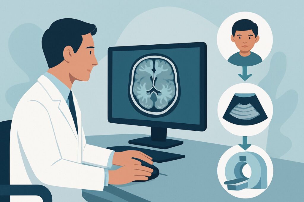
It’s late in your pediatric clinic day, and you’re seeing an 8-year-old whose parents report a slow but steady decline in coordination over the past six months. Once agile on the playground, the child now has a wide-based, unsteady gait and increasing trouble with fine motor tasks like writing. The neurologic exam confirms truncal and appendicular ataxia. You suspect a central nervous system process, but the differential is broad. The immediate clinical question is what initial imaging study will provide the most diagnostic information safely and efficiently. For this specific presentation, the American College of Radiology (ACR) rates MRI head without and with IV contrast as “Usually Appropriate.”
Who Fits This Clinical Scenario?
This guidance applies specifically to a child presenting with chronic progressive ataxia. This clinical pattern is defined by an insidious onset and a gradual, persistent worsening of ataxic symptoms over weeks, months, or even years. The key features are the chronicity (not acute) and the progression (not static or episodic). A thorough history and serial examinations are crucial to establish this trajectory.
This workflow is distinct from other pediatric ataxia presentations. It is essential to differentiate this scenario from:
- Acute Ataxia: This involves a sudden onset of ataxia, often over hours to days. The differential is different, frequently pointing toward post-infectious cerebellitis, intoxication, or acute trauma. This presentation is covered in a separate ACR variant.
- Chronic Nonprogressive Ataxia: In this case, the ataxia is long-standing but stable. The deficit is often present from early childhood and does not worsen over time, suggesting a congenital or static structural anomaly like cerebellar hypoplasia.
- Recurrent or Episodic Ataxia: This involves distinct episodes of ataxia with a return to a normal or near-normal neurologic baseline between events. This pattern suggests channelopathies or metabolic disorders.
Applying this article’s imaging strategy to these other scenarios may lead to a delayed or incorrect diagnosis.
What Diagnoses Are You Working Up in This Scenario?
The insidious and progressive nature of the ataxia in this scenario narrows the differential diagnosis toward structural, degenerative, or metabolic etiologies. The primary goal of initial imaging is to identify treatable structural lesions and provide clues for the subsequent diagnostic workup.
Posterior Fossa Tumors: This is a primary and urgent consideration in any child with progressive ataxia. Tumors such as medulloblastoma, ependymoma, and pilocytic astrocytoma arise in or near the cerebellum and brainstem, directly disrupting motor coordination pathways. Imaging is critical for detection, characterization, and surgical planning.
Leukodystrophies and Metabolic Disorders: Many inherited metabolic diseases present with progressive neurologic decline, including ataxia. Conditions like metachromatic leukodystrophy, Alexander disease, or mitochondrial disorders can cause characteristic patterns of white matter signal abnormality (leukoencephalopathy) or cerebellar atrophy that are well-visualized on MRI.
Congenital Structural Abnormalities: While often presenting as nonprogressive ataxia, some structural anomalies like a Chiari I malformation can become symptomatic and progressive during childhood due to factors like altered cerebrospinal fluid (CSF) dynamics or the development of a syrinx. Imaging definitively identifies these anatomical issues.
Inherited Neurodegenerative Disorders: Conditions like Friedreich ataxia or ataxia-telangiectasia are classic causes of chronic progressive ataxia. While the diagnosis is often confirmed with genetic testing, neuroimaging is essential to rule out structural mimics. In later stages, these disorders may show characteristic patterns of cerebellar and spinal cord atrophy.
Why Is MRI Head without and with IV Contrast the Recommended Study?
The ACR designates MRI head without and with IV contrast as “Usually Appropriate” because it offers the highest diagnostic yield for the key differential diagnoses in this scenario, all without using ionizing radiation.
The superior soft-tissue contrast of Magnetic Resonance Imaging (MRI) is unmatched for evaluating the cerebellum, brainstem, and cerebral white matter—the primary locations of pathology in chronic progressive ataxia. It can clearly delineate tumor margins, identify subtle white matter signal changes seen in leukodystrophies, and define structural anomalies like a Chiari malformation.
The addition of intravenous gadolinium-based contrast is crucial. Many posterior fossa tumors, particularly malignant ones like medulloblastoma, demonstrate avid enhancement, which helps in their detection and characterization. Contrast can also highlight areas of active inflammation or demyelination, which may be relevant in less common inflammatory etiologies. While an MRI head without IV contrast is also rated “Usually Appropriate,” proceeding directly to a contrast-enhanced study often prevents the need for a second scan if a suspicious non-enhancing lesion is found, saving time and reducing the burden on the patient and family.
Alternative studies are rated lower for specific reasons:
- CT head without IV contrast: Rated “May be appropriate,” a Computed Tomography (CT) scan is significantly less sensitive than MRI for this indication. Beam-hardening artifacts often obscure detail in the posterior fossa, potentially missing non-calcified, non-hemorrhagic tumors or subtle white matter disease. While faster and more accessible, its use of ionizing radiation (pediatric dose ☢☢☢ 0.3-3 mSv) and lower diagnostic utility make it a suboptimal first choice unless MRI is contraindicated or unavailable.
- MRA head and neck without IV contrast: Rated “Usually not appropriate,” a Magnetic Resonance Angiography (MRA) is designed to visualize blood vessels. Since the primary differential for chronic progressive ataxia is parenchymal (tumors, degenerative changes) rather than vascular, an MRA is not the correct initial study and would fail to assess the relevant anatomy properly.
What’s Next After MRI Head without and with IV contrast? Downstream Workflow
The results of the initial MRI will guide the subsequent clinical pathway. The workflow branches based on whether the findings are positive, negative, or indeterminate.
If the MRI is positive for a mass lesion: An urgent referral to pediatric neuro-oncology and neurosurgery is the immediate next step. Further imaging, such as an MRI of the complete spine with and without contrast, is often required for staging, particularly to look for drop metastases common with tumors like medulloblastoma or ependymoma. Biopsy or surgical resection will provide a definitive histopathologic diagnosis.
If the MRI is negative (structurally normal): A normal brain MRI significantly narrows the differential but does not end the workup. The focus shifts toward non-structural causes. The next steps typically involve a comprehensive metabolic and genetic evaluation. This may include testing for specific inherited ataxias (e.g., Friedreich ataxia gene panel), whole-exome sequencing, and screening for metabolic disorders with serum and urine studies.
If the MRI shows nonspecific findings: Results like diffuse cerebellar volume loss or nonspecific white matter signal changes require further investigation. These findings may be consistent with a neurodegenerative or metabolic process. In this situation, an MRI of the complete spine (rated “May be appropriate”) could be considered to look for associated spinal cord atrophy. Consultation with a pediatric neurologist or geneticist is essential to correlate the imaging pattern with the clinical picture and guide further specialized testing.
Pitfalls to Avoid (and When to Get Help)
Navigating the workup for chronic progressive ataxia requires careful consideration to avoid common diagnostic errors.
- Misclassifying the Temporal Profile: Mistaking a chronic progressive course for an acute or episodic one can lead to an inappropriate and low-yield workup. A detailed history is paramount.
- Accepting a “Normal” CT Scan: Given the low sensitivity of CT for posterior fossa pathology, a normal CT scan does not adequately rule out a tumor or white matter disease in this clinical context. Persisting with the workup to obtain an MRI is critical.
- Omitting IV Contrast: Ordering a non-contrast MRI can miss enhancing lesions. If a tumor is on the differential, a contrast-enhanced study should be the default initial examination.
- Delaying the Imaging: While the progression is chronic, the underlying cause could be a malignancy that requires timely intervention. Unexplained progressive ataxia warrants prompt neuroimaging.
If the clinical picture is complex, the imaging is equivocal, or the diagnosis remains elusive after initial steps, early consultation with a pediatric neurologist is strongly recommended.
Related ACR Topics and Tools
For a comprehensive overview of all clinical scenarios related to pediatric ataxia, please refer to our parent topic hub article. For further exploration of imaging guidelines, protocols, and radiation safety, the following resources are available.
- For breadth across all scenarios in Ataxia-Child, see our parent guide: Ataxia-Child: ACR Appropriateness Decoded.
- ACR Appropriateness Criteria Lookup — for adjacent scenarios
- Imaging Protocol Library — for technique on the recommended study
- Radiation Dose Calculator — for cumulative dose conversations
Frequently Asked Questions
Is an MRI of the spine also needed for the initial workup?
Not usually for the initial workup. The ACR rates ‘MRI complete spine without and with IV contrast’ as ‘May be appropriate.’ It is typically reserved as a downstream study. If the initial brain MRI identifies a posterior fossa tumor known to cause spinal ‘drop’ metastases (like medulloblastoma), a spine MRI becomes essential for staging. It may also be considered if the brain MRI is normal but a spinal cord process or a condition like Friedreich ataxia (which causes cord atrophy) is suspected.
Why is CT rated so much lower than MRI for chronic progressive ataxia?
CT is rated lower primarily due to its limited ability to visualize the soft tissues of the posterior fossa (cerebellum and brainstem). Beam-hardening artifacts from the surrounding dense skull base often obscure detail, making it difficult to detect non-calcified tumors, inflammation, or the subtle white matter changes of leukodystrophy. MRI provides far superior contrast and detail of these structures without using ionizing radiation, making it the clear first choice.
Does a normal brain MRI rule out all serious causes of progressive ataxia?
No. A normal brain MRI is a crucial step that effectively rules out most structural causes like tumors or Chiari malformations. However, it does not rule out many genetic and metabolic neurodegenerative disorders. Conditions like Friedreich ataxia may show a normal or near-normal brain MRI in the early stages. A normal MRI result shifts the diagnostic focus toward genetic and metabolic testing.
What if my patient cannot tolerate an MRI without sedation?
The need for sedation is a common and important consideration in pediatric imaging. The diagnostic superiority of MRI for this specific clinical question is so significant that proceeding with the study under sedation is standard practice and strongly recommended. The risks associated with anesthesia are generally considered lower than the risks of missing a potentially life-threatening diagnosis like a brain tumor by choosing a less sensitive test like CT.
Should I order an MR spectroscopy (MRS) with the initial MRI?
The ACR rates ‘MR spectroscopy head without IV contrast’ as ‘Usually not appropriate’ for the initial evaluation. While MRS can provide metabolic information about brain tissue and can sometimes help differentiate tumor types or identify metabolic disorders, it is considered an advanced, problem-solving sequence. It is not part of the routine initial imaging protocol and is typically added by the radiologist or ordered in follow-up if the initial MRI findings are indeterminate.
Reviewed by Pouyan Golshani, MD, Interventional Radiologist — May 30, 2026