Should You Order MRI or CT for a Child with a Post-Traumatic Seizure?

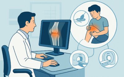
It’s 10 p.m. in the emergency department, and you’re evaluating a 9-year-old who fell off his scooter an hour ago. He was wearing a helmet and had no initial loss of consciousness, but while waiting to be seen, he had a two-minute generalized tonic-clonic seizure. He is now postictal but stable. The immediate clinical question is clear: what is the right imaging study to order first to evaluate for a structural cause of this post-traumatic seizure? This article provides a detailed workflow for this specific scenario, guiding you through the differential diagnosis, imaging rationale, and downstream decisions. For this presentation, the American College of Radiology (ACR) rates both `MRI head without IV contrast` and `CT head without IV contrast` as Usually Appropriate.
Who Fits This Clinical Scenario for Post-Traumatic Seizures?
This guidance applies to a specific patient population: children aged 1 month to 17 years who present for initial imaging after experiencing a seizure following a known, non-abusive traumatic event. The key inclusion criteria are the clear temporal link between a physical trauma and the subsequent seizure, and the fact that this is the first imaging workup for this event.
It is critical to distinguish this scenario from others that require a different diagnostic approach:
- Abusive Head Trauma (AHT): This workflow explicitly excludes suspected non-accidental trauma. AHT has a different pattern of injury and a distinct, often more extensive, imaging protocol that may include skeletal surveys and specific MRI sequences.
- Neonatal Seizures (0-29 days): Seizures in the first month of life have a unique differential diagnosis, including hypoxic-ischemic injury, metabolic disorders, and congenital anomalies. This requires a separate evaluation detailed in its own ACR variant.
- Seizures Without a Traumatic Precedent: If a child presents with a new-onset focal or generalized seizure without a clear history of recent trauma, the workup shifts. The evaluation would then follow the guidance for focal seizures or generalized seizures, which prioritizes different potential etiologies like epilepsy syndromes, tumors, or infections.
Correctly identifying your patient’s presentation ensures the imaging choice is tailored to the most likely and most critical underlying causes.
What Diagnoses Are You Working Up in a Child with a Post-Traumatic Seizure?
When a child has a seizure after head trauma, the primary goal of imaging is to identify an acute structural brain injury that may be causing cortical irritation. The differential diagnosis is centered on traumatic brain injury (TBI) and its immediate sequelae.
Intracranial Hemorrhage
This is a primary and urgent concern. Blood products are highly irritating to the cerebral cortex and are a common cause of acute seizures. Imaging must be able to detect several types of hemorrhage:
- Epidural Hematoma (EDH): Often associated with an overlying skull fracture and arterial injury.
- Subdural Hematoma (SDH): Results from tearing of bridging veins.
- Subarachnoid Hemorrhage (SAH): Blood in the sulci and cisterns.
- Intraparenchymal Hemorrhage / Contusion: Bleeding directly into the brain tissue, often at sites of coup-contrecoup injury.
Cortical Contusion and Gliosis
Direct bruising of the brain parenchyma, even without significant hemorrhage, can create an epileptogenic focus. These contusions represent areas of neuronal and glial injury. Over time, these areas can evolve into gliosis (scarring), which can become a nidus for chronic post-traumatic epilepsy.
Skull Fracture
While not an injury to the brain itself, a skull fracture, particularly a depressed fracture, can directly irritate or injure the underlying cortex, leading to a seizure. Identifying the presence and type of fracture is crucial for management and potential neurosurgical intervention.
Cerebral Edema
Significant trauma can cause focal or diffuse cerebral edema (swelling). This can increase intracranial pressure and contribute to neuronal dysfunction and seizures. Imaging helps assess the severity of edema and look for signs of herniation.
Why Are MRI or CT Recommended for a Child’s Post-Traumatic Seizure?
For the initial imaging of a child with a post-traumatic seizure, the ACR Appropriateness Criteria rate both `MRI head without IV contrast` and `CT head without IV contrast` as Usually Appropriate. The choice between them often depends on clinical stability, institutional resources, and the specific information needed.
MRI head without IV contrast is often considered the superior study if the patient is stable and it can be performed in a timely manner. Its primary advantage is its exceptional soft-tissue contrast, which allows for the detailed evaluation of the brain parenchyma. MRI is more sensitive than CT for detecting subtle cortical contusions, non-hemorrhagic diffuse axonal injury, and small amounts of extra-axial blood. Crucially, MRI achieves this without using ionizing radiation (Pediatric Relative Radiation Level: O, 0 mSv), a significant consideration in the pediatric population.
CT head without IV contrast is an excellent and often more practical first-line option, especially in the emergency setting. Its main advantages are speed and accessibility. A non-contrast head CT can be completed in minutes, which is critical for an unstable patient. It is highly sensitive for detecting acute intracranial hemorrhage, significant cerebral edema, and skull fractures—the most life-threatening traumatic injuries. However, this speed comes with the trade-off of exposure to ionizing radiation (Pediatric Relative Radiation Level: ☢☢☢, 0.3-3 mSv).
Why are other studies rated lower for this scenario?
- MRI head without and with IV contrast is rated Usually not appropriate. In the acute trauma setting, intravenous contrast does not typically add diagnostic value for identifying the primary injuries (hemorrhage, contusion, fracture). Contrast is reserved for later when there is a specific concern for secondary complications like infection or vascular injury, which are not part of the initial workup.
- US head is rated Usually not appropriate. While it avoids radiation, head ultrasound is only useful in infants with an open anterior fontanelle, providing a limited acoustic window to the brain. It lacks the sensitivity and global view needed to rule out the key traumatic injuries in most children and is inadequate for this scenario.
What’s Next After Initial Imaging? Downstream Workflow
The results of the initial CT or MRI will dictate the immediate next steps in management, which almost always involve consultation with pediatric neurology and potentially neurosurgery.
If the study is POSITIVE for acute injury:
A finding of significant intracranial hemorrhage, a depressed skull fracture, or severe cerebral edema is a neurosurgical emergency. The immediate next step is an urgent consultation with neurosurgery to determine if surgical intervention (e.g., craniotomy for hematoma evacuation) is required. The child will require admission to an intensive care unit for close neurologic monitoring, management of intracranial pressure, and initiation of anti-epileptic drug (AED) therapy to prevent further seizures.
If the study is NEGATIVE:
A normal head CT or MRI is reassuring, as it rules out an immediate life-threatening structural injury. However, it does not eliminate the possibility of a concussion or microscopic injury causing the seizure. The downstream workflow includes:
- Neurology Consultation: To guide decisions on the need for and duration of AED therapy.
- Electroencephalogram (EEG): An EEG is often obtained to evaluate for epileptiform discharges, which can help risk-stratify the patient for future seizures and guide treatment.
- Observation: The patient will typically be admitted for a period of observation to monitor for any further seizures or change in neurologic status.
If the study is INDETERMINATE:
Findings like a subtle cortical contusion or a small, non-operative hemorrhage require careful management. This typically involves admission, neurology consultation, and serial neurologic exams. A follow-up imaging study (often MRI, if the initial study was CT) may be performed in 24-48 hours to ensure the injury is not evolving.
Common Pitfalls to Avoid in Pediatric Post-Traumatic Seizures
Navigating this clinical scenario requires avoiding several common pitfalls to ensure optimal patient care.
- Delaying Imaging in an Unstable Patient: While MRI offers superior detail, its longer acquisition time is a risk for a clinically unstable child. Prioritize a rapid non-contrast head CT to quickly rule out a life-threatening bleed that requires immediate intervention.
- Over-reassurance from a Normal CT: A normal head CT is good news, but it does not fully exclude underlying parenchymal injury. If seizures recur or the child has a persistent focal neurologic deficit, a follow-up MRI is warranted to look for subtle contusions or other injuries that CT may miss.
- Forgetting Radiation Dose: In a stable child where both CT and MRI are viable options, defaulting to CT without considering the radiation-free alternative of MRI is a potential pitfall. Always apply the As Low As Reasonably Achievable (ALARA) principle.
- Dismissing a Single Seizure: A single, brief post-traumatic seizure is a significant event. It signals a potentially serious underlying brain injury and warrants a full diagnostic workup and consultation, not premature discharge from the emergency department.
If there are any signs of rapidly worsening neurologic status, such as a declining Glasgow Coma Scale (GCS) score or new focal deficits, escalate immediately to neurosurgery and anesthesiology for airway protection and potential surgical management.
Related ACR Topics and Tools
For a comprehensive overview of all pediatric seizure scenarios and for help with ordering the right study, the following resources are available:
- For breadth across all scenarios in Seizures-Child, see our parent guide: Seizures-Child: ACR Appropriateness Decoded.
- Imaging Appropriateness Selector: Access the full ACR guidelines for this and other clinical presentations.
- Imaging Protocol Library: Review detailed institutional imaging protocols for head CT and MRI.
- Radiation Dose Calculator: Estimate and discuss cumulative radiation exposure with families when considering CT.
Frequently Asked Questions
If both CT and MRI are ‘Usually Appropriate’, which one should I choose?
The choice depends on patient stability and resource availability. For an unstable child or in an emergency setting where speed is critical, a non-contrast head CT is the preferred first step to rapidly detect life-threatening hemorrhage or fractures. If the child is clinically stable and MRI is readily available, it is often favored for its superior ability to detect subtle brain injuries without using ionizing radiation.
Is a single, brief seizure after minor head trauma enough to warrant imaging?
Yes. An immediate post-traumatic seizure is a significant clinical event, regardless of the apparent severity of the initial trauma. It is considered a marker of moderate to severe traumatic brain injury and substantially increases the likelihood of finding an intracranial injury on imaging. Therefore, neuroimaging is indicated.
If the initial non-contrast head CT is negative, is any further imaging needed?
Not always, but it depends on the clinical course. If the child recovers well with no further seizures or neurologic deficits, a negative CT may be sufficient. However, if seizures recur, if a new focal deficit develops, or if there is a prolonged alteration of consciousness, a follow-up MRI is strongly recommended to look for injuries that may not be visible on CT, such as non-hemorrhagic contusions or diffuse axonal injury.
Why is contrast not recommended for the initial imaging in this scenario?
In the acute setting of a post-traumatic seizure, the primary goal is to identify traumatic injuries like hemorrhage, fracture, and edema. Intravenous contrast does not improve the detection of these specific findings. Contrast is typically used to evaluate for breakdown of the blood-brain barrier, which is more relevant for concerns like tumor, infection, or subacute/chronic inflammation—diagnoses that are not the primary focus of the initial post-traumatic workup.
Does this guidance apply to a seizure that occurs days or weeks after a head injury?
This guidance is for ‘initial imaging’ in the acute or early post-traumatic period. A seizure that occurs more than one week after the injury is classified as a late post-traumatic seizure. While the underlying cause is still the trauma, the imaging workup may be different, often focusing on MRI to detect gliosis (scarring) or encephalomalacia that could be an epileptic focus. The urgency may also be lower compared to an immediate post-traumatic seizure.
Reviewed by Pouyan Golshani, MD, Interventional Radiologist — May 26, 2026