What Is the Best Initial Imaging for a Resolved Transient Ischemic Attack (TIA)?

A 68-year-old man presents to the emergency department after an episode of slurred speech and right-hand weakness that lasted approximately 20 minutes. By the time he arrived for evaluation, his symptoms had completely resolved. His neurologic examination is now entirely normal. You diagnose a clinical transient ischemic attack (TIA) and must decide on the initial imaging workup to assess his risk for a future, potentially disabling stroke. This is a critical decision point where imaging directly informs secondary prevention strategies, from antiplatelet therapy to surgical intervention. This article provides a focused workflow for this specific scenario, guided by the American College of Radiology (ACR) Appropriateness Criteria, which rates several studies as appropriate, with US duplex Doppler carotid artery being a key non-invasive component of the initial evaluation, rated Usually Appropriate.
Who Fits This Clinical Scenario for a Resolved TIA?
This guidance applies specifically to an adult patient who has experienced a transient ischemic attack (TIA), defined as a temporary episode of neurologic dysfunction caused by focal brain, spinal cord, or retinal ischemia, without acute infarction. The crucial inclusion criteria for this workflow are:
- Transient Symptoms: The patient presented with focal neurologic deficits consistent with a vascular territory.
- Complete Resolution: All symptoms have fully resolved at the time of clinical evaluation.
- Initial Imaging: This is the first imaging workup for this specific event.
It is critical to distinguish this scenario from similar but distinct clinical presentations that require different imaging pathways. This guidance does not apply if:
- The patient has persistent neurologic deficits. A patient with ongoing symptoms is managed under the acute ischemic stroke protocol, which prioritizes immediate brain imaging to assess for infarction and eligibility for thrombolysis or thrombectomy.
- The primary suspicion is venous sinus thrombosis. If the patient presents with headache, seizures, or non-arterial-territory deficits, a dedicated workup for venous thrombosis is indicated.
- Symptoms are non-focal. Presentations like syncope, generalized weakness, or lightheadedness without focal findings are less likely to represent a TIA and may require a different diagnostic approach.
What Diagnoses Are You Working Up in a Patient with Resolved TIA Symptoms?
The primary goal of imaging after a TIA is to identify the underlying cause to prevent a subsequent stroke. The differential diagnosis guides the choice of studies, focusing on identifying treatable sources of ischemia.
Cervical Carotid Artery Stenosis
This is a primary and highly actionable diagnosis. Atherosclerotic plaque in the internal carotid artery is a common source of artery-to-artery emboli that can cause TIA or stroke. Identifying a hemodynamically significant stenosis (typically >50-70%) is critical, as it may warrant intervention with carotid endarterectomy or stenting for secondary stroke prevention.
Evidence of Ischemic Injury
Even with resolved symptoms, a significant portion of patients with a clinical TIA will have evidence of acute infarction on diffusion-weighted MRI. Identifying a small infarct confirms the ischemic origin of the event, heightens the urgency of the workup, and solidifies the diagnosis over TIA mimics.
Intracranial Atherosclerotic Disease (ICAD)
Stenosis of major intracranial arteries (e.g., middle cerebral artery, basilar artery) is another important cause of ischemic events, particularly in certain patient populations. While management is typically medical, identifying ICAD is crucial for risk stratification and tailoring antiplatelet or statin therapy.
Other Embolic Sources or TIA Mimics
While imaging focuses on vascular pathology, it also helps rule out other conditions. A cardioembolic source (e.g., from atrial fibrillation) is a major cause, and while brain imaging doesn’t identify the cardiac source itself, it can show a pattern of multiple infarcts in different vascular territories suggestive of an embolic shower. Imaging also helps exclude non-vascular mimics like a small tumor, seizure focus, or complex migraine.
Why Is a Combined Brain and Vascular Workup Recommended for a TIA?
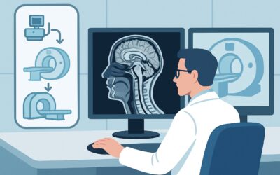
For a patient with a resolved TIA, the ACR rates several imaging studies as Usually Appropriate, reflecting the need to evaluate both the brain parenchyma and the cervicocerebral vasculature. The optimal initial workup often involves a combination of non-invasive studies to achieve these two goals efficiently and safely.
1. Brain Parenchymal Imaging: MRI or CT
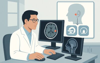
The first step is to image the brain. MRI head without IV contrast is highly sensitive for detecting acute ischemia via diffusion-weighted imaging (DWI) and is considered a superior study. It can identify small infarcts that are invisible on CT, confirming the diagnosis and increasing the estimated short-term stroke risk. CT head without IV contrast is also rated Usually Appropriate. It is faster and more accessible, making it a practical first choice in many emergency settings. Its primary role is to exclude hemorrhage or a non-ischemic structural lesion, though its sensitivity for small acute infarcts is low.
2. Cervical Vascular Imaging: US, CTA, or MRA
The second, equally important step is to assess the carotid arteries. US duplex Doppler carotid artery is rated Usually Appropriate and is an excellent non-invasive initial study. It involves no radiation (0 mSv) or contrast and is highly effective at identifying and grading stenosis at the carotid bifurcation, the most common site of symptomatic plaque. CTA neck with IV contrast is another Usually Appropriate option that provides a comprehensive view of the aortic arch through the intracranial vessels. It is fast and accurate but involves both radiation (1-10 mSv) and iodinated contrast.
Rationale for Lower-Rated Alternatives
Studies like Arteriography cervicocerebral are rated Usually not appropriate for initial evaluation. While it is the gold standard for vessel imaging, this invasive catheter-based procedure carries a small but real risk of causing a stroke itself and is reserved for problem-solving or planning interventions after non-invasive imaging has identified a significant lesion. Similarly, perfusion studies (MRI head perfusion or CT head perfusion) are Usually not appropriate in this scenario because the patient’s symptoms have resolved, and the clinical question is about etiology and risk, not identifying a penumbra in an acute stroke.
Once you’ve decided on US duplex Doppler carotid artery as part of the workup, our protocol guide covers the technique and interpretation principles in detail: US Carotid Doppler.
What’s Next After Initial Imaging? Downstream Workflow
The results of your initial brain and vascular imaging will guide the subsequent clinical pathway. The workflow branches based on whether a clear etiology is found.
If a significant carotid stenosis is found:
If US Doppler or CTA identifies a high-grade stenosis (e.g., >70% symptomatic stenosis), the patient requires urgent consultation with vascular surgery or neurointerventional specialists to be considered for carotid endarterectomy or stenting. The timing of intervention is critical, with maximal benefit seen when performed within two weeks of the initial event.
If brain imaging is positive but vascular imaging is negative:
If an acute infarct is seen on MRI but the cervical and intracranial vascular imaging is unremarkable, the focus shifts to other potential causes. The highest priority is investigating a cardioembolic source. This typically involves an EKG, telemetry monitoring, and often an echocardiogram to look for atrial fibrillation, valvular disease, or intracardiac thrombus.
If all initial imaging is negative:
If both brain imaging (ideally MRI with DWI) and comprehensive vascular imaging are negative, the patient is considered to have a lower short-term risk, but still requires aggressive medical management. This includes optimizing antiplatelet therapy, high-intensity statin treatment, and strict blood pressure and glucose control. Further investigation for less common causes, such as a hypercoagulable state, may be considered based on the patient’s age and clinical context.
Pitfalls to Avoid (and When to Get Help)
Navigating the TIA workup requires avoiding several common pitfalls to ensure patient safety and an accurate diagnosis.
- Underestimating Urgency: A TIA is a neurologic emergency. Delaying the diagnostic workup significantly increases the risk of a completed stroke in the hours and days following the event. The workup should ideally be completed within 24-48 hours.
- Incomplete Vascular Imaging: Evaluating only the cervical carotids can miss other etiologies. If initial carotid ultrasound is negative, consider imaging the intracranial vessels with CTA or MRA, as intracranial atherosclerotic disease is a significant cause of stroke.
- Over-reliance on Non-contrast CT: While useful for ruling out hemorrhage, a negative non-contrast head CT does not rule out an ischemic event. If clinical suspicion for TIA is high, an MRI with DWI should be strongly considered.
- Forgetting the Heart: A negative vascular workup does not mean the workup is over. Always consider and investigate for a cardioembolic source, as this is one of the most common causes of ischemic events.
If the non-invasive imaging results are equivocal or contradictory (e.g., US and CTA disagree on stenosis severity), escalate to a specialist or consider a problem-solving study like conventional arteriography.
Related ACR Topics and Tools
This article focuses on a single clinical scenario. For a comprehensive overview of imaging for all stroke and stroke-related conditions, from acute stroke to venous thrombosis, refer to the parent topic guide. For tools to help with ordering, protocoling, and explaining studies, see the resources below.
- For breadth across all scenarios in Cerebrovascular Diseases-Stroke and Stroke-Related Conditions, see our parent guide: Cerebrovascular Diseases-Stroke and Stroke-Related Conditions: ACR Appropriateness Decoded.
- ACR Appropriateness Criteria Lookup — for adjacent scenarios
- Imaging Protocol Library — for technique on the recommended study
- Radiation Dose Calculator — for cumulative dose conversations
Frequently Asked Questions
Is a non-contrast head CT sufficient for the initial TIA workup?
A non-contrast head CT is rated ‘Usually Appropriate’ by the ACR and is an excellent first test to rule out hemorrhage or a large structural lesion. However, its sensitivity for small, acute infarcts is low. An MRI of the head with diffusion-weighted imaging (DWI) is far more sensitive and is preferred for confirming an ischemic event if available in a timely manner.
If the carotid ultrasound is normal, is the vascular workup complete?
Not necessarily. A normal carotid ultrasound effectively rules out significant stenosis in the cervical internal carotid arteries, but it does not evaluate the vertebral arteries or the intracranial circulation. If no other cause is found, CTA or MRA of the head and neck may be needed to look for intracranial atherosclerotic disease or other vascular pathologies.
Why isn’t catheter angiography the first test if it’s the ‘gold standard’?
Digital subtraction angiography (DSA), or catheter angiography, is an invasive procedure that carries a small but significant risk of causing a stroke, bleeding, or vessel dissection. For this reason, it is rated ‘Usually not appropriate’ for the initial diagnosis. It is reserved for cases where non-invasive tests are inconclusive or when an intervention like stenting is being planned.
What is the role of perfusion imaging (CTP/MRP) in a resolved TIA?
Perfusion imaging is designed to identify the ischemic penumbra—brain tissue at risk of infarction—in an acute stroke. Since the symptoms in this scenario have already resolved, there is no ongoing ischemic event to assess with perfusion. Therefore, both CT and MR perfusion are rated ‘Usually not appropriate’ for the initial workup of a resolved TIA.
Do all patients with a clinical TIA need imaging?
Yes. Modern guidelines strongly recommend urgent imaging for all patients with a suspected TIA. Imaging helps confirm the diagnosis (by identifying an infarct on DWI), determines the underlying cause (like carotid stenosis), and critically informs risk stratification (e.g., using the ABCD2 score in conjunction with imaging findings) to guide urgent secondary prevention.
Reviewed by Pouyan Golshani, MD, Interventional Radiologist — May 30, 2026