What Is the Best Initial Imaging for Acquired Sensorineural Hearing Loss?
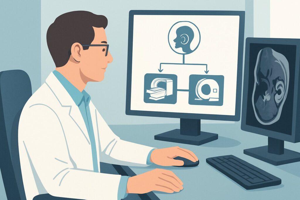
A 55-year-old patient presents to your outpatient clinic with a six-month history of worsening hearing in his left ear, accompanied by a persistent, high-pitched ringing. An audiogram confirms asymmetric sensorineural hearing loss (SNHL). Your primary goal is to evaluate for a retrocochlear cause, and you need to decide on the most appropriate initial imaging study. This scenario requires a specific, high-yield imaging approach to visualize the delicate structures of the inner ear and cranial nerves. This article provides a focused workflow for this exact presentation, explaining why the American College of Radiology (ACR) designates ‘MRI head and internal auditory canal without and with IV contrast’ as Usually Appropriate for the initial imaging of acquired sensorineural hearing loss.
Who Fits This Clinical Scenario for Acquired Sensorineural Hearing Loss?
This imaging workflow is designed for patients with an audiometrically confirmed diagnosis of acquired sensorineural hearing loss. The key features that place a patient in this category include hearing loss that is not present from birth and originates from the inner ear (cochlea) or the auditory nerve pathway to the brain. This guidance is most applicable when the SNHL is:
- Asymmetric or Unilateral: Hearing loss is significantly worse in one ear compared to the other. This is the classic indication for imaging to rule out a structural lesion.
- Progressive: The hearing loss has gradually worsened over weeks, months, or years.
- Associated with other neurologic symptoms: The patient also reports unilateral tinnitus (ringing in the ear), vertigo, or disequilibrium, raising suspicion for pathology affecting the vestibulocochlear nerve complex.
It is crucial to distinguish this presentation from similar, but distinct, clinical scenarios that require different imaging pathways. This guidance does not apply to:
- Acquired Conductive Hearing Loss: If the hearing loss is due to a problem in the outer or middle ear (e.g., otosclerosis, ossicular chain disruption), the primary imaging modality is often different. This presentation is covered in the ACR variant for conductive hearing loss.
- Episodic Vertigo without Significant Hearing Loss: Patients with classic symptoms of peripheral vertigo (e.g., benign paroxysmal positional vertigo or Meniere’s disease) often do not require initial imaging, as outlined in the ACR variant for episodic vertigo.
- Congenital Hearing Loss: The workup for hearing loss present at birth, especially in the context of cochlear implant candidacy, involves a different set of imaging priorities focused on inner ear anatomy.
What Diagnoses Are You Working Up with Imaging for Acquired SNHL?
When ordering imaging for acquired, asymmetric SNHL, the primary goal is to identify or exclude structural lesions along the auditory pathway, from the inner ear to the brainstem. The differential diagnosis is focused and drives the choice of imaging modality.
The most critical diagnosis to exclude is a vestibular schwannoma, also known as an acoustic neuroma. These are benign, slow-growing tumors arising from the Schwann cells of the vestibulocochlear nerve (cranial nerve VIII). While they account for a small percentage of SNHL cases, their classic presentation is progressive, unilateral hearing loss and tinnitus. Early detection is key to preserving hearing and preventing complications from brainstem compression.
Less commonly, other tumors can arise in the same location—the internal auditory canal (IAC) and the cerebellopontine angle (CPA). These include meningiomas, which arise from the meninges, and epidermoid cysts, which are congenital lesions that can grow to compress adjacent nerves. Metastatic disease to this region is rare but possible.
Inflammatory or demyelinating conditions are also on the differential. Multiple sclerosis (MS) can present with SNHL if a demyelinating plaque involves the cochlear nerve root entry zone in the brainstem, though this is an uncommon initial symptom. Other inflammatory processes like neurosarcoidosis can also cause cranial neuropathies, including hearing loss.
Finally, while less frequent, vascular abnormalities can cause these symptoms. A prominent vascular loop, typically from the anterior inferior cerebellar artery (AICA), can compress the vestibulocochlear nerve, leading to neurovascular conflict. Aneurysms or arteriovenous malformations in the CPA are rare causes but are important to identify.
Why Is MRI of the Head and IAC the Recommended Initial Study for Acquired SNHL?
The ACR designates MRI head and internal auditory canal without and with IV contrast as Usually Appropriate because it provides the highest diagnostic yield for the key pathologies in the differential diagnosis. Magnetic Resonance Imaging offers superior soft-tissue contrast, which is essential for visualizing the small cranial nerves, the fluid-filled structures of the inner ear, and the brainstem.
The sensitivity of contrast-enhanced MRI for detecting vestibular schwannomas, even small intracanalicular tumors measuring only a few millimeters, is exceptionally high. These tumors, along with meningiomas, typically demonstrate avid enhancement after the administration of gadolinium-based contrast, making them highly conspicuous. A non-contrast MRI is also rated as Usually Appropriate and can be sufficient in many cases, especially with modern high-resolution sequences (like FIESTA or CISS). However, the addition of contrast increases confidence in excluding small enhancing lesions and helps characterize any abnormality that is found.
Alternative studies are rated lower for specific reasons. For instance, CT temporal bone without IV contrast is rated as May be appropriate. While excellent for evaluating the bony anatomy of the temporal bone and middle ear, its ability to detect small, soft-tissue tumors within the IAC or CPA is significantly limited compared to MRI. A small vestibular schwannoma can be easily missed on CT. CT is a reasonable alternative only when MRI is contraindicated (e.g., incompatible implanted device, severe claustrophobia) or unavailable.
Other studies like a standard CT head without IV contrast are rated Usually not appropriate. This study lacks the resolution and soft-tissue detail to adequately assess the IAC and CPA, and it would almost certainly miss the primary pathologies being investigated in this scenario. Furthermore, CT involves ionizing radiation (☢☢☢ 1-10 mSv), whereas MRI does not (O 0 mSv), a key consideration, especially in younger patients.
What’s Next After MRI? Downstream Workflow
The results of the MRI will guide the subsequent clinical pathway. The downstream workflow depends on whether the findings are positive, negative, or indeterminate.
If the study is positive for a vestibular schwannoma or other CPA mass: The next step is a referral to Otolaryngology (Neurotology) and/or Neurosurgery. Management options for a vestibular schwannoma include observation with serial imaging, stereotactic radiosurgery, or surgical resection. The choice depends on the tumor size, patient age, degree of hearing loss, and other symptoms. The initial MRI provides the critical information needed for this specialist consultation.
If the study is negative: A negative, high-quality MRI effectively rules out a structural retrocochlear cause for the SNHL. In this case, the hearing loss is typically considered idiopathic or cochlear in origin (e.g., presbycusis, noise-induced, or viral). The patient’s care continues under the management of an audiologist and otolaryngologist. Treatment focuses on hearing amplification (hearing aids), aural rehabilitation, and monitoring for any changes in hearing status. No further imaging is typically required unless new, localizing neurologic symptoms develop.
If the study is indeterminate or shows an unexpected finding: In some cases, the MRI may reveal findings of uncertain significance, such as a prominent vascular loop contacting the nerve or subtle, non-specific enhancement. If a vascular loop is suspected to be the cause of symptoms (neurovascular conflict), an MRA may be considered. If inflammatory or demyelinating disease is suspected based on other findings, a full brain MRI with contrast may be warranted to look for characteristic lesions elsewhere.
Pitfalls to Avoid (and When to Get Help)
In the workup of acquired SNHL, several common pitfalls can delay diagnosis or lead to unnecessary testing. First, avoid ordering a standard “MRI brain” without specifying the need for thin-section imaging through the internal auditory canals. A routine brain protocol may not have the slice thickness or resolution to detect small intracanalicular tumors. Always specify “IAC protocol” or “for asymmetric SNHL.” Second, do not default to CT as a screening tool; its low sensitivity for the primary differential makes it an inadequate first-line test unless MRI is absolutely contraindicated. Finally, attributing asymmetric SNHL to aging without a proper workup can lead to a significant delay in diagnosing a treatable condition like a vestibular schwannoma. Any significant asymmetry warrants investigation. If a patient presents with sudden-onset SNHL accompanied by acute, persistent vertigo and other brainstem signs (e.g., diplopia, ataxia, facial numbness), this constitutes a neurologic emergency, and the patient should be evaluated immediately for a posterior circulation stroke.
Related ACR Topics and Tools
This article focuses on a single, common clinical scenario. For a comprehensive overview of imaging for all related presentations, from conductive hearing loss to central vertigo, refer to the parent guide. For help with ordering, protocoling, and discussing radiation dose, the following GigHz resources are available.
- For breadth across all scenarios in Hearing Loss and/or Vertigo, see our parent guide: Hearing Loss and/or Vertigo: ACR Appropriateness Decoded.
- Imaging Appropriateness Selector — for adjacent scenarios
- Imaging Protocol Library — for technique on the recommended study
- Radiation Dose Calculator — for cumulative dose conversations
Frequently Asked Questions
Is an MRI without contrast sufficient for working up acquired sensorineural hearing loss?
According to the ACR, an MRI of the head and internal auditory canals without contrast is also rated as ‘Usually Appropriate.’ Modern high-resolution, heavily T2-weighted sequences (such as FIESTA or CISS) are highly sensitive for detecting non-enhancing lesions like epidermoid cysts and can often identify even small vestibular schwannomas by visualizing the nerve displacement within the CSF-filled canal. However, adding gadolinium contrast increases diagnostic confidence, especially for very small enhancing tumors, and is generally preferred if there are no contraindications.
My patient has a pacemaker. Can I still order an MRI?
The presence of a pacemaker or other implantable electronic device requires careful consideration. Many modern devices are ‘MR-conditional,’ meaning an MRI can be performed safely under specific protocols and with cardiology or device representative supervision. You must confirm the exact make and model of the device and consult with the radiology department’s safety officer before ordering. If the device is ‘MR-unsafe’ or the required conditions cannot be met, a high-resolution CT of the temporal bone with contrast is the next best alternative, though it is less sensitive for small tumors.
What if the hearing loss is bilateral and symmetric?
Bilateral, symmetric sensorineural hearing loss is most commonly due to presbycusis (age-related hearing loss), noise exposure, or ototoxicity. In the absence of other neurologic signs or symptoms, imaging is generally not indicated for this presentation. The workup is typically focused on audiology and management with hearing aids.
How should I order the MRI to ensure the correct protocol is used?
When ordering, be specific. The order should state ‘MRI Head and Internal Auditory Canals (IACs) without and with contrast.’ In the indication or clinical history field, clearly write ‘Asymmetric sensorineural hearing loss, rule out vestibular schwannoma’ or ‘Unilateral tinnitus and SNHL.’ This clinical information is critical for the radiologist and technologist to select the appropriate high-resolution, thin-section sequences focused on the cerebellopontine angles and IACs.
Does a vascular loop contacting the nerve on MRI explain the patient’s symptoms?
The finding of a vascular loop (commonly the AICA) in contact with the vestibulocochlear nerve is common in asymptomatic individuals, making its clinical significance controversial. While it can be a cause of symptoms in a minority of patients (neurovascular conflict), it is often an incidental finding. The diagnosis is one of exclusion, made after ruling out other pathologies and correlating the imaging finding closely with the clinical symptoms. This finding alone does not automatically confirm the cause of the hearing loss.
Reviewed by Pouyan Golshani, MD, Interventional Radiologist — June 27, 2026