What Is the Best Initial Imaging for Acute Scrotal Pain Without Trauma?
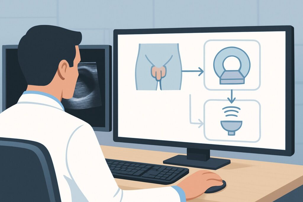
A 16-year-old boy arrives in the emergency department at 2 a.m. with sudden, severe left-sided scrotal pain and nausea that woke him from sleep. There was no injury, and he has no prior history of a scrotal mass. On exam, the testicle is high-riding and exquisitely tender. The immediate clinical concern is testicular torsion, a time-sensitive urologic emergency. You need to confirm the diagnosis and assess testicular viability, but which imaging study provides the fastest, most accurate answer? This article provides a detailed workflow for this specific scenario: the initial imaging workup for an adult or child with acute, non-traumatic scrotal pain. For this presentation, the American College of Radiology (ACR) rates US duplex Doppler scrotum as *Usually Appropriate*.
Who Fits This Clinical Scenario?
This guidance applies to a specific and urgent clinical presentation: a patient, either a child or an adult, who presents with the acute onset of scrotal pain. The key inclusion criteria are the suddenness of the pain and the absence of two important confounders: significant trauma and a known, pre-existing scrotal mass. This workflow is designed for the initial diagnostic step when the cause of pain is unknown.
This article is *not* intended for patients with:
- Recent Scrotal Trauma: A history of a direct blow, straddle injury, or penetrating trauma changes the differential diagnosis to include testicular rupture, hematoma, or contusion, which may alter the imaging approach.
- A Known Scrotal Mass: If a patient has a previously identified or palpable mass that is now painful, the workup is focused on characterizing the mass and evaluating for complications like hemorrhage or malignant change.
- Chronic or Subacute Pain: This guidance is for acute pain (typically less than 24-48 hours). Patients with pain lasting for weeks or months require a different diagnostic algorithm, often focused on varicocele, chronic epididymitis, or post-vasectomy pain syndrome.
Correctly identifying your patient’s presentation as acute and non-traumatic is crucial for applying this imaging pathway effectively.
What Diagnoses Are You Working Up in This Scenario?
When a patient presents with acute scrotal pain, the imaging choice is driven by the need to rapidly differentiate between surgical emergencies and conditions managed medically. The differential diagnosis is broad, but a few key possibilities guide the workup.
The most critical diagnosis to exclude is **testicular torsion**. This occurs when the spermatic cord twists, cutting off blood supply to the testicle. It is a true urologic emergency, as irreversible ischemic damage can occur within 4 to 6 hours. It is most common in neonates and peripubertal boys but can occur at any age. The classic presentation is abrupt, severe, unilateral pain, often associated with nausea and vomiting.
A more common cause of acute scrotal pain, particularly in postpubertal males, is **epididymitis or epididymo-orchitis**. This is an inflammatory condition, usually infectious, of the epididymis and/or testis. The onset of pain may be slightly more gradual than in torsion, and patients may have associated urinary symptoms or fever. While not a surgical emergency, it requires prompt medical treatment with antibiotics.
In prepubertal boys, **torsion of the appendix testis or appendix epididymis** is a frequent cause. These are small, vestigial remnants of embryonic ducts that can twist on their stalk. The pain can mimic testicular torsion, though it is often less severe. The classic “blue dot” sign on the scrotum is pathognomonic but not always present. This is a self-limiting condition managed with supportive care.
Other important, though less common, considerations include an **incarcerated inguinal hernia**, where a loop of bowel becomes trapped and extends into the scrotum, and **Fournier’s gangrene**, a rare but life-threatening necrotizing fasciitis of the perineum that requires emergent surgical debridement.
Why Is US duplex Doppler scrotum the Recommended Study for This Presentation?
For the initial imaging of acute, non-traumatic scrotal pain, the ACR designates both US scrotum and US duplex Doppler scrotum as *Usually Appropriate*. However, the addition of duplex Doppler is critical for evaluating the primary concern: testicular torsion. This makes it the de facto first-line study.
The rationale is based on several key advantages:
- Assessment of Blood Flow: Duplex Doppler ultrasound is highly sensitive and specific for detecting arterial and venous blood flow within the testicle. The complete absence of flow on the affected side is the hallmark of testicular torsion, while increased flow (hyperemia) is characteristic of epididymitis or orchitis. This single capability allows the imager to directly answer the most urgent clinical question.
- Excellent Anatomic Detail: Beyond blood flow, ultrasound provides detailed images of the scrotal contents. It can identify an enlarged, inflamed epididymis in epididymitis, detect hydroceles or pyoceles, characterize testicular echotexture, and identify a torsed testicular appendage.
- Safety and Accessibility: Ultrasound uses no ionizing radiation (adult and pediatric radiation relative level: O, 0 mSv) and does not require IV contrast. It is fast, relatively inexpensive, and widely available in most emergency settings, making it ideal for a time-sensitive workup.
Alternative imaging modalities are rated *Usually not appropriate* for this initial evaluation.
- CT Pelvis (with or without IV contrast): This study exposes the patient to significant ionizing radiation (adult RRL: ☢☢☢ 1-10 mSv; pediatric RRL: ☢☢☢☢ 3-10 mSv) and offers poor soft-tissue resolution of the testicular parenchyma compared to ultrasound. It cannot reliably assess intratesticular blood flow and is not the correct tool for this indication.
- Nuclear Medicine Scrotal Scan: While historically used to assess testicular perfusion, this modality has been largely replaced by Doppler ultrasound. It is less available, takes longer to perform, involves radiation exposure (adult RRL: ☢☢☢ 1-10 mSv), and provides far less anatomic information.
The clear diagnostic superiority, safety profile, and accessibility of duplex Doppler ultrasound make it the undisputed initial imaging test for this clinical scenario.
What’s Next After US duplex Doppler scrotum? Downstream Workflow
The results of the scrotal ultrasound will guide your immediate next steps, branching into distinct clinical pathways. The goal is to act decisively based on the findings to preserve testicular viability or initiate appropriate medical therapy.
If the study is positive for testicular torsion: This is a surgical emergency. The report will note an absence of detectable blood flow in the affected testicle. The immediate next step is an urgent urology consultation for surgical exploration. Do not delay consultation while waiting for a formal report if the preliminary findings from the sonographer or radiologist indicate torsion. The mantra “time is testis” is paramount, with the highest salvage rates achieved when detorsion occurs within 6 hours of symptom onset.
If the study is negative for torsion but positive for epididymitis/orchitis: The ultrasound will show a thickened, hyperemic epididymis, often with increased blood flow to the adjacent testicle. This confirms an inflammatory process. The patient can be managed medically with appropriate antibiotics (covering common pathogens like *Chlamydia trachomatis* and *Neisseria gonorrhoeae* in sexually active men, or enteric organisms in others), analgesics, and scrotal support.
If the study is negative or indeterminate: A technically adequate ultrasound that shows normal, symmetric blood flow to both testes effectively rules out complete torsion. If the patient’s symptoms resolve, they may be discharged with close follow-up. However, if a high clinical suspicion for torsion persists despite a non-diagnostic or negative ultrasound (e.g., in cases of intermittent torsion-detorsion), an urgent urology consultation is still warranted. The clinical exam remains the gold standard, and surgical exploration may be pursued based on suspicion alone.
Pitfalls to Avoid (and When to Get Help)
Navigating the workup for acute scrotal pain requires vigilance to avoid common diagnostic traps.
- Delaying the Ultrasound: For suspected torsion, imaging should be obtained without delay. Every minute counts toward testicular salvage. Do not wait for lab results or other studies if torsion is the primary concern.
- Ignoring Intermittent Torsion: A patient may describe severe pain that spontaneously resolved. This could represent a torsion that has temporarily detorted. The ultrasound may be normal between episodes, but the patient remains at high risk for a recurrent, complete torsion. These cases require urologic evaluation.
- Over-reliance on a “Normal” Report: If your clinical suspicion for torsion is high based on the physical exam (e.g., a high-riding, horizontally-oriented testis), but the ultrasound is reported as normal, do not be falsely reassured. Discuss the case directly with the radiologist and consult urology. An equivocal study in a high-suspicion patient should prompt surgical exploration.
- Missing the “Whirlpool Sign”: This specific sonographic sign, showing the twisted spermatic cord, is a direct visualization of the pathology. Ensure the sonographer actively looks for it, as it can confirm the diagnosis even if some residual flow is present.
If there is any diagnostic uncertainty or if the findings do not align with the clinical picture, escalate immediately by speaking with the radiologist and consulting a urologist.
Related ACR Topics and Tools
For a comprehensive overview of all clinical variants related to this topic, please consult the parent guide. Additional GigHz tools can help you navigate adjacent scenarios and understand imaging protocols.
- Parent Topic Hub: For breadth across all scenarios in Acute Onset of Scrotal Pain-Without Trauma, Without Antecedent Mass, see our parent guide: Acute Onset of Scrotal Pain-Without Trauma, Without Antecedent Mass: ACR Appropriateness Decoded.
- ACR Criteria Lookup: For other clinical presentations, you can search the full ACR Appropriateness Criteria Lookup.
- Imaging Protocols: To understand the technical details of how scrotal ultrasound is performed, explore our Imaging Protocol Library.
- Dose Conversations: For discussions about radiation exposure from other modalities, use the Radiation Dose Calculator.
Frequently Asked Questions
What is the difference between a ‘US scrotum’ and a ‘US duplex Doppler scrotum’?
A standard ‘US scrotum’ (or grayscale ultrasound) provides anatomical images of the testicles, epididymis, and surrounding structures. A ‘US duplex Doppler scrotum’ adds a critical component: the evaluation of blood flow. Doppler uses sound waves to measure the direction and velocity of blood in the testicular arteries and veins. For acute scrotal pain, the Doppler component is essential to rule out testicular torsion, which is diagnosed by the absence of blood flow.
How quickly do I need to get the ultrasound for suspected torsion?
Immediately. Testicular torsion is a time-sensitive emergency. The highest rates of testicular salvage (over 90%) occur when surgical detorsion is performed within 6 hours of symptom onset. This rate drops dramatically to below 50% after 12 hours and near 10% after 24 hours. The ultrasound should be considered a STAT order.
Can ultrasound reliably diagnose torsion of the appendix testis?
Yes, in many cases. Ultrasound can often identify the torsed appendage as a small, avascular nodule located between the testis and the epididymis. There may be an associated reactive hydrocele and increased scrotal wall blood flow. Importantly, the ultrasound will also confirm normal blood flow to the testicle itself, ruling out the more dangerous testicular torsion.
What if the ultrasound is negative but the patient’s pain persists or is severe?
Clinical judgment supersedes imaging in high-suspicion cases. If a patient’s history and physical exam are highly suggestive of testicular torsion (e.g., acute onset, high-riding testis, absent cremasteric reflex), a negative or equivocal ultrasound should not prevent an urgent urology consultation. The urologist may decide to proceed with surgical exploration based on clinical findings alone, as ultrasound is not 100% sensitive and intermittent torsion can be missed.
Is an MRI ever useful for acute scrotal pain?
Rarely, and almost never as the initial study. The ACR rates MRI of the pelvis/scrotum as ‘Usually not appropriate’ for this scenario. It is time-consuming, expensive, and less available than ultrasound. It may have a limited role as a problem-solving tool in complex or indeterminate cases after an ultrasound has been performed, such as distinguishing a complex abscess from a tumor, but it is not part of the primary emergency workflow.
Reviewed by Pouyan Golshani, MD, Interventional Radiologist — May 21, 2026