What Is the Best Initial Imaging for an Adult with Acute Left Upper Quadrant Pain and Fever?
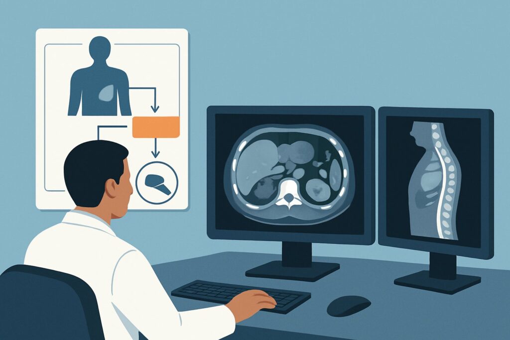
A 58-year-old male presents to the emergency department with a two-day history of sharp, constant left upper quadrant (LUQ) pain and a subjective fever. On examination, he is tender in the LUQ and his temperature is 38.6°C (101.5°F). His white blood cell count is elevated. You suspect an infectious or inflammatory process, but the differential is broad, spanning splenic, renal, pancreatic, and colonic etiologies. The immediate clinical question is which imaging study will most efficiently and accurately diagnose the cause to guide treatment. According to the American College of Radiology (ACR) Appropriateness Criteria, for an adult with acute left upper quadrant pain and fever, CT abdomen and pelvis with IV contrast is rated Usually appropriate as the initial imaging study.
Who Fits This Clinical Scenario?
This diagnostic workflow is specifically for an adult patient presenting with an acute onset of pain localized to the left upper quadrant, accompanied by a fever. The presence of fever is a critical component, as it shifts the differential diagnosis heavily toward infectious and inflammatory conditions, such as abscess, infarction with secondary infection, or pyelonephritis. This guidance applies when you are ordering the initial imaging study, meaning no prior relevant imaging has been performed for this episode of pain.
This pathway should be distinguished from similar but distinct clinical presentations:
- No Fever Present: If the patient has acute LUQ pain but is afebrile, the clinical scenario is different. The workup would follow the guidance for “Acute left upper quadrant pain, not otherwise specified,” which has a slightly different set of imaging considerations.
- Suspected Splenomegaly: If the primary physical exam finding is a palpable spleen or there is a strong clinical suspicion of splenomegaly (e.g., in a patient with known hematologic malignancy or portal hypertension), the imaging approach is tailored to that specific question.
- Known Trauma: This guidance does not apply to patients with a recent history of abdominal trauma, as the workup for splenic or renal injury follows a separate, trauma-focused protocol.
What Diagnoses Are You Working Up in This Scenario?
The combination of localized LUQ pain and fever points toward a limited but critical set of potential diagnoses. The choice of imaging is driven by the need to differentiate among these possibilities, as their management varies significantly.
Splenic Infarct or Abscess: The spleen is a primary resident of the LUQ. A splenic infarct, often caused by an embolic event (e.g., from atrial fibrillation), can present with acute pain and may be associated with a low-grade fever. A splenic abscess is a less common but life-threatening condition, which can arise from hematogenous spread of infection, contiguous infection, or a superinfected infarct. Both require urgent diagnosis and management.
Acute Pyelonephritis and Perinephric Abscess: The upper pole of the left kidney and the left adrenal gland are located in the LUQ. A complicated urinary tract infection ascending to the kidney (pyelonephritis) can cause flank and LUQ pain with high fevers. If left untreated, it can progress to a perinephric abscess, a collection of pus around the kidney, which often requires drainage in addition to antibiotics.
Splenic Flexure Diverticulitis or Colitis: While diverticulitis more commonly affects the left lower quadrant (sigmoid colon), inflammation can occur at the splenic flexure of the large intestine. This can present with localized pain, fever, and leukocytosis, mimicking other LUQ pathologies.
Pancreatitis (Tail): The tail of the pancreas extends into the LUQ, terminating near the splenic hilum. While pancreatitis often presents as epigastric pain radiating to the back, inflammation localized to the pancreatic tail can manifest as isolated LUQ pain. Fever may be present due to systemic inflammation or a complication like a pancreatic abscess.
Why Is CT Abdomen and Pelvis with IV Contrast the Recommended Study?
The ACR designates CT of the abdomen and pelvis with intravenous (IV) contrast as Usually appropriate because it provides a comprehensive and detailed evaluation of all potential etiologies in this scenario. It excels at simultaneously assessing the solid organs, bowel, vasculature, and retroperitoneum.
The IV contrast is essential. It enhances the solid organs, allowing for detailed assessment of their perfusion and parenchyma. For the key differential diagnoses:
- Splenic Infarct: Appears as a wedge-shaped, non-enhancing area within the spleen. This diagnosis is nearly impossible to make confidently on a non-contrast CT.
- Splenic Abscess: Typically seen as a complex fluid collection with a thick, enhancing rim after contrast administration.
- Pyelonephritis: May show as patchy, delayed, or striated enhancement of the renal parenchyma (a striated nephrogram). A perinephric abscess is clearly delineated as a rim-enhancing fluid collection.
- Diverticulitis/Colitis: Contrast helps identify mural thickening and enhancement of the affected colonic segment, as well as surrounding inflammatory changes (fat stranding) and potential abscess formation.
Alternative studies are rated lower for specific reasons in this context:
- US abdomen is rated May be appropriate. While it is excellent for detecting fluid collections like an abscess and uses no ionizing radiation, it is often limited by overlying bowel gas, which can obscure the spleen, pancreatic tail, and left kidney. It is also highly operator-dependent and less sensitive for subtle pyelonephritis or small infarcts.
- CT abdomen and pelvis without IV contrast is also rated May be appropriate. It can identify some findings like free air, perinephric gas, or calcified kidney stones, but it cannot assess organ perfusion. It will miss splenic infarcts and makes the characterization of abscesses and inflammatory changes significantly more difficult.
The recommended CT study carries a moderate radiation dose (ACR RRL ☢☢☢, 1-10 mSv), a consideration that is generally outweighed by the high diagnostic yield in a patient with fever and acute pain, where a timely and accurate diagnosis is critical. Once you’ve decided on CT abdomen and pelvis with IV contrast, our protocol guide covers the technique, contrast, and reading principles: CT Chest/Abdomen/Pelvis with IV Contrast.
What’s Next After CT Abdomen and Pelvis with IV Contrast? Downstream Workflow
The results of the CT scan will dictate the immediate next steps in patient management. The workflow branches based on whether the findings are positive, negative, or indeterminate.
If the study is positive for a splenic abscess or large infarct: This is a critical finding requiring urgent consultation. The next steps typically involve admitting the patient for IV antibiotics and consulting Interventional Radiology (IR) for potential percutaneous drainage of an abscess. An infectious disease consultation is also highly recommended. For a large infarct, hematology and cardiology consults may be needed to investigate an underlying embolic source or hypercoagulable state.
If the study is positive for pyelonephritis or a perinephric abscess: The patient will require admission for IV antibiotics. If a drainable perinephric abscess is identified, a urology or IR consultation is necessary for percutaneous drainage.
If the study is negative: A negative high-quality CT scan makes a serious intra-abdominal infectious process much less likely. The clinical team should reconsider the differential diagnosis. Could the pain be referred from a basilar pneumonia or pulmonary embolism? Is a musculoskeletal cause possible? If the pain persists without a diagnosis, the workup may shift toward the “Acute left upper quadrant pain, not otherwise specified” ACR variant, which might involve different imaging or endoscopic evaluation depending on the evolving clinical picture.
If the study is indeterminate: Occasionally, a CT may reveal a finding that is not definitive (e.g., a complex splenic cyst that could represent an early abscess). In these cases, a follow-up study may be warranted. MRI abdomen without and with IV contrast, rated May be appropriate, can be an excellent problem-solving tool, offering superior soft-tissue contrast to better characterize complex fluid collections or subtle parenchymal abnormalities without additional ionizing radiation.
Pitfalls to Avoid (and When to Get Help)
In the workup of acute LUQ pain with fever, several common pitfalls can delay diagnosis or lead to suboptimal care. Be mindful of the following:
- Omitting IV Contrast: Ordering a non-contrast CT in a patient with adequate renal function is a major pitfall. It severely limits the diagnostic utility for the most likely and most serious conditions, particularly splenic infarct and abscess.
- Ignoring Extra-Abdominal Causes: Pain in the LUQ can be referred from the chest. Always consider a lower-lobe pneumonia, pleurisy, or even a pulmonary embolism in your differential, especially if abdominal findings are absent.
- Delayed Imaging: In a patient who appears septic or is hemodynamically unstable, imaging should not be delayed. Stabilize the patient concurrently with arranging for an expedited CT scan.
- Anchoring Bias: Avoid fixating on one diagnosis before imaging. The strength of CT is its ability to survey the entire region for a wide range of pathologies.
If the patient shows signs of hemodynamic instability, peritonitis, or sepsis, escalate immediately. This involves early consultation with surgery or critical care services, often while the diagnostic workup is still in progress.
Related ACR Topics and Tools
This article focuses on a single, specific clinical scenario. For a comprehensive overview of all variants related to this symptom, or to explore the technical details of the recommended imaging studies, the following resources are essential:
- Parent Topic Hub: For breadth across all scenarios in Acute Left Upper Quadrant Pain, see our parent guide: Acute Left Upper Quadrant Pain: ACR Appropriateness Decoded.
- ACR Appropriateness Criteria Lookup: To review the official ACR guidelines for this and thousands of other clinical scenarios, use the ACR Appropriateness Criteria Lookup tool.
- Imaging Protocol Library: For detailed technical specifications on how imaging studies are performed, consult the Imaging Protocol Library.
- Radiation Dose Calculator: To discuss cumulative radiation exposure with patients, the Radiation Dose Calculator can help estimate and contextualize dose.
Frequently Asked Questions
Why is CT preferred over ultrasound for initial imaging in a patient with LUQ pain and fever?
While ultrasound is a valuable tool without radiation, CT with IV contrast is rated higher (*Usually appropriate*) in this scenario because it provides a more comprehensive and reliable evaluation. Ultrasound can be limited by bowel gas obscuring the spleen, pancreatic tail, and left kidney. CT offers superior visualization of the entire abdomen and retroperitoneum, and IV contrast is critical for diagnosing key conditions like splenic infarcts, abscesses, and pyelonephritis, which may be missed or poorly characterized on ultrasound.
What should I order if my patient has a severe contrast allergy or significant renal insufficiency?
If IV contrast is contraindicated, the decision becomes more complex. An MRI of the abdomen, either without contrast or with a gadolinium-based contrast agent (if safe for the patient’s renal function), is rated *May be appropriate* and can be an excellent alternative. It provides superb soft-tissue detail for evaluating for abscesses or inflammation. A non-contrast CT is also rated *May be appropriate* but is significantly less sensitive for the primary concerns in this scenario. The best choice depends on institutional availability and the specific suspected diagnosis after clinical evaluation.
Does a normal CT scan rule out all serious causes of LUQ pain with fever?
A high-quality negative CT abdomen and pelvis with IV contrast makes a major intra-abdominal process like an abscess, infarct, or significant diverticulitis highly unlikely. However, it does not rule out all causes. The pain could be from an extra-abdominal source (e.g., basilar pneumonia, shingles) or an early inflammatory process not yet visible on CT. Clinical re-evaluation is crucial if symptoms persist despite a negative scan.
Is an abdominal radiograph (X-ray) ever useful in this scenario?
According to the ACR, an abdominal radiograph is *Usually not appropriate* for the initial workup of acute LUQ pain with fever. Its diagnostic yield is very low. It cannot visualize the solid organs (spleen, kidney, pancreas) or soft tissues effectively. While it could potentially show free air from a perforated viscus, CT is far more sensitive for this finding and for all the other primary diagnostic considerations.
How does this workup change if the patient is pregnant?
The workup changes significantly in a pregnant patient to avoid ionizing radiation. The ACR guidelines for pregnant patients with abdominal pain would apply. Typically, ultrasound is the first-line imaging modality. If ultrasound is non-diagnostic and there is high suspicion for a serious condition, MRI without contrast is the preferred next step, as it provides excellent soft-tissue detail without using radiation. CT is reserved for situations where MRI is unavailable or contraindicated and the risk of not making a diagnosis is felt to outweigh the radiation risk to the fetus.
Reviewed by Pouyan Golshani, MD, Interventional Radiologist — May 30, 2026