What Is the Best Initial Imaging for an Infant Under 4 Months With Signs of DDH?
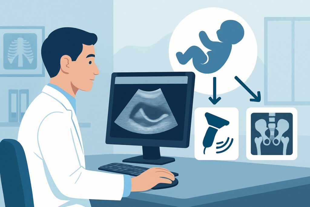
You are in your pediatric clinic on a busy afternoon, evaluating a 2-month-old infant for a routine well-child visit. During the physical examination, you elicit a positive Ortolani sign, a distinct “clunk” as the dislocated femoral head reduces back into the acetabulum. The finding is concerning for Developmental Dysplasia of the Hip (DDH), and you know that timely diagnosis is critical for successful non-operative treatment. The immediate clinical question is which imaging study to order to confirm the diagnosis and guide management. For an infant younger than 4 months with positive physical findings, the American College of Radiology (ACR) rates US hips as Usually appropriate.
Who Fits This Clinical Scenario?
This clinical workflow is specifically for an infant who is younger than 4 months of age and has positive physical findings suggestive of Developmental Dysplasia of the Hip. These findings are the cornerstone of the initial suspicion and include:
- A positive Ortolani test (reduction of a dislocated hip)
- A positive Barlow test (provocation of a dislocatable hip)
- Significant leg-length discrepancy (a positive Galeazzi sign)
- Asymmetric thigh or gluteal skin folds
- Limited hip abduction (less than 60 degrees)
This guidance does not apply to patients who fall into slightly different clinical categories, which have their own distinct imaging recommendations. Key exclusions from this specific scenario include:
- Infants with only risk factors: An infant younger than 4 months with risk factors for DDH (e.g., breech presentation, positive family history) but an equivocal or normal physical exam follows a different ACR variant.
- Older infants: Once an infant is between 4 and 6 months of age, or older than 6 months, the ossification of the femoral head changes, and the recommended initial imaging modality shifts from ultrasound to radiography.
- Surveillance imaging: This workflow is for initial diagnosis. An infant with a known diagnosis of DDH undergoing surveillance imaging while in a treatment device, like a Pavlik harness, is a separate clinical question.
What Diagnoses Are You Working Up in This Scenario?
When ordering imaging for an infant with a positive hip exam, you are primarily evaluating for a spectrum of abnormalities that fall under the umbrella of Developmental Dysplasia of the Hip. The differential is narrow but represents a continuum of severity.
Developmental Dysplasia of the Hip (DDH)
This is the primary diagnosis of concern. DDH is not a single entity but a spectrum ranging from mild acetabular dysplasia (a shallow socket) with a stable hip, to a subluxatable hip (partially dislocates), to a fully dislocatable or dislocated hip. The positive physical exam findings in this scenario raise the suspicion for a more significant form of DDH, such as a subluxatable or dislocated hip, which requires prompt intervention.
Physiologic Ligamentous Laxity
In the neonatal period, residual maternal hormones can cause generalized ligamentous laxity. This can sometimes manifest as a “clicky” hip on exam that is not a true Ortolani or Barlow sign. While often benign and self-resolving, a hip that feels unstable on exam warrants imaging to differentiate this physiologic laxity from true structural dysplasia. Ultrasound is excellent for making this distinction.
Teratologic Hip Dislocation
This is a much less common but more severe condition where the hip is rigidly dislocated at birth. It is often associated with other neuromuscular conditions like arthrogryposis or myelomeningocele. While the physical exam is typically more dramatic than in standard DDH, it remains on the differential for a dislocated hip in a newborn. Ultrasound can confirm the dislocation and assess the anatomy, though the management pathway is significantly different.
Why Is US hips the Recommended Study for This Presentation?
For an infant under four months of age, ultrasound is the ideal imaging modality for evaluating the hip joint, providing a dynamic, radiation-free assessment of the unossified anatomy. The ACR designates US hips as Usually appropriate for this clinical scenario for several key reasons.
The primary advantage of ultrasound is its ability to visualize the cartilaginous structures of the infant hip. In this age group, the femoral head and much of the acetabulum are composed of cartilage, which is nearly invisible on a plain radiograph. Ultrasound provides excellent visualization of the cartilaginous femoral head, its position relative to the bony acetabulum, and the morphology of the acetabular roof. This allows for precise measurement of key indicators of stability and development, such as the alpha and beta angles in the Graf method.
Furthermore, hip ultrasound includes a dynamic component. The sonographer or radiologist performs stress maneuvers, similar to the Barlow test, under direct visualization to assess the stability of the femoral head within the acetabulum. This functional information is critical for distinguishing a stable, well-formed hip from one that is subluxatable or dislocatable, which a static image cannot provide.
In contrast, the ACR rates Radiography pelvis as Usually not appropriate. The reason is straightforward: without a well-ossified femoral head, the key landmarks used to assess hip alignment on an X-ray are absent or unreliable. This makes radiography an uninformative and potentially misleading study in this age group. It also exposes the infant to ionizing radiation, even if the dose is low (pediatric RRL ☢☢ 0.03-0.3 mSv), which is unnecessary when a superior, radiation-free alternative exists. Ultrasound carries a radiation dose of 0 mSv.
What’s Next After US hips? Downstream Workflow
The results of the hip ultrasound will directly guide your next steps, typically leading to either orthopedic referral or clinical reassurance. The downstream workflow is generally clear-cut.
If the ultrasound is positive for DDH:
A report indicating acetabular dysplasia, hip subluxation, or dislocation confirms the diagnosis. The next step is a prompt referral to a pediatric orthopedic surgeon. For infants in this age group, the standard treatment is typically a dynamic positioning device, most commonly the Pavlik harness, which holds the hips in a flexed and abducted position to encourage the femoral head to mold the acetabulum. Early treatment initiation leads to high rates of success with non-operative management.
If the ultrasound is negative (normal hips):
A normal ultrasound, showing a well-covered femoral head and mature acetabular angles with no instability on dynamic assessment, effectively rules out significant DDH. No further imaging is required. The recommendation is to continue routine clinical follow-up at subsequent well-child visits to monitor for any new clinical signs, though the risk of developing later problems is very low.
If the ultrasound is indeterminate or equivocal:
Occasionally, the findings may be borderline, such as mild physiologic immaturity of the acetabulum without instability. This is more common in very young infants (e.g., under 6 weeks). In these cases, the typical recommendation is a short-interval follow-up ultrasound in 4 to 6 weeks. This allows time for the hip to mature naturally. Most cases of mild immaturity will resolve on their own, but a follow-up study is necessary to ensure progression to a normal state and not toward dysplasia.
Pitfalls to Avoid (and When to Get Help)
In the workup of suspected DDH in an infant, a few common pitfalls can delay diagnosis or lead to unnecessary testing. Being aware of them can help streamline patient care.
- Ordering the wrong study: The most common error is ordering a pelvic radiograph for an infant under 4 months old. This provides no useful information about hip morphology and exposes the infant to unnecessary radiation. Always order an ultrasound in this age group.
- Delaying the imaging: While DDH is not a surgical emergency, timely diagnosis is key. Treatment with a Pavlik harness is most effective when initiated before 6 months of age. A positive physical exam finding should prompt an ultrasound order within a few weeks.
- Not specifying a dynamic study: Ensure the imaging center is capable of performing a dynamic pediatric hip ultrasound with stress maneuvers. A static-only ultrasound provides incomplete information.
If the physical exam reveals a hip that is dislocated and cannot be reduced (an “irreducible dislocation”), this represents a more urgent situation. An immediate consultation with a pediatric orthopedic surgeon is warranted, often before imaging is even obtained.
Related ACR Topics and Tools
For a comprehensive overview of imaging for DDH across all pediatric age groups and clinical presentations, this article is best used in conjunction with its parent topic guide. The tools below can also help you navigate adjacent scenarios and understand the technical aspects of the recommended imaging.
- For breadth across all scenarios in Developmental Dysplasia of the Hip-Child, see our parent guide: Developmental Dysplasia of the Hip-Child: ACR Appropriateness Decoded.
- To explore other clinical scenarios, consult the Imaging Appropriateness Selector.
- For details on how a hip ultrasound is performed, visit the Imaging Protocol Library.
- To discuss radiation exposure with families for other studies, use the Radiation Dose Calculator.
Frequently Asked Questions
Why can’t I just order an X-ray for an infant with a positive hip exam?
In infants younger than 4-6 months, the femoral head (the ‘ball’ of the hip joint) is made of cartilage and has not yet started to form bone. Because cartilage is not visible on an X-ray, the study cannot be used to accurately assess the position and stability of the hip. Ultrasound is the required modality because it directly visualizes these cartilaginous structures.
What if the physical exam is unclear but the infant has risk factors for DDH?
That represents a different clinical scenario. For an infant younger than 4 months with an equivocal exam or only risk factors (like breech birth or family history), hip ultrasound is also the recommended study, but the timing and interpretation may differ slightly. This article specifically addresses infants with definitive positive physical findings.
Is sedation required for a pediatric hip ultrasound?
No, sedation is not required. The procedure is non-invasive and painless. It is typically performed with the infant lying on an examination table, and the parent can often be present to help comfort the child. Feeding the infant just before the scan can sometimes help keep them calm.
What is the difference between a static and a dynamic hip ultrasound?
A static ultrasound involves taking images and measurements of the hip at rest. A dynamic ultrasound, which is the standard for DDH evaluation, adds a functional component. The sonographer or radiologist applies gentle stress to the hip (similar to a Barlow maneuver) while watching on the screen to see if the femoral head moves out of the socket. This directly assesses hip stability, which is a critical piece of information.
How urgently should I order the ultrasound after finding a positive Ortolani sign?
While a positive finding for DDH is not a medical emergency requiring a trip to the emergency department (unless the hip is irreducible), it does warrant prompt action. The ultrasound should be scheduled within one to two weeks to facilitate a timely referral to pediatric orthopedics if the diagnosis is confirmed. Early intervention dramatically improves outcomes.
Reviewed by Pouyan Golshani, MD, Interventional Radiologist — May 26, 2026