What Is the Best Initial Imaging for Early Postoperative Dysphagia?
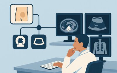
It’s 4 PM on a Tuesday, and you’re covering the surgical service. A patient who is three days post-anterior cervical discectomy and fusion (ACDF) reports new-onset difficulty swallowing both solids and liquids, accompanied by retrosternal discomfort. The immediate concern is a potential postoperative complication, but the differential is broad. Deciding on the right initial imaging study is critical to rule out urgent pathology without causing unnecessary delays or radiation exposure. This article provides a focused, evidence-based workflow for this exact scenario, guiding you to the most appropriate first step. For early postoperative dysphagia, the American College of Radiology (ACR) rates Fluoroscopy single contrast esophagram as Usually Appropriate.
Who Fits This Clinical Scenario?
This guidance applies specifically to patients experiencing new dysphagia—either oropharyngeal (trouble initiating a swallow) or retrosternal (a sensation of food getting stuck)—in the early postoperative period. “Early” is typically defined as occurring within the first 30 days following a surgical procedure known to carry a risk of esophageal or pharyngeal injury.
Common preceding surgeries include:
- Anterior cervical spine surgery (e.g., ACDF)
- Thoracic surgery (e.g., esophagectomy, lung resection)
- Cardiac surgery (e.g., coronary artery bypass grafting, valve replacement)
- Head and neck surgery
- Thyroidectomy
This workflow is distinct from other clinical presentations. It does not apply to patients with delayed postoperative dysphagia, where symptoms arise more than one month after surgery, suggesting a different set of causes like chronic stricture or fibrosis. It also excludes patients with unexplained oropharyngeal dysphagia without a recent surgical history or those with retrosternal dysphagia in an immunocompromised state, as these scenarios have their own dedicated ACR workups.
What Diagnoses Are You Working Up in This Scenario?
In the early postoperative setting, the differential diagnosis for dysphagia is weighted toward acute surgical complications. The choice of initial imaging is driven by the need to identify or exclude these time-sensitive conditions.
Anastomotic Leak or Esophageal Perforation: This is the most urgent and feared complication. A breach in the esophageal wall can lead to mediastinitis or abscess, conditions with high morbidity and mortality. It is the primary reason a water-soluble contrast study is the first-line investigation. Any recent instrumentation or surgical manipulation of the esophagus or adjacent structures puts the patient at risk.
Postoperative Edema or Hematoma: A more common and often self-limiting cause of dysphagia, particularly after anterior neck surgery. Swelling or a contained collection of blood can cause extrinsic compression on the pharynx or esophagus, leading to mechanical obstruction. While less emergent than a leak, it must be distinguished from more severe pathology.
Early Stricture or Stenosis: While significant fibrotic strictures take longer to develop, early inflammation and healing at a surgical anastomosis can cause luminal narrowing. This is a key consideration after procedures like an esophagectomy with primary anastomosis.
Fistula Formation: A less common but consequential diagnosis is the formation of an abnormal connection, such as a tracheoesophageal or bronchoesophageal fistula. This can present with coughing after swallowing in addition to dysphagia and requires prompt diagnosis to prevent aspiration pneumonia and sepsis.
Iatrogenic Nerve Injury: Damage to the recurrent laryngeal nerve or pharyngeal plexus can cause neuromuscular dysfunction, leading to oropharyngeal dysphagia. While the nerve itself isn’t visualized, a dynamic swallow study can demonstrate the functional consequences, such as aspiration or pharyngeal residue.
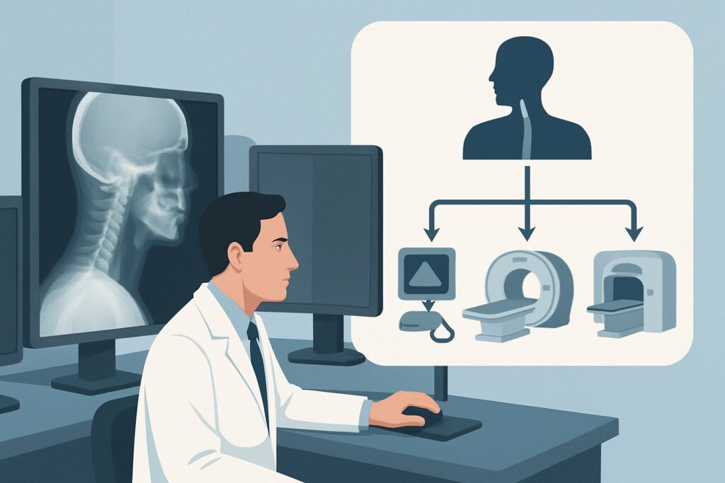
Why Is a Fluoroscopy Single Contrast Esophagram the Recommended Study?
The ACR designates a Fluoroscopy single contrast esophagram as Usually Appropriate for evaluating early postoperative dysphagia. This dynamic, real-time study is uniquely suited to assess both the anatomy and function of the esophagus, making it the ideal first step for addressing the most critical differential diagnoses.
The primary rationale is its high sensitivity for detecting an esophageal leak or perforation. For this indication, the study must be performed with a water-soluble contrast agent (e.g., Gastrografin or iohexol). If a leak is present, this contrast can extravasate into the mediastinum or pleural space without causing the severe inflammatory reaction associated with barium. If the initial water-soluble study is negative and clinical suspicion for a subtle leak remains high, it may be followed by thin barium, which provides better mucosal coating and can reveal smaller abnormalities.
Another key advantage is its ability to evaluate motility and identify mechanical obstruction from extrinsic compression (e.g., a hematoma) or early stricture formation. The radiologist can observe the entire swallowing process, providing crucial functional information that a static imaging modality like CT cannot.
Why are other studies rated lower for this specific scenario?
- CT neck and chest with IV contrast is also rated Usually Appropriate but is often considered a complementary or second-line study. While excellent for evaluating extraluminal pathology like a mediastinal abscess, fluid collection, or large hematoma, it is less sensitive than an esophagram for detecting small mucosal leaks. It also involves a higher radiation dose (adult RRL ☢☢☢☢, 10-30 mSv) compared to the esophagram (adult RRL ☢☢☢, 1-10 mSv).
- A modified barium swallow (MBS) is rated Usually not appropriate. The MBS is a detailed functional study of the oropharyngeal phase of swallowing, typically performed with a speech-language pathologist. While useful for unexplained oropharyngeal dysphagia, it is not designed to evaluate for an esophageal leak, which is the primary concern in the early postoperative period. Using barium in a patient with a potential perforation is contraindicated.
What’s Next After a Fluoroscopy Single Contrast Esophagram? Downstream Workflow
The results of the esophagram will guide your next steps in a branching clinical pathway. The goal is to move quickly from diagnosis to management.
If the study is positive for a leak or perforation: This is a surgical emergency. The immediate next step is urgent consultation with the surgical service (e.g., thoracic or general surgery) for operative management. A CT scan with IV contrast may be ordered preoperatively to delineate the extent of any associated fluid collection or abscess in the mediastinum or neck, which helps in planning the surgical approach.
If the study is negative for a leak but shows extrinsic compression or stenosis: If a significant hematoma or fluid collection is suspected as the cause of compression, a CT with IV contrast is the appropriate next step to characterize the collection and guide potential drainage. If a high-grade stenosis is seen, endoscopic evaluation and potential dilation may be required, though this is often deferred until the acute postoperative inflammation has subsided.
If the study is completely negative: If the esophagram is normal and the patient’s symptoms persist, the focus may shift toward a potential neuromuscular cause (e.g., nerve injury) or a subtle motility disorder. Depending on the nature of the dysphagia (oropharyngeal vs. esophageal), further evaluation could include a formal modified barium swallow with speech pathology, esophageal manometry, or direct visualization with endoscopy once the patient is further out from their surgery and the risk of perforation from instrumentation is lower.
Pitfalls to Avoid (and When to Get Help)
Navigating the workup for early postoperative dysphagia requires careful attention to a few common pitfalls. First, never order a barium study as the initial test if there is any suspicion of an esophageal perforation; always start with water-soluble contrast. Second, do not mistake a normal CT scan for a clean bill of health; CT can miss small mucosal leaks that are readily apparent on a fluoroscopic esophagram. Third, be mindful of the patient’s ability to cooperate. A patient with an altered mental status or who cannot protect their airway is at high risk of aspiration during a swallow study, and the risks and benefits must be carefully weighed. If a patient presents with systemic signs of sepsis, such as fever, tachycardia, and hypotension, in the setting of postoperative dysphagia, escalate immediately to the surgical team and consider a broad-spectrum antibiotic while initiating the diagnostic workup, as this may signal mediastinitis from a perforation.
Related ACR Topics and Tools
This article focuses on one specific clinical scenario. For a comprehensive overview of imaging for all types of dysphagia, from oropharyngeal to retrosternal, and across different patient populations, please consult our parent guide. For further exploration of appropriateness criteria or technical protocols, the following resources are available.
- For breadth across all scenarios in Dysphagia, see our parent guide: Dysphagia: ACR Appropriateness Decoded.
- ACR Appropriateness Criteria Lookup — for adjacent scenarios
- Imaging Protocol Library — for technique on the recommended study
- Radiation Dose Calculator — for cumulative dose conversations
Frequently Asked Questions
Why must I use water-soluble contrast instead of barium for the initial esophagram?
Barium is an inert substance within the gastrointestinal tract but causes a severe inflammatory reaction (barium peritonitis or mediastinitis) if it leaks into the chest or abdominal cavity. In the early postoperative setting, the primary concern is an esophageal leak or perforation. Water-soluble contrast is readily absorbed by surrounding tissues and does not cause this intense inflammation, making it the safe and required first choice.
The esophagram was negative, but my patient still can’t swallow. What should I do?
If the esophagram rules out a leak, stenosis, or significant extrinsic compression, the differential shifts. Consider a CT with IV contrast to look for a non-compressive fluid collection or abscess. If the dysphagia is primarily oropharyngeal (choking, coughing), a neuromuscular cause like recurrent laryngeal nerve injury is more likely, and a formal modified barium swallow with speech pathology may be warranted once the immediate risk of perforation is deemed low.
Can I just order a CT scan with oral contrast instead of the fluoroscopic esophagram?
While a CT with oral contrast can sometimes show a large leak, it is significantly less sensitive than a dynamic fluoroscopic esophagram for detecting small or contained perforations. Fluoroscopy allows for real-time visualization of the contrast bolus moving through the esophagus under different patient positions (e.g., supine, prone), which maximizes the chance of identifying a subtle leak. A CT is a static ‘snapshot’ and can easily miss these findings.
Is there a role for endoscopy in the early postoperative period?
Upper endoscopy is generally avoided in the immediate postoperative period if a leak or perforation is suspected. The insufflation of air and the mechanical pressure of the endoscope can worsen a small, contained perforation, turning it into a free perforation. Endoscopy is typically reserved for later in the course to evaluate for strictures or other mucosal abnormalities once the acute phase has passed and anastomoses have had time to heal.
My patient had cardiac surgery, not neck surgery. Is an esophagram still the right first test?
Yes. Although less common than after neck or esophageal surgery, dysphagia after cardiac surgery can occur due to compression from a posterior hematoma, mediastinal inflammation, or iatrogenic injury to the vagus or recurrent laryngeal nerves. The diagnostic algorithm remains the same: the primary goal is to rule out a rare but catastrophic esophageal injury, making the water-soluble esophagram the appropriate initial imaging test.
Reviewed by Pouyan Golshani, MD, Interventional Radiologist — May 29, 2026