Why Is MRI the Top Choice for Staging Distant Pancreatic Cancer Metastases?
It’s 4 PM on a Tuesday, and you are reviewing the pathology report for a 68-year-old patient: pancreatic ductal adenocarcinoma. The diagnosis is confirmed. The next critical question for the multidisciplinary tumor board, and for your patient, is the extent of the disease. Before any discussion of surgery or local therapy, you must confidently rule in or rule out distant metastases, which would fundamentally change the treatment plan from curative-intent to palliative. The imaging you order now is pivotal. This article details the clinical workflow for evaluating distant metastatic disease in an adult with newly diagnosed or post-treatment pancreatic ductal adenocarcinoma, explaining why the American College of Radiology (ACR) has rated MRI abdomen without and with hepatobiliary contrast as Usually Appropriate for this specific scenario.
Who Fits This Clinical Scenario?
This guidance applies to adult patients with a confirmed diagnosis of pancreatic ductal adenocarcinoma (PDAC) who require imaging for one of two purposes: initial staging to assess for distant metastatic disease, or post-procedure surveillance to monitor for the development of new metastatic disease. The primary goal is to identify metastases outside the locoregional confines of the pancreas, most commonly in the liver, peritoneum, or distant lymph nodes.
This workflow is not intended for:
- Initial diagnosis: Patients with abdominal symptoms clinically suspicious for pancreatic cancer but without a tissue diagnosis yet. That presentation falls under the ACR Appropriateness Criteria for initial evaluation of suspected PDAC.
- High-risk screening: Asymptomatic individuals with a strong family history or genetic predisposition who are undergoing screening. This is a distinct clinical scenario with its own imaging recommendations.
- Detailed locoregional staging: Patients who need a detailed assessment of tumor involvement with local structures like the superior mesenteric artery or portal vein to determine surgical resectability. While distant staging is part of that process, the locoregional question requires specific protocols, often with multiphasic CT, and is covered in a separate variant.
Correctly identifying your patient’s clinical question—in this case, the search for distant disease—is the first step to selecting the most effective imaging study.
What Diagnoses Are You Working Up in This Scenario?
With a known primary PDAC, the imaging “differential” is focused on identifying the presence, location, and extent of metastatic spread. The findings will determine the patient’s stage (specifically, M0 vs. M1 disease) and guide therapy.
Liver Metastases: This is the most common site of distant spread for pancreatic cancer. The liver’s dual blood supply and filtration role make it highly susceptible to hematogenous seeding. Lesions can be solitary or multifocal, and their detection is a primary objective of staging imaging. Small, sub-centimeter liver metastases can be particularly challenging to identify but are critical for accurate staging.
Peritoneal Carcinomatosis: This refers to the diffuse spread of tumor implants along the surfaces of the peritoneum. It can be a subtle finding on imaging, sometimes manifesting only as small nodules, omental caking, or new-onset ascites. Detecting peritoneal disease is crucial as it signifies advanced, unresectable cancer.
Distant Lymphadenopathy: While regional lymph node involvement is part of locoregional staging, the evaluation for distant metastases includes searching for abnormal nodes outside the typical peripancreatic, celiac, and superior mesenteric artery basins. This includes para-aortic, pelvic, or mediastinal lymph nodes.
Less Common Sites: Although less frequent, PDAC can metastasize to the lungs, adrenal glands, or bones. While abdominal imaging is the primary focus, findings may prompt dedicated imaging of other body parts if suspicion is high or symptoms are present.
Why Is MRI of the Abdomen the Recommended Study for This Presentation?
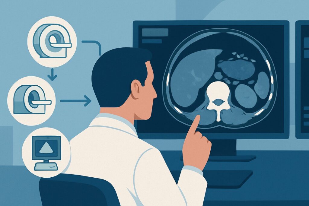
For evaluating distant metastatic disease in pancreatic cancer, particularly in the liver, the ACR designates MRI abdomen without and with hepatobiliary contrast as Usually Appropriate. This recommendation is driven by MRI’s superior soft-tissue contrast resolution, which provides a distinct advantage in detecting and characterizing liver lesions.
The key rationale is MRI’s high sensitivity for small hepatic metastases. Hepatobiliary contrast agents, such as gadoxetate disodium, are taken up by healthy hepatocytes but not by metastatic tumor cells. During the delayed hepatobiliary phase (typically 15-20 minutes post-injection), normal liver parenchyma enhances while metastases appear as dark, conspicuous defects. This significantly improves the detection of small lesions that might be missed or indeterminate on other modalities. An MRI abdomen without and with IV contrast using standard extracellular gadolinium agents is also rated Usually Appropriate and offers excellent lesion detection, though it may be slightly less sensitive for certain small lesions compared to a hepatobiliary agent.
Let’s compare this to other common imaging choices:
- CT chest abdomen pelvis with IV contrast: This study is also rated Usually Appropriate. Its main advantages are speed, wide availability, and the ability to simultaneously evaluate the chest for pulmonary metastases. However, its sensitivity for small (<1 cm) liver and peritoneal metastases is lower than that of MRI. It remains a very reasonable and widely used alternative, especially when MRI is contraindicated or unavailable.
- FDG-PET/CT skull base to mid-thigh: Rated as May be appropriate, this modality excels at identifying metabolically active disease anywhere in the body. It can be valuable for detecting unexpected distant metastases or for clarifying equivocal findings on CT or MRI. However, it is not the primary staging tool because its spatial resolution is lower, potentially missing very small or non-FDG-avid peritoneal or liver lesions. It also involves a significant radiation dose (☢☢☢☢ 10-30 mSv).
A significant advantage of MRI is the lack of ionizing radiation (adult relative radiation level of O), which is an important consideration for patients who may require multiple surveillance scans over time. In contrast, CT-based options carry radiation levels from ☢☢☢ (1-10 mSv) to ☢☢☢☢ (10-30 mSv).
What’s Next After MRI? Downstream Workflow
The results of the staging MRI will direct the subsequent management pathway. The decision tree branches based on whether distant metastatic disease is identified.
If the MRI is positive for distant metastases:
The patient is classified as having Stage IV disease. The primary treatment shifts from local control to systemic therapy, typically with chemotherapy. The goal becomes palliative—to extend survival and manage symptoms. A biopsy of an accessible metastatic lesion (e.g., a liver lesion) may be performed to confirm the M1 stage and potentially for molecular profiling to guide targeted therapies. The patient will generally not be a candidate for surgical resection of the primary tumor.
If the MRI is negative for distant metastases:
The patient is considered to have M0 (non-metastatic) disease. The focus then shifts to detailed locoregional staging to determine resectability. This often requires a dedicated multiphase pancreatic protocol CT to precisely evaluate the tumor’s relationship with critical blood vessels. If the tumor is deemed resectable, borderline resectable, or locally advanced, the patient may proceed to neoadjuvant therapy followed by surgery, or definitive chemoradiation. This workflow routes the patient to the “Locoregional disease staging” clinical scenario.
If the MRI is indeterminate:
An equivocal finding, such as a tiny liver lesion that is too small to characterize, presents a clinical challenge. Management options include a short-interval follow-up MRI to assess for stability or growth, a problem-solving FDG-PET/CT to assess for metabolic activity, or percutaneous biopsy if the lesion is safely accessible.
Pitfalls to Avoid (and When to Get Help)
Accurate staging is critical, and several pitfalls can lead to misclassification and inappropriate treatment.
- Misinterpreting Benign Lesions: The liver is host to many benign lesions (cysts, hemangiomas, focal nodular hyperplasia). Mistaking these for metastases can lead to over-staging a patient and incorrectly denying them a chance for curative surgery. This is an area where MRI with hepatobiliary contrast excels.
- Overlooking Peritoneal Disease: Small-volume peritoneal carcinomatosis can be subtle on imaging. Carefully scrutinize the omentum, peritoneal reflections, and for any new or unexplained ascites.
- Incomplete Chest Evaluation: While abdominal MRI is superior for liver evaluation, it does not assess the lungs. If a CT of the chest is not performed as part of the initial workup, small pulmonary metastases may be missed. A baseline chest CT is often performed in conjunction with abdominal imaging.
- Ignoring MRI Contraindications: Always screen for contraindications to MRI (e.g., certain implants, severe claustrophobia) or gadolinium-based contrast agents (e.g., severe renal impairment, known allergy). If contraindications exist, a high-quality contrast-enhanced CT is the appropriate alternative.
If findings are equivocal or the clinical picture does not match the imaging results, discussion at a multidisciplinary tumor board with experienced radiologists, oncologists, and surgeons is the essential next step.
Related ACR Topics and Tools
For a comprehensive overview of imaging across all clinical presentations of pancreatic cancer, from screening to surveillance, please refer to our parent guide. Additional tools from GigHz can help you apply these criteria in your daily practice.
- For breadth across all scenarios in Screening, Locoregreal Assessment, and Surveillance of Pancreatic Ductal Adenocarcinoma, see our parent guide: Screening, Locoregional Assessment, and Surveillance of Pancreatic Ductal Adenocarcinoma: ACR Appropriateness Decoded.
- To explore other clinical scenarios and see the full evidence tables, use the ACR Appropriateness Criteria Lookup.
- For detailed technical specifications on how to perform the recommended studies, consult the Imaging Protocol Library.
- To discuss cumulative radiation exposure with your patients, especially when considering CT, the Radiation Dose Calculator can be a useful aid.
Frequently Asked Questions
Why is MRI specifically preferred over CT for detecting liver metastases in pancreatic cancer?
MRI, particularly with a hepatobiliary contrast agent, has higher sensitivity for detecting small (<1 cm) liver metastases. The contrast agent is taken up by healthy liver cells but not cancer cells, making metastases stand out as dark spots on a bright background in delayed imaging phases. This superior contrast resolution can detect lesions that may be invisible or indeterminate on a contrast-enhanced CT.
If MRI is best for the abdomen, does the patient still need a CT of the chest?
Yes, typically. The lungs are another potential site for metastases from pancreatic cancer. An abdominal MRI does not adequately evaluate the lung parenchyma. Therefore, a dedicated CT of the chest (often performed without contrast if the primary goal is to find nodules) is a standard part of the initial staging workup to complete the assessment for distant disease.
When is an FDG-PET/CT scan the right choice for staging pancreatic cancer?
According to the ACR, FDG-PET/CT is rated ‘May be appropriate.’ It is not typically the first-line imaging modality for initial staging. Its primary roles are to clarify equivocal findings seen on CT or MRI, to detect unexpected sites of distant disease when clinical suspicion is high, or to assess treatment response. Its lower spatial resolution makes it less reliable for detecting very small liver or peritoneal metastases compared to MRI.
What is the best imaging alternative if my patient cannot have an MRI or receive gadolinium contrast?
If a patient has a contraindication to MRI (e.g., an incompatible medical device) or gadolinium contrast (e.g., severe renal failure or allergy), a high-quality, contrast-enhanced CT of the abdomen and pelvis is the best alternative. This study is also rated as ‘Usually Appropriate’ by the ACR and provides excellent information, though with slightly lower sensitivity for small liver lesions compared to MRI.
How does imaging for post-procedure surveillance differ from initial staging?
The imaging modality and goals are often the same: to detect any new metastatic disease. The choice between CT and MRI for surveillance may depend on institutional preference, prior imaging findings, and patient factors. The key difference is the context. In surveillance, you are looking for new disease in a patient who has already undergone treatment, and the findings are compared to a prior post-treatment baseline scan to assess for recurrence or progression.
Reviewed by Pouyan Golshani, MD, Interventional Radiologist — May 30, 2026