Which Imaging Is Best for Locoregional Staging of Pancreatic Cancer? An ACR-Guided Workflow
A 68-year-old male with a new diagnosis of pancreatic ductal adenocarcinoma, confirmed by endoscopic ultrasound with fine-needle aspiration, is discussed at your multidisciplinary tumor board. The consensus is to proceed with neoadjuvant FOLFIRINOX chemotherapy followed by potential surgical resection. As the ordering physician, your immediate task is to obtain the most accurate locoregional staging to establish a baseline, define the tumor’s relationship to critical vascular structures, and guide the treatment plan. The choice of imaging will directly influence the determination of resectability and the subsequent surgical approach. This article details the clinical workflow for this specific scenario, explaining why the American College of Radiology (ACR) rates MRI abdomen without and with IV contrast as Usually Appropriate.
Who Fits This Clinical Scenario for Pancreatic Cancer Staging?
This guidance applies to a specific subset of adult patients: those with a histologically confirmed diagnosis of pancreatic ductal adenocarcinoma (PDAC). The clinical question is not about initial diagnosis but about defining the extent of the known cancer for treatment planning. This includes several key decision points:
- Initial locoregional staging: Precisely mapping the primary tumor and its relationship to adjacent organs and, most critically, the major mesenteric and celiac vessels.
- Pretreatment planning: Determining resectability status (resectable, borderline resectable, or locally advanced/unresectable) to guide decisions between upfront surgery and neoadjuvant therapy.
- Post-neoadjuvant evaluation: Re-staging the tumor after chemotherapy or radiation to assess treatment response and reconsider surgical eligibility.
This workflow is distinct from other related clinical situations. This article does not apply if:
- The diagnosis is suspected but not confirmed: A patient presenting with new-onset jaundice or abdominal pain without a tissue diagnosis falls under the ACR Appropriateness Criteria for “Clinically suspected pancreatic ductal adenocarcinoma.”
- The patient is asymptomatic but high-risk: Individuals with a strong family history or known genetic predisposition undergoing surveillance are covered by the “High-risk screening” scenario.
- The primary goal is to find distant metastases: While locoregional staging studies may identify liver metastases, a dedicated workup for widespread disease is addressed in the “Distant metastatic evaluation” scenario.
What Are You Assessing with Locoregional Staging Imaging?
Once PDAC is diagnosed, imaging is no longer about identifying the mass but about characterizing its behavior and extent. The primary goal is to stratify the disease into surgical categories, which hinges on its relationship with adjacent vascular structures. The key staging questions you are working up are:
Resectable Disease: This is the ideal finding. Imaging would show a clear fat plane between the pancreatic tumor and the critical surrounding vessels, including the superior mesenteric artery (SMA), celiac axis, common hepatic artery, superior mesenteric vein (SMV), and portal vein. There is no evidence of distant metastatic disease.
Borderline Resectable Disease: This is a crucial and nuanced category. Imaging may show tumor abutment (contacting less than 180 degrees of the vessel circumference) or limited encasement of the SMV/portal vein. It may also show abutment of the SMA or celiac axis. Identifying this status is vital, as these patients often benefit most from neoadjuvant therapy to “downstage” the tumor and increase the likelihood of a successful R0 (negative margin) resection.
Locally Advanced (Unresectable) Disease: This diagnosis is made when imaging demonstrates extensive vascular involvement that precludes a safe and effective surgical resection. This includes solid tumor encasement (more than 180 degrees) of the SMA or celiac axis, or un-reconstructible occlusion of the SMV or portal vein. The goal of therapy in this case shifts from curative resection to systemic control.
Post-Neoadjuvant Response: After chemotherapy or chemoradiation, imaging is used to assess treatment effect. The challenge here is differentiating residual viable tumor from post-treatment fibrosis and inflammation. A decrease in tumor size and reduced contact with vessels may indicate a positive response, potentially converting a borderline resectable or even locally advanced tumor to a resectable one.
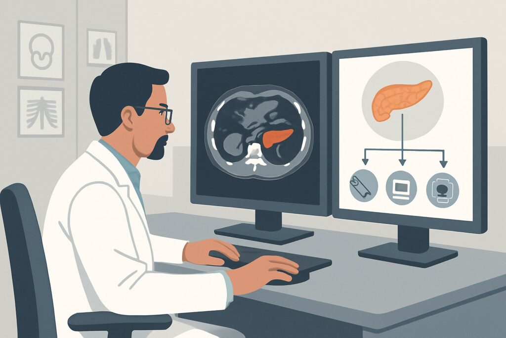
Why Is MRI of the Abdomen the Top Recommended Study for Locoregional Staging?
For the detailed soft-tissue and vascular assessment required in this scenario, the ACR designates MRI abdomen without and with IV contrast as Usually Appropriate. This recommendation is driven by MRI’s distinct physical advantages for characterizing pancreatic tumors and their local environment.
The primary strength of MRI is its superior soft-tissue contrast resolution. It can often better delineate the margins of the tumor against the background of normal pancreatic parenchyma, which is crucial for assessing subtle extension into the peripancreatic fat. Furthermore, sequences like diffusion-weighted imaging (DWI) provide functional information about tissue cellularity, which can help in identifying the tumor and assessing its response to neoadjuvant therapy. For evaluating potential liver metastases, contrast-enhanced MRI is considered the most sensitive modality.
While MRI is highly rated, it is not the only appropriate option. The ACR also rates CT abdomen with IV contrast multiphase as Usually Appropriate. A high-quality, pancreas-protocol CT is a powerful and widely available tool that provides excellent spatial resolution and robust vascular mapping. It is often the institutional standard and a perfectly acceptable first choice. The selection between pancreas-protocol MRI and CT often depends on local expertise, scanner availability, and specific patient factors (e.g., contraindications to gadolinium contrast or iodinated contrast).
Other modalities are rated lower for this specific task. For example, FDG-PET/CT skull base to mid-thigh is rated May be appropriate. While valuable for detecting occult distant metastases and assessing metabolic response to therapy, its spatial resolution is inferior to dedicated CT or MRI for defining the precise tumor-vessel interface, which is the central question in locoregional staging.
From a safety perspective, MRI offers a significant advantage by avoiding ionizing radiation (0 mSv), a key consideration for patients who will require multiple follow-up scans. This contrasts with a multiphase CT, which carries a radiation dose of ☢☢☢☢ (10-30 mSv). When ordering, specify a “pancreas protocol MRI” to ensure the appropriate sequences, including thin-slice, post-contrast arterial and venous phases, and DWI, are performed.
What Happens After the Staging MRI? Downstream Workflow
The results of the locoregional staging MRI are a critical input for the multidisciplinary tumor board and will dictate the next steps in management. The workflow branches based on the findings:
- If the study shows clearly resectable disease: The patient may be referred directly for surgical consultation for upfront pancreatectomy. In some centers, neoadjuvant therapy is increasingly considered even for resectable disease, a decision made by the tumor board.
- If the study shows borderline resectable disease: This is the most common indication for neoadjuvant chemotherapy (like FOLFIRINOX) or chemoradiation. The goal is to shrink the tumor away from the involved vessels. The patient will undergo a course of therapy, followed by a repeat staging scan (either MRI or CT) to evaluate for response and determine if they have become a candidate for surgery.
- If the study shows locally advanced/unresectable disease: The patient is generally not a surgical candidate. The primary treatment will be systemic chemotherapy, sometimes with the addition of radiation for local control. Palliative interventions for biliary or duodenal obstruction may be necessary.
- If the study is indeterminate: In rare cases where the tumor-vessel interface is ambiguous, endoscopic ultrasound (EUS) may provide additional high-resolution detail of the portal vein or SMV. However, for arterial involvement, high-quality cross-sectional imaging with CT or MRI remains the standard.
This staging and re-staging process is iterative. Patients will undergo serial imaging throughout their treatment course to monitor response and adjust the therapeutic plan accordingly.
Pitfalls to Avoid (and When to Get Help)
Accurate locoregional staging of pancreatic cancer is unforgiving. Small errors in imaging acquisition or interpretation can lead to major changes in management. Here are common pitfalls to avoid:
- Ordering a routine abdomen scan: A standard, single-phase “CT Abdomen/Pelvis with contrast” is inadequate. You must order a dedicated, multiphase “Pancreas Protocol” CT or MRI to acquire the necessary thin-slice arterial and venous phase images.
- Misinterpreting post-treatment changes: Fibrosis and inflammation after neoadjuvant therapy can mimic residual tumor on imaging. This can lead to over-calling unresectability. Interpretation should be done by an experienced abdominal radiologist, often in the context of tumor marker trends (e.g., CA 19-9).
- Ignoring the venous anatomy variant: Anatomic variations of the portal or superior mesenteric veins are common. Failure to recognize these can impact the surgical plan.
- Underestimating subtle metastatic disease: Meticulously review the liver, peritoneum, and distant lymph nodes on every scan, as the presence of any metastatic disease upstages the patient and changes the treatment intent from curative to palliative.
If imaging findings are equivocal or do not align with the clinical picture, always bring the case back to your multidisciplinary tumor board for discussion with radiologists, surgeons, oncologists, and gastroenterologists.
Related ACR Topics and Tools
For a comprehensive overview of imaging across all clinical presentations of pancreatic adenocarcinoma, from screening to surveillance, please consult our parent topic hub article. For specific questions about imaging protocols or radiation dose, the tools below provide direct access to valuable data.
- For breadth across all scenarios in this topic, see our parent guide: Screening, Locoregional Assessment, and Surveillance of Pancreatic Ductal Adenocarcinoma: ACR Appropriateness Decoded.
- To explore adjacent clinical scenarios, use the ACR Appropriateness Criteria Lookup.
- For technical details on the recommended study, browse the Imaging Protocol Library.
- To discuss cumulative exposure with patients undergoing serial scans, consult the Radiation Dose Calculator.
Frequently Asked Questions
Is a pancreas-protocol CT just as good as an MRI for locoregional staging?
Yes, for this specific scenario, the ACR rates both pancreas-protocol CT and pancreas-protocol MRI as ‘Usually Appropriate.’ A high-quality multiphase CT is an excellent and often more accessible alternative to MRI. The choice between them may depend on institutional preference, radiologist expertise, and patient-specific factors like renal function or contrast allergies. MRI may offer a slight advantage in soft-tissue contrast and detection of small liver metastases, and it avoids ionizing radiation.
Why isn’t PET/CT the primary study for staging pancreatic cancer?
While FDG-PET/CT is rated ‘May be appropriate’ and is very useful for detecting distant metastatic disease, its spatial resolution is lower than that of dedicated CT or MRI. For the critical task of locoregional staging—defining the precise relationship between the tumor and adjacent blood vessels—the anatomical detail provided by a high-quality MRI or CT is superior.
What if the patient has a contraindication to gadolinium-based contrast for an MRI?
If a patient has a severe allergy to gadolinium-based contrast agents or severely impaired renal function (low eGFR), a non-contrast MRI is ‘Usually not appropriate’ for this indication as it lacks the necessary vascular and parenchymal detail. In this case, a multiphase pancreas-protocol CT with iodinated contrast would be the preferred alternative, assuming no contraindication to iodine exists.
How soon after neoadjuvant therapy should re-staging imaging be performed?
Re-staging imaging is typically performed 4 to 6 weeks after the completion of neoadjuvant therapy. This interval allows for acute inflammatory effects from treatment (especially radiation) to subside, providing a more accurate assessment of the true residual tumor burden before a final decision on surgery is made.
Should I add MRCP to the MRI order for locoregional staging?
Adding MRCP (Magnetic Resonance Cholangiopancreatography) sequences is also rated ‘Usually Appropriate.’ MRCP is a non-invasive technique that provides detailed images of the biliary and pancreatic ducts. It is particularly useful if there is a question of biliary obstruction or pancreatic duct involvement that could influence the surgical plan, such as the need for biliary stenting or the specific type of pancreatic resection required.
Reviewed by Pouyan Golshani, MD, Interventional Radiologist — May 30, 2026