What Is the Best Initial Imaging for First Trimester Vaginal Bleeding?
A patient presents to the emergency department on a Tuesday morning. She is 8 weeks pregnant by her last menstrual period, and this morning she noticed a small amount of vaginal spotting. She is anxious, and you need to determine the status and location of the pregnancy. This scenario is one of the most common presentations in early pregnancy, and the clinical question is direct: what is the most appropriate initial imaging study to order? This article provides a detailed workflow for this specific situation, grounded in the American College of Radiology (ACR) Appropriateness Criteria, which rate transabdominal and transvaginal pelvic ultrasound as ‘Usually Appropriate’ for the initial evaluation of first trimester vaginal bleeding.
Who Fits This Clinical Scenario?
This guidance applies to a specific and common patient presentation: a pregnant individual in the first trimester (up to 13 weeks and 6 days gestation) who presents with vaginal bleeding and is hemodynamically stable. The core objective of imaging is to confirm the presence, viability, and location of the pregnancy. This patient is typically alert, has normal vital signs, and may report symptoms ranging from light spotting to moderate bleeding, with or without mild cramping.
It is crucial to distinguish this scenario from others that require a different approach. This workflow does not apply if the patient is:
- Hemodynamically Unstable: A patient with hypotension, tachycardia, or other signs of shock requires immediate resuscitation and an emergent obstetrics/gynecology (OB/Gyn) consultation. Imaging should not delay definitive management, which may be surgical.
- Experiencing Severe, Focal Pelvic Pain: While mild cramping can be normal, severe or unilateral pain raises the suspicion for a ruptured ectopic pregnancy or ovarian torsion, necessitating a more urgent evaluation.
- In the Second or Third Trimester: Vaginal bleeding later in pregnancy has a different differential diagnosis (e.g., placenta previa, placental abruption) and is evaluated under separate clinical guidelines.
What Diagnoses Are You Working Up in This Scenario?
When ordering an ultrasound for first trimester bleeding, you are primarily investigating several potential diagnoses, ranging from benign to life-threatening. The imaging study is designed to differentiate among these possibilities.
Viable Intrauterine Pregnancy (IUP): This is the most reassuring and often the most common finding. The ultrasound aims to identify a gestational sac within the uterus containing a yolk sac and/or an embryo with detectable cardiac activity. The presence of a subchorionic hematoma, a collection of blood between the chorion and the uterine wall, is also a frequent cause of bleeding in an otherwise viable pregnancy.
Spontaneous Abortion (Miscarriage): Ultrasound is critical for diagnosing and classifying a pregnancy loss. This can be categorized as a threatened abortion (bleeding with a closed cervix and viable IUP), an inevitable abortion (dilated cervix), an incomplete abortion (retained products of conception), or a complete abortion (an empty uterus with a previously confirmed IUP).
Ectopic Pregnancy: This is the most critical, can’t-miss diagnosis. An ectopic pregnancy occurs when a fertilized egg implants outside the uterine cavity, most commonly in the fallopian tube. An ultrasound evaluation that shows an empty uterus in the setting of a positive pregnancy test, particularly with a quantitative beta-hCG level above the discriminatory zone, is highly suspicious for an ectopic pregnancy until proven otherwise. The sonographer will carefully evaluate the adnexa for any suspicious mass.
Gestational Trophoblastic Disease (GTD): A less common but important consideration is a molar pregnancy, a rare condition where abnormal tissue grows inside the uterus instead of an embryo. Ultrasound may reveal a characteristic “snowstorm” or “cluster of grapes” appearance within the uterus, with no viable fetus.
Why Is Pelvic Ultrasound the Recommended Initial Study?
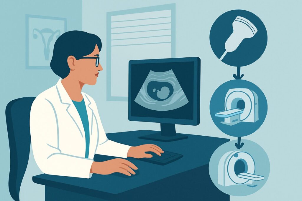
The ACR designates both US pelvis transabdominal and US pelvis transvaginal as ‘Usually Appropriate’ for the initial imaging of first trimester vaginal bleeding. These two examinations are complementary and are almost always performed together as a single, comprehensive pelvic ultrasound study. There is no ionizing radiation (0 mSv) involved, making it the safest imaging modality for the developing fetus.
The transabdominal approach provides a wide field of view, allowing for a general survey of the entire pelvis, including the uterus, cervix, and adnexa. It is useful for identifying large abnormalities and orienting the anatomy. However, its resolution is often insufficient for the fine details of an early pregnancy. The transvaginal approach uses a higher-frequency transducer placed in the vaginal fornix, providing superior spatial resolution. This is essential for visualizing the key early pregnancy landmarks: the gestational sac, the yolk sac, the fetal pole, and, crucially, fetal cardiac activity, which can be detected as early as 5.5 to 6 weeks gestation.
Alternative imaging modalities are rated lower for this initial evaluation for clear reasons:
- CT of the Pelvis: This is rated ‘Usually not appropriate’. CT exposes the fetus to significant ionizing radiation (☢☢☢ to ☢☢☢☢) and provides poor visualization of intrauterine contents compared to ultrasound. Its use is reserved for trauma or other indications unrelated to the pregnancy itself.
- MRI of the Pelvis: MRI without contrast is rated ‘May be appropriate’ but is not a first-line tool. It is used as a problem-solving modality when ultrasound is inconclusive, for example, to better characterize a complex adnexal mass or confirm a rare type of ectopic pregnancy (e.g., cornual, cervical). MRI with IV contrast is ‘Usually not appropriate’ as gadolinium-based contrast agents are generally avoided during pregnancy due to theoretical fetal risks.
What’s Next After Pelvic Ultrasound? Downstream Workflow
The results of the pelvic ultrasound will guide your next clinical steps. The workflow branches based on one of three primary outcomes: a confirmed intrauterine pregnancy, a confirmed ectopic pregnancy, or a pregnancy of unknown location.
If a viable IUP is confirmed: The patient can typically be reassured. If a subchorionic hematoma is identified, she should be counseled about the potential for continued bleeding and the slightly increased risk of pregnancy loss, with instructions for close follow-up with her OB/Gyn provider.
If a nonviable pregnancy (miscarriage) is diagnosed: The patient requires counseling on management options, which include expectant management (waiting for the tissue to pass naturally), medical management (e.g., with misoprostol), or surgical management (dilation and curettage). The choice depends on clinical stability, gestational age, and patient preference.
If an ectopic pregnancy is diagnosed: This constitutes a medical emergency. An immediate OB/Gyn consultation is required to determine the best course of action, which may be medical treatment with methotrexate (for stable patients meeting specific criteria) or surgical intervention.
If the study is indeterminate (Pregnancy of Unknown Location – PUL): This is a common and important outcome. It means no definitive IUP or ectopic pregnancy was identified on ultrasound. The next step is to correlate the imaging findings with a quantitative serum beta-hCG level. If the hCG level is below the institution’s “discriminatory zone” (typically 1,500-3,000 mIU/mL), the pregnancy may simply be too early to visualize. The standard management is to repeat the hCG level in 48 hours and schedule a follow-up ultrasound. An abnormally rising hCG in the absence of an IUP increases the suspicion for an ectopic pregnancy.
Pitfalls to Avoid (and When to Get Help)
Several common pitfalls can complicate the evaluation of first trimester bleeding. Awareness of these can help prevent diagnostic errors.
- Not correlating with beta-hCG: Interpreting an ultrasound showing an “empty uterus” without knowing the quantitative hCG level is a major pitfall. An empty uterus with an hCG of 500 is very different from an empty uterus with an hCG of 5,000.
- Mistaking a pseudosac for a true gestational sac: A small fluid collection in the endometrium, known as a pseudosac, can be seen in up to 20% of ectopic pregnancies. A true gestational sac will have a thick, echogenic rim (the double decidual sign) and should eventually contain a yolk sac.
- Incomplete adnexal evaluation: A thorough search of the adnexa, cul-de-sac, and upper abdomen is mandatory if an IUP is not seen. An ectopic pregnancy can be located anywhere from the cervix to the cornua to the ovary.
- Over-relying on transabdominal imaging: In gestations under 7-8 weeks, a transvaginal scan is almost always necessary for a definitive diagnosis. Ordering only a transabdominal ultrasound can lead to an indeterminate result and delay care.
If you identify a suspected ectopic pregnancy, a heterotopic pregnancy (co-existing IUP and ectopic), or the patient becomes hemodynamically unstable, escalate immediately with an urgent OB/Gyn consultation.
Related ACR Topics and Tools
For a comprehensive overview of all clinical variants related to this topic, please see our parent guide. For further exploration of imaging guidelines, protocols, and safety, the following resources are available:
- For breadth across all scenarios in First Trimester Vaginal Bleeding, see our parent guide: First Trimester Vaginal Bleeding: ACR Appropriateness Decoded.
- To review adjacent clinical scenarios and their respective imaging recommendations, use the ACR Appropriateness Criteria Lookup tool.
- For detailed procedural techniques on the recommended study, consult the Imaging Protocol Library.
- To discuss radiation exposure from alternative studies like CT, the Radiation Dose Calculator can help frame conversations about cumulative dose.
Frequently Asked Questions
Why are both transabdominal and transvaginal ultrasound performed?
They are complementary. The transabdominal scan provides a broad overview of the entire pelvis and can identify large masses or free fluid that might be missed on the focused transvaginal view. The transvaginal scan offers much higher resolution, which is essential for visualizing the fine details of an early pregnancy, such as the yolk sac, fetal pole, and cardiac activity, often before they are visible transabdominally.
What is the ‘discriminatory zone’ for beta-hCG and why does it matter?
The discriminatory zone is the level of serum beta-hCG above which a normal intrauterine pregnancy should be visible with transvaginal ultrasound. While the exact number varies by institution (often 1,500-3,000 mIU/mL), its significance is critical. If the hCG level is above this zone and the ultrasound shows an empty uterus, the suspicion for an ectopic pregnancy is very high.
Is an ultrasound necessary if the bleeding is very light?
Yes. The amount of bleeding does not reliably correlate with the underlying diagnosis. A life-threatening ectopic pregnancy can present with only light spotting, while a benign subchorionic hematoma can cause heavy bleeding. Imaging is necessary to determine the location and viability of the pregnancy regardless of the volume of bleeding.
Can ultrasound determine the exact cause of the bleeding?
Sometimes. Ultrasound can identify potential sources like a subchorionic hematoma or signs of an impending miscarriage. However, in many cases of a threatened abortion with a viable intrauterine pregnancy, no specific source of bleeding is identified on the scan. The primary role of the ultrasound is to confirm viability and rule out an ectopic pregnancy.
If the first ultrasound is inconclusive, when should it be repeated?
If the initial ultrasound results in a diagnosis of Pregnancy of Unknown Location (PUL), the standard of care is to obtain serial quantitative beta-hCG levels. A repeat ultrasound is typically scheduled once the hCG level is expected to be above the discriminatory zone or after a 48-72 hour interval to assess for appropriate development of an early IUP.
Reviewed by Pouyan Golshani, MD, Interventional Radiologist — May 21, 2026