When to Order Imaging for First Trimester Vaginal Bleeding: ACR Appropriateness Decoded

It’s 11 p.m. in the emergency department. A patient presents with vaginal bleeding at an estimated 8 weeks of gestation. Their vitals are stable, but the clinical picture is uncertain. Is this a threatened abortion, an incomplete abortion, or the first sign of a potentially life-threatening ectopic pregnancy? The immediate next step is imaging, but the specific choice and sequence matter for diagnostic accuracy and patient safety. The American College of Radiology (ACR) Appropriateness Criteria provide an evidence-based framework for this exact scenario, guiding clinicians away from unnecessary radiation and toward the most direct diagnostic path. This article decodes those recommendations to help you make the right call, every time.
What Does ACR First Trimester Vaginal Bleeding Cover?
The ACR Appropriateness Criteria for “First Trimester Vaginal Bleeding” focus on a specific and common clinical presentation: a pregnant patient, up to 13 weeks and 6 days of gestation, who presents with vaginal bleeding. The primary goals of imaging in this context are to confirm the presence of an intrauterine pregnancy (IUP), assess fetal viability (e.g., presence of a heartbeat), and identify the cause of the bleeding. This includes diagnosing a normal IUP, a failed IUP (threatened, incomplete, or complete abortion), an ectopic pregnancy, or a gestational trophoblastic disease.
These guidelines do not apply to patients with bleeding in the second or third trimesters, postpartum hemorrhage, or non-pregnant patients with vaginal bleeding. Each of those scenarios has its own distinct diagnostic algorithm and corresponding ACR guidelines. The criteria outlined here are specifically tailored for the initial imaging evaluation of a hemodynamically stable patient in early pregnancy.
What Imaging Should I Order for First Trimester Vaginal Bleeding? Recommendations by Clinical Scenario
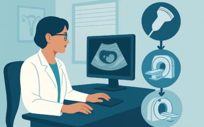
For the initial imaging of a patient with first-trimester vaginal bleeding, the ACR guidelines are clear and heavily favor ultrasound as the primary modality due to its safety and high diagnostic yield.
For the clinical scenario of First trimester vaginal bleeding, initial imaging, the ACR rates both US pelvis transabdominal and US pelvis transvaginal as Usually appropriate. These are the cornerstone examinations. A transabdominal ultrasound is typically performed first to provide a broad overview of the pelvis, identify large abnormalities, and orient the examiner. The transvaginal ultrasound follows, offering superior spatial resolution for detailed evaluation of the endometrium, gestational sac, yolk sac, embryo, and adnexa. This combination is essential for confirming an intrauterine pregnancy and detecting subtle signs of an ectopic pregnancy.
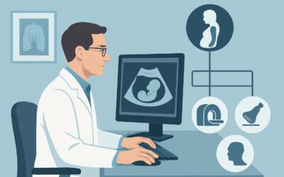
Two other studies are rated as May be appropriate in specific contexts. US duplex Doppler of the pregnant uterus can be a valuable adjunct to standard grayscale ultrasound. Its primary role is to confirm fetal cardiac activity, which is a key indicator of viability. It can also help characterize blood flow in complex adnexal masses or evaluate for suspected gestational trophoblastic disease. MRI of the pelvis without IV contrast is considered a problem-solving tool. It is not a first-line test but may be used when ultrasound is inconclusive, such as in cases of a suspected but non-visualized ectopic pregnancy or to better characterize a complex pelvic mass found incidentally on ultrasound.
Modalities involving ionizing radiation or gadolinium contrast are strongly discouraged. CT of the pelvis (with, without, or with and without IV contrast) is rated Usually not appropriate due to the radiation dose to the developing fetus. Similarly, MRI of the pelvis without and with IV contrast is Usually not appropriate because gadolinium-based contrast agents are generally avoided during pregnancy unless the potential benefit unequivocally outweighs the potential risks. CT is typically reserved for emergent, life-threatening situations where the clinical suspicion for a non-gynecologic cause of symptoms (like appendicitis or renal colic) is high and ultrasound is non-diagnostic. For more details on CT protocols, see our guide on CT Chest/Abdomen/Pelvis with IV Contrast.
ACR Imaging Recommendations Table
| Clinical Scenario | Procedure | ACR Rating | Adult RRL | Pediatric RRL |
|---|---|---|---|---|
| First trimester vaginal bleeding. Initial imaging. | US pelvis transabdominal | Usually appropriate | O 0 mSv | O 0 mSv [ped] |
| First trimester vaginal bleeding. Initial imaging. | US pelvis transvaginal | Usually appropriate | O 0 mSv | O 0 mSv [ped] |
| First trimester vaginal bleeding. Initial imaging. | US duplex Doppler pregnant uterus | May be appropriate | O 0 mSv | O 0 mSv [ped] |
| First trimester vaginal bleeding. Initial imaging. | MRI pelvis without IV contrast | May be appropriate | O 0 mSv | O 0 mSv [ped] |
| First trimester vaginal bleeding. Initial imaging. | MRI pelvis without and with IV contrast | Usually not appropriate | O 0 mSv | O 0 mSv [ped] |
| First trimester vaginal bleeding. Initial imaging. | CT pelvis with IV contrast | Usually not appropriate | ☢ ☢ ☢ 1-10 mSv | ☢ ☢ ☢ ☢ 3-10 mSv [ped] |
| First trimester vaginal bleeding. Initial imaging. | CT pelvis without IV contrast | Usually not appropriate | ☢ ☢ ☢ 1-10 mSv | ☢ ☢ ☢ ☢ 3-10 mSv [ped] |
| First trimester vaginal bleeding. Initial imaging. | CT pelvis without and with IV contrast | Usually not appropriate | ☢ ☢ ☢ ☢ 10-30 mSv | ☢ ☢ ☢ ☢ 3-10 mSv [ped] |
Adult vs. Pediatric First Trimester Vaginal Bleeding Imaging: Radiation Dose Tradeoffs
The clinical scenario of first-trimester vaginal bleeding primarily involves adult or adolescent patients. While the ACR provides pediatric relative radiation level (RRL) estimates for completeness across all imaging procedures, the practical application here is for younger pregnant patients, such as teenagers. The core principle of ALARA (As Low As Reasonably Achievable) is paramount in this population.
The strong preference for non-ionizing radiation modalities like ultrasound (transabdominal, transvaginal, and Doppler) and non-contrast MRI is even more critical in younger patients. These studies have an RRL of zero, posing no radiation risk to the patient or the fetus. For the rare and exceptional cases where CT is deemed medically necessary, it’s important to note the higher pediatric RRL symbol (☢ ☢ ☢ ☢) for CT scans. This reflects the increased lifetime attributable risk of cancer from radiation exposure in younger individuals. Any consideration of CT in a pregnant adolescent must involve a careful risk-benefit discussion and consultation with a radiologist to ensure the protocol is optimized for dose reduction.
Imaging Protocol Details for First Trimester Vaginal Bleeding
Once you’ve decided on the right study, the specific imaging protocol is crucial for diagnostic quality. Our protocol guides provide detailed, actionable information on technique, contrast parameters, and key interpretation principles for the studies recommended by the ACR.
Tools to Help You Order the Right Study
Navigating imaging guidelines efficiently is key to modern clinical practice. GigHz offers a suite of free reference tools designed to support evidence-based ordering and enhance patient care.
For clinical scenarios beyond first-trimester bleeding, our comprehensive ACR Appropriateness Criteria Lookup tool provides instant access to the full library of ACR guidelines, covering hundreds of clinical variants across all specialties.
To dive deeper into the technical aspects of any imaging study, the Imaging Protocol Library offers detailed, step-by-step protocols used by leading academic centers, helping ensure you order a high-quality, diagnostically valuable exam.
When discussing radiation exposure with patients or tracking cumulative dose, the Radiation Dose Calculator is an invaluable resource for translating complex dosimetry into understandable terms and reinforcing your commitment to radiation safety.
Frequently Asked Questions
Here are some common questions clinicians have when ordering imaging for first-trimester vaginal bleeding.
When is a transvaginal ultrasound absolutely necessary?
A transvaginal ultrasound is almost always necessary if the transabdominal ultrasound does not definitively confirm a viable intrauterine pregnancy. It provides the high-resolution detail needed to visualize the gestational sac, yolk sac, and fetal pole in very early gestations (as early as 5 weeks). It is also superior for evaluating the adnexa for an ectopic pregnancy and assessing for subtle findings in the endometrium.
What should I do if ultrasound is inconclusive for ectopic pregnancy?
If the ultrasound is inconclusive but there is a high clinical suspicion for an ectopic pregnancy (e.g., adnexal pain, rising beta-hCG levels without a visible IUP), the next step is typically serial beta-hCG monitoring and a repeat ultrasound in 48-72 hours. In select, complex cases where a non-adnexal ectopic pregnancy (e.g., cornual, cervical, or abdominal) is suspected, a non-contrast MRI may be considered as a problem-solving tool.
Can I order a CT scan if I suspect appendicitis in a pregnant patient?
Yes, but with caution. If appendicitis is suspected, the recommended initial imaging is a graded-compression ultrasound of the right lower quadrant. If the ultrasound is non-diagnostic, MRI without IV contrast is the preferred next step to avoid radiation. A low-dose CT scan is reserved for situations where MRI is unavailable or contraindicated and the clinical suspicion remains high, as the risk of a missed, ruptured appendix can be catastrophic. This decision should be made in consultation with a radiologist.
Is Doppler ultrasound safe in the first trimester?
Yes, when used appropriately. While there are theoretical concerns about thermal effects from prolonged Doppler use, the brief application needed to confirm fetal cardiac activity is considered safe. The principle of ALARA still applies: use the lowest possible power output and limit the dwell time to the minimum necessary to obtain the diagnostic information.
What beta-hCG level should prompt visualization of an IUP on ultrasound?
This is known as the “discriminatory zone.” While it can vary by institution, an intrauterine gestational sac should typically be visible on transvaginal ultrasound when the serum beta-hCG level is between 1,500 and 2,000 mIU/mL. If the hCG level is above this zone and no IUP is seen, the suspicion for an ectopic pregnancy is significantly elevated.
Reviewed by Pouyan Golshani, MD, Interventional Radiologist — May 21, 2026