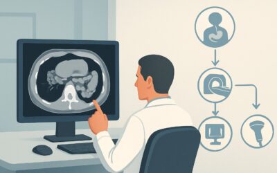
What Is the Best Initial Imaging for Hyperbilirubinemia and Acute Cholestasis?
A 62-year-old woman presents to the emergency department with a three-day history of yellowing skin, intense itching, and dark-colored urine. Her initial lab work reveals a total bilirubin of 8.2 mg/dL with a direct bilirubin of 6.5 mg/dL, consistent with conjugated hyperbilirubinemia. The clinical picture suggests an acute cholestatic process, but the immediate challenge is to distinguish between an obstructive cause, like a gallstone, and an intrahepatic cause, like drug-induced liver injury. This distinction is critical, as the former may require urgent intervention while the latter is managed medically. This article provides a focused workflow for this exact scenario—acute or subacute cholestasis with hyperbilirubinemia—explaining why the American College of Radiology (ACR) designates abdominal ultrasound as Usually appropriate for initial imaging.
Who Fits This Clinical Scenario for Acute Cholestasis?
This guidance is specifically for patients presenting with acute or subacute (days to weeks) signs and symptoms of cholestasis, supported by laboratory findings of hyperbilirubinemia. The bilirubin can be either conjugated (direct) or unconjugated (indirect), though a cholestatic picture typically involves a significant conjugated component.
Inclusion criteria for this workflow:
- Clinical signs of cholestasis: jaundice, scleral icterus, pruritus, dark urine, pale stools.
- Laboratory evidence of hyperbilirubinemia.
- Acute or subacute onset (less than a few months).
- The clinical question is the initial workup to determine the etiology, primarily to differentiate obstructive from non-obstructive causes.
It is crucial to distinguish this presentation from related but distinct clinical scenarios that follow different imaging pathways. This workflow does not apply if:
- The pattern is primarily hepatocellular: If the lab work shows a predominant elevation of aminotransferases (AST/ALT) with only mild or no hyperbilirubinemia, the workup shifts toward parenchymal liver injury.
- The pattern is isolated alkaline phosphatase elevation: If alkaline phosphatase is significantly elevated with normal or near-normal bilirubin, the differential diagnosis and imaging strategy are different, focusing on infiltrative diseases or partial biliary obstruction.
- The patient has known chronic liver disease: In a patient with established cirrhosis presenting with jaundice, the workup is for acute-on-chronic liver failure, and the imaging goals may differ (e.g., screening for hepatocellular carcinoma).
What Diagnoses Are You Working Up in This Scenario?
The primary goal of initial imaging in acute cholestasis is to identify or exclude a mechanical obstruction of the biliary tree (extrahepatic cholestasis). Answering this question dictates the entire subsequent management plan, separating patients who may need a procedural intervention from those who require medical management and further hepatological workup.
Choledocholithiasis: This is the most common cause of extrahepatic biliary obstruction. A gallstone migrates from the gallbladder into the common bile duct, causing a blockage. Imaging is focused on identifying the stone itself or, more commonly, the secondary sign of biliary ductal dilation proximal to the obstruction.
Malignant Obstruction: A less common but critical diagnosis to consider is obstruction from a tumor. A pancreatic head adenocarcinoma is a classic cause, but cholangiocarcinoma (cancer of the bile ducts) or an ampullary tumor can also present this way. Imaging looks for a discrete mass, ductal wall thickening, or an abrupt cutoff of the bile duct.
Benign Biliary Stricture: Narrowing of the bile ducts can occur from non-malignant causes. This is often seen in patients with a history of biliary surgery (e.g., cholecystectomy), pancreatitis, or inflammatory conditions like primary sclerosing cholangitis. Imaging aims to characterize the location and length of the stricture.
Intrahepatic Cholestasis: If imaging shows no evidence of biliary ductal dilation or obstruction, the cause is likely intrahepatic. This broad category includes viral hepatitis, drug-induced liver injury (DILI), alcoholic hepatitis, and autoimmune conditions. In these cases, imaging plays a crucial role by confidently ruling out a surgical problem, allowing the clinical team to focus on medical causes.
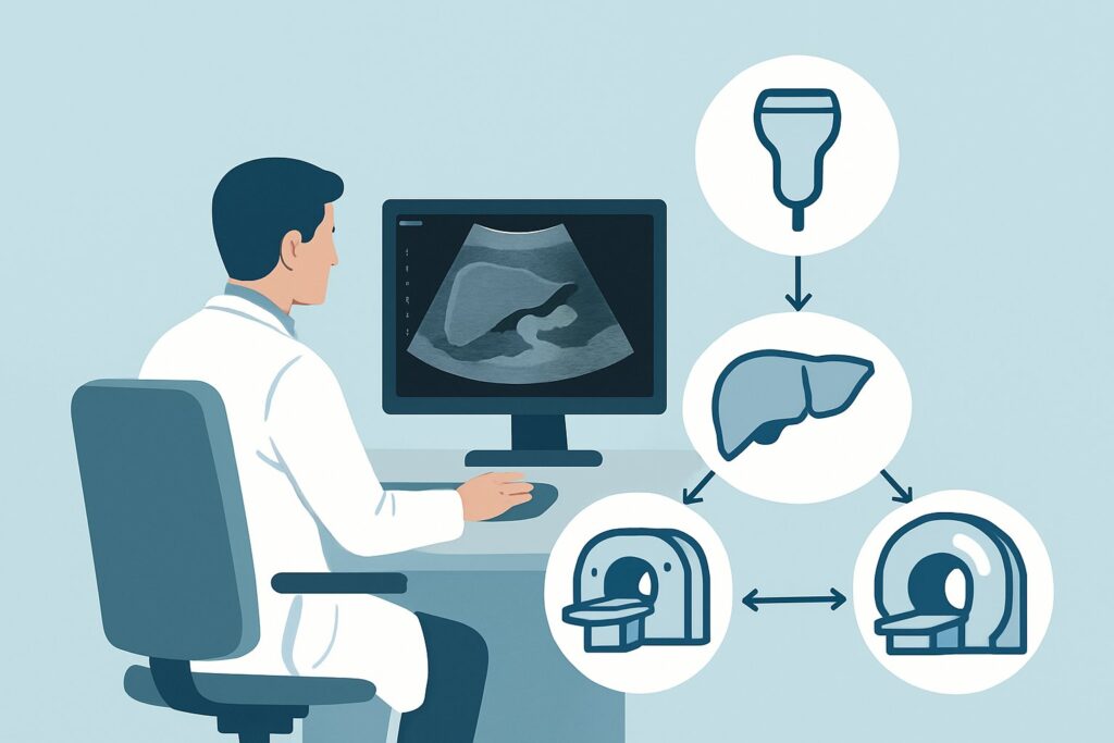
Why Is Abdominal Ultrasound the Recommended First Study for This Presentation?
For the initial evaluation of a patient with suspected acute cholestasis, the ACR designates US abdomen as Usually appropriate. This recommendation is based on its high diagnostic utility, safety profile, and accessibility.
The primary strength of ultrasound is its excellent ability to detect biliary ductal dilation, the key indicator of an obstructive process. A normal common bile duct diameter (typically <6 mm, with some increase allowed for age and prior cholecystectomy) effectively rules out significant downstream obstruction. Ultrasound is also highly sensitive for detecting stones within the gallbladder (cholelithiasis) and can often directly visualize stones within the common bile duct (choledocholithiasis), especially in the proximal portion.
From a safety and practical standpoint, ultrasound is the ideal first-line test. It involves no ionizing radiation (adult_rrl=O 0 mSv) and does not require IV contrast, avoiding risks of nephrotoxicity or allergic reaction. It is relatively inexpensive, widely available in most clinical settings, and can be performed quickly at the bedside if necessary.
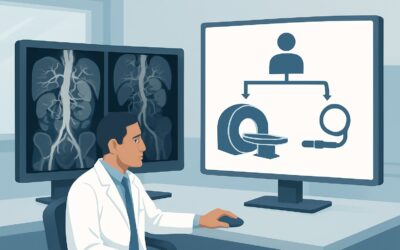
Why are other advanced imaging modalities not the first choice?
- CT abdomen and pelvis with IV contrast is also rated Usually appropriate but is typically considered a second-line or problem-solving tool in this specific scenario. While excellent for detecting pancreatic masses and assessing for metastatic disease, it involves significant ionizing radiation (adult_rrl=☢☢☢ 1-10 mSv) and is less sensitive than ultrasound for identifying gallstones. It becomes the preferred study if the initial ultrasound is equivocal or if there is a high clinical suspicion for malignancy.
- MRI abdomen with MRCP (Magnetic Resonance Cholangiopancreatography) is also Usually appropriate and is the non-invasive gold standard for detailed evaluation of the biliary tree. It provides exquisite anatomical detail of the ducts, identifying stones, strictures, and small tumors with very high accuracy. However, its higher cost, longer acquisition time, and limited availability make it unsuitable as a first-line screening tool. It is best reserved for cases where ultrasound is non-diagnostic or when a detailed biliary map is required to plan for a therapeutic procedure like Endoscopic Retrograde Cholangiopancreatography (ERCP).
What’s Next After US abdomen? Downstream Workflow
The results of the initial abdominal ultrasound will guide the subsequent diagnostic and therapeutic pathway. The workflow typically branches based on the presence or absence of biliary ductal dilation.
If the ultrasound shows biliary ductal dilation (Positive for Obstruction):
- This finding strongly suggests an extrahepatic, obstructive cause. The next step is to define the level and nature of the obstruction.
- MRI with MRCP is often the next logical imaging study. It can precisely locate the obstruction (e.g., a stone in the distal common bile duct vs. a pancreatic head mass) and provides the detailed anatomical map needed to plan for intervention.
- If a stone is confirmed or highly suspected, the patient will likely be referred to gastroenterology for ERCP, a procedure that can both diagnose and treat the obstruction by removing the stone.
- If a suspicious mass is seen, a contrast-enhanced CT or MRI may be performed for staging, followed by a biopsy.
If the ultrasound is negative (No Biliary Dilation):
- A normal-caliber biliary tree makes a significant mechanical obstruction highly unlikely. The workup should pivot to investigate intrahepatic causes of cholestasis.
- This involves a comprehensive serological workup, including a viral hepatitis panel, autoimmune markers (e.g., ANA, AMA), and a thorough review of the patient’s medications for potential drug-induced liver injury.
- Further imaging is typically not required unless the clinical picture changes or the serologic workup is unrevealing, in which case a liver biopsy may be considered.
If the ultrasound is indeterminate or equivocal:
- This can occur in cases of early or partial obstruction where ductal dilation is minimal, or if patient body habitus limits visualization.
- In this situation, proceeding to MRI with MRCP is the most appropriate next step to definitively evaluate the biliary system.
Pitfalls to Avoid (and When to Get Help)
Navigating the workup for acute cholestasis requires careful interpretation of both clinical and imaging findings. Here are a few common pitfalls to avoid:
- Stopping the workup after a “negative” ultrasound: A normal ultrasound rules out significant obstruction but does not provide a final diagnosis. It is a critical branch point, not an endpoint. The workup must continue to identify the intrahepatic cause.
- Over-reliance on ductal diameter alone: While ductal dilation is a key sign, its absence does not completely exclude a small, non-obstructing stone or an early obstruction. Correlate with the degree of hyperbilirubinemia and clinical symptoms.
- Delaying advanced imaging when indicated: If the ultrasound is equivocal but clinical suspicion for obstruction remains high (e.g., severe pain, rising bilirubin), do not delay proceeding to MRCP or CT.
- Forgetting about acalculous cholecystitis: In a critically ill patient, consider acalculous cholecystitis as a cause of abnormal liver tests, which may present with gallbladder wall thickening and pericholecystic fluid on ultrasound without stones.
If the patient shows signs of ascending cholangitis (fever, right upper quadrant pain, and jaundice—Charcot’s triad), this is a medical emergency. Escalate immediately for surgical and gastroenterology consultation for urgent biliary decompression.
Related ACR Topics and Tools
The ACR Appropriateness Criteria are a powerful resource for evidence-based imaging decisions. For a comprehensive overview of all clinical variants related to liver function tests, please see our parent guide. For tools to help with ordering, protocoling, and patient communication, see the resources below.
- For breadth across all scenarios in Abnormal Liver Function Tests, see our parent guide: Abnormal Liver Function Tests: ACR Appropriateness Decoded.
- To explore other clinical scenarios and their recommended imaging pathways, use the ACR Appropriateness Criteria Lookup.
- For detailed procedural techniques for the recommended studies, consult the Imaging Protocol Library.
- To help discuss radiation exposure with patients for studies like CT, use the Radiation Dose Calculator.
Frequently Asked Questions
Is there a role for HIDA scan in the initial workup of acute cholestasis and jaundice?
A HIDA scan (hepatobiliary iminodiacetic acid scan) is primarily used to evaluate for acute cholecystitis or to assess biliary motility and is not a first-line test for the initial workup of jaundice. Its main role is to confirm or exclude cystic duct obstruction. In the setting of high bilirubin levels (>5 mg/dL), the liver’s ability to excrete the radiotracer is impaired, making the test less reliable or uninterpretable. Ultrasound is the preferred initial test to assess for biliary ductal dilation.
If the ultrasound shows a dilated common bile duct but no stone, what is the next step?
This is a common and important clinical scenario. A dilated duct without a visible stone on ultrasound necessitates further investigation to find the cause of the obstruction. The most appropriate next step is an MRI with MRCP. MRCP is highly sensitive for detecting non-calcified stones, small tumors, or benign strictures that may not be visible on ultrasound. This will guide whether the patient needs an ERCP for stone removal or a different workup for a suspected mass.
Can I order a CT scan instead of an ultrasound as the first test?
While CT with IV contrast is also rated as ‘Usually appropriate’ by the ACR for this scenario, ultrasound is generally preferred as the initial test. Ultrasound avoids ionizing radiation, is more sensitive for detecting gallstones, and is more cost-effective. CT is an excellent alternative if ultrasound is unavailable or non-diagnostic, or if there is a high suspicion of a pancreatic or hepatic mass, as CT provides superior evaluation of solid organ parenchyma and vascular structures.
What if the patient’s hyperbilirubinemia is primarily unconjugated?
While this workflow applies to both conjugated and unconjugated hyperbilirubinemia, a predominantly unconjugated pattern shifts the differential diagnosis away from biliary obstruction and towards causes like hemolysis or impaired bilirubin conjugation (e.g., Gilbert’s syndrome). While an initial ultrasound is still reasonable to exclude underlying liver parenchymal disease or biliary pathology, the primary workup will be hematologic (e.g., checking LDH, haptoglobin, and a peripheral smear) rather than procedural.
Does a patient need to be NPO (nothing by mouth) for the initial abdominal ultrasound?
Yes, for an optimal elective study, patients should be NPO for 6-8 hours before an abdominal ultrasound. This allows the gallbladder to be fully distended and reduces the amount of bowel gas, both of which significantly improve visualization of the gallbladder, biliary ducts, and pancreas. In an emergent setting, the study should not be delayed, but the report should note any limitations due to the patient not being NPO.
Reviewed by Pouyan Golshani, MD, Interventional Radiologist — May 30, 2026