What Is the Best Initial Imaging for Multiple Middle Cranial Nerve Palsies (CN V-VII)?
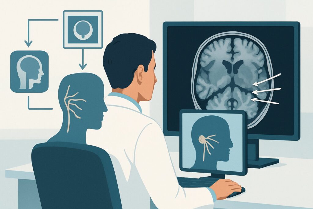
A 58-year-old patient presents to your neurology clinic with a three-week history of progressive right-sided facial numbness, a new facial droop, and difficulty chewing. The constellation of symptoms points away from an isolated nerve issue and toward a single, unifying lesion affecting the trigeminal (CN V) and facial (CN VII) nerves. The anatomical localization could be the brainstem, the cerebellopontine angle, the petrous apex, or the skull base. You need to visualize these complex structures to uncover the cause. This clinical workflow article addresses the American College of Radiology (ACR) Appropriateness Criteria for this specific scenario: initial imaging for multiple different middle cranial nerve palsies. For this presentation, the ACR rates MRI head without and with IV contrast as Usually appropriate.
Who Fits This Clinical Scenario?
This guidance applies to patients presenting with new-onset deficits involving two or more of the middle cranial nerves: the trigeminal (CN V), abducens (CN VI), and facial (CN VII) nerves. The key feature is a polyneuropathy within this specific group, suggesting a lesion localized to a region where these nerves travel in close proximity, such as the cavernous sinus, petrous apex, or pons.
This workflow is intended for the initial diagnostic imaging workup in patients without a clear traumatic cause. It is crucial to distinguish this scenario from similar but distinct clinical presentations that follow different diagnostic pathways:
- Isolated Unilateral Facial Weakness (Bell’s Palsy): This involves only the facial nerve (CN VII). While imaging may eventually be considered if the condition is atypical or fails to resolve, it is a separate clinical variant with its own ACR recommendations.
- Isolated Trigeminal Neuralgia or Weakness: When symptoms are confined to the trigeminal nerve (CN V), the imaging focus and differential diagnosis are narrower.
- Lower Cranial Nerve Palsies (CN IX, X, XI, XII): Deficits in swallowing, voice, or shoulder/tongue movement point to pathology at the jugular foramen or hypoglossal canal, requiring a different imaging protocol.
Applying this workflow to the correct patient—one with a pattern of multiple V, VI, and/or VII palsies—is essential for a high-yield diagnostic evaluation.
What Diagnoses Are You Working Up in This Scenario?
The combination of middle cranial nerve palsies points to pathology along their intricate paths from the brainstem through the skull base. The imaging study is ordered to investigate a specific and serious differential diagnosis. The goal is to find a single lesion that can explain the entire clinical picture.
A primary concern is a neoplastic process. A benign tumor like a schwannoma (arising from the nerve sheath of CN V or VII) or a meningioma can compress multiple adjacent nerves as it grows. Malignant processes are also a major consideration, including perineural spread of a head and neck cancer (like squamous cell carcinoma), direct invasion from a nasopharyngeal carcinoma, or metastases to the skull base or leptomeninges from a distant primary cancer.
Inflammatory and infectious causes are also high on the differential. Sarcoidosis can present with cranial neuropathies due to granulomatous inflammation of the meninges. Tolosa-Hunt syndrome is an idiopathic granulomatous inflammation specifically affecting the cavernous sinus. Infections such as tuberculosis, fungal disease (especially in immunocompromised patients), or Lyme disease can also manifest with multiple cranial nerve deficits.
Less commonly, a vascular abnormality can be the culprit. An aneurysm of the internal carotid artery within the cavernous sinus can compress surrounding nerves. Cavernous sinus thrombosis, though rare, can produce a similar clinical picture, often accompanied by orbital signs like proptosis and chemosis.
Why Is MRI Head Without and With IV Contrast the Recommended Study for This Presentation?
The ACR designates MRI head without and with IV contrast as Usually appropriate because it provides the most comprehensive evaluation for the suspected pathologies in this scenario. Its superior soft-tissue resolution is unmatched for visualizing the cranial nerves, brainstem parenchyma, meninges, and complex anatomy of the skull base.
The protocol’s two components are both critical:
- Without IV Contrast: Pre-contrast sequences (like T1, T2, and FLAIR) establish an anatomical baseline. They are excellent for detecting edema in the brainstem, identifying cystic components of a mass, and visualizing the cerebrospinal fluid spaces. Diffusion-weighted imaging (DWI) is highly sensitive for acute ischemic stroke, which could be a rare cause.
- With IV Contrast: The administration of gadolinium-based contrast is essential. It causes abnormal tissues with increased vascularity or a disrupted blood-brain barrier to enhance. This is the key to identifying inflammatory processes (like sarcoidosis), meningeal disease (carcinomatosis or meningitis), and most tumors (schwannomas, meningiomas, metastases). Subtle perineural tumor spread may only be visible on fat-suppressed, post-contrast images.
Why are other studies rated lower?
- CT head without IV contrast is rated May be appropriate. While it is fast and excellent for evaluating bone destruction at the skull base, its ability to visualize the nerves themselves and detect subtle soft-tissue or meningeal enhancement is poor. It is a reasonable alternative if MRI is contraindicated or unavailable, but it may miss the underlying diagnosis. It also involves ionizing radiation (ACR Relative Radiation Level ☢☢☢).
- MRA head without IV contrast is rated Usually not appropriate as the initial study. MRA is optimized to visualize arteries and is used to diagnose aneurysms or dissections. While a vascular cause is on the differential, it is less common than neoplastic or inflammatory conditions. A standard MRI with contrast is far more comprehensive for evaluating the full range of potential causes and can often detect large aneurysms or flow voids suggestive of vascular pathology.
For this indication, MRI provides the highest diagnostic yield with no ionizing radiation (ACR Relative Radiation Level O). When ordering, consider specifying “include high-resolution, thin-section T2 sequences (e.g., CISS, FIESTA) and fat-suppressed post-contrast T1 sequences through the posterior fossa and skull base” to maximize visualization of the cranial nerves.
What’s Next After MRI Head Without and With IV Contrast? Downstream Workflow
The results of the MRI will guide the subsequent clinical pathway. The goal is to move from radiologic findings to a definitive diagnosis, which often requires tissue sampling or other tests.
- If the study is positive for a discrete mass (e.g., schwannoma, meningioma): The next step is typically a referral to neurosurgery and/or neuro-oncology for consideration of biopsy or resection. Depending on the location and extent, a more extensive study like an MRI orbits face neck without and with IV contrast (also rated Usually appropriate) may be needed to fully stage the lesion.
- If the study is negative or non-specific: A negative MRI is reassuring but may not end the workup if clinical suspicion remains high. The next step is often a lumbar puncture to analyze cerebrospinal fluid (CSF). CSF analysis can detect signs of infection, inflammation (e.g., elevated ACE for sarcoidosis), or malignant cells (leptomeningeal carcinomatosis) that may not be apparent on imaging.
- If the study shows diffuse meningeal or perineural enhancement: This finding is suggestive of an inflammatory, infectious, or infiltrative malignant process. A lumbar puncture is almost always the next step. Depending on the results and clinical context, a biopsy of an accessible enhancing area (e.g., meninges) or referral to rheumatology or infectious disease may be warranted.
In some cases of a negative initial MRI, if symptoms progress, a repeat MRI in several weeks or a more specialized study like a PET/CT may be considered to look for an occult primary malignancy or systemic inflammatory disease.
Pitfalls to Avoid (and When to Get Help)
Navigating the workup for multiple cranial neuropathies requires careful attention to detail to avoid common diagnostic errors.
- Inadequate MRI Protocol: Ordering a routine “MRI brain” without specifying the need for thin sections through the skull base or post-contrast fat-suppressed sequences can lead to a non-diagnostic study that misses subtle nerve enhancement or small lesions.
- Stopping at a “Normal” CT: Accepting a normal non-contrast CT of the head as the end of the workup is a significant pitfall. CT lacks the sensitivity for the most common causes in this differential, and a follow-up MRI is almost always necessary if the CT is unrevealing.
- Misinterpreting Enhancement: Normal structures like venous plexuses around the skull base foramina can enhance. Differentiating this from pathologic perineural enhancement requires experienced radiologic interpretation.
- Delaying the Workup: While some causes are indolent, others like infection or aggressive malignancy are time-sensitive. A prompt and logical diagnostic sequence is crucial.
If the imaging findings are complex or do not fit the clinical picture, a multidisciplinary discussion involving neurology, neuroradiology, and potentially neurosurgery or otolaryngology is the best path forward.
Related ACR Topics and Tools
This article focuses on a single clinical scenario. For a broader view of imaging for all cranial nerve palsies, or to explore the tools and data behind these recommendations, the following resources are available.
- For breadth across all scenarios in Cranial Neuropathy, see our parent guide: Cranial Neuropathy: ACR Appropriateness Decoded.
- ACR Appropriateness Criteria Lookup — for adjacent scenarios
- Imaging Protocol Library — for technique on the recommended study
- Radiation Dose Calculator — for cumulative dose conversations
Frequently Asked Questions
Why is contrast essential for the initial MRI in this scenario?
Intravenous contrast is critical because many of the potential causes, including tumors (meningioma, schwannoma, metastasis), inflammatory conditions (sarcoidosis), and infections, are most clearly identified by their pattern of enhancement. A non-contrast MRI may miss these pathologies entirely, leading to a false-negative result and a delayed diagnosis.
What if my patient has a contraindication to MRI, like a non-compatible pacemaker?
If MRI is absolutely contraindicated, the next best option is a high-resolution CT of the head and skull base, both without and with IV contrast. While CT has lower soft-tissue resolution than MRI, a contrast-enhanced CT can still identify bone destruction, large masses, and some vascular abnormalities. It is important to acknowledge its limitations and understand that it may not be as sensitive as an MRI.
Does this guidance apply if the patient only has an isolated CN VI (abducens) palsy?
No, this specific guidance is for patients with palsies of *multiple different* middle cranial nerves (V-VII). An isolated CN VI palsy has a different differential diagnosis, with microvascular ischemia being a common cause in older adults with vascular risk factors. While imaging is often still performed, the pre-test probability and workup strategy differ from the scenario of a polyneuropathy.
Should I order an ‘MRI orbits face neck’ instead of an ‘MRI head’ initially?
The ACR rates both ‘MRI head without and with IV contrast’ and ‘MRI orbits face neck without and with IV contrast’ as ‘Usually appropriate’. The choice depends on clinical localization. If symptoms are purely intracranial (e.g., facial sensation, facial motor weakness), an MRI of the head focused on the posterior fossa and skull base is sufficient. If there is any suspicion of pathology extending into the face, orbits, or neck (e.g., a palpable neck mass, proptosis, or signs of perineural spread from a skin cancer), the more extensive ‘orbits face neck’ protocol is the better initial choice.
What is the role of PET/CT in this workup?
FDG-PET/CT is rated ‘Usually not appropriate’ for the *initial* imaging workup. Its primary role is downstream. If an MRI is unrevealing but there is high suspicion for an occult malignancy or a systemic inflammatory disease like sarcoidosis, a PET/CT can be very useful to survey the entire body for a primary tumor or other sites of active inflammation that could be more accessible for biopsy.
Reviewed by Pouyan Golshani, MD, Interventional Radiologist — May 29, 2026