What Imaging Is Best for Oropharyngeal Pain from Suspected Glossopharyngeal Neuropathy?

A 62-year-old patient presents to your clinic with a three-month history of severe, lancinating pain in his left throat and the base of his tongue, triggered every time he swallows. The episodes are brief but excruciating, and he has started losing weight due to fear of eating. The neurologic exam is otherwise normal. You suspect a glossopharyngeal neuropathy, specifically glossopharyngeal neuralgia, but need to rule out a structural cause. This is the critical decision point for initial imaging: which study provides the most definitive information without exposing the patient to unnecessary radiation or repeat testing? This article details the clinical workflow for this specific scenario, explaining why the American College of Radiology (ACR) rates MRI head without and with IV contrast as Usually Appropriate.
Who Fits This Clinical Scenario?
This guidance applies to patients presenting with symptoms attributable to isolated dysfunction of the glossopharyngeal nerve (Cranial Nerve IX). The two primary presentations are:
- Oropharyngeal Neurogenic Pain: This typically manifests as glossopharyngeal neuralgia, characterized by paroxysmal, severe, stabbing pain localized to the ear, base of the tongue, tonsillar fossa, or posterior pharynx. The pain is often triggered by specific actions like swallowing, chewing, talking, or coughing.
- Oropharyngeal Neurogenic Dysphagia: This involves difficulty initiating a swallow or a sensation of food sticking in the throat, directly related to impaired motor or sensory function of the pharynx supplied by CN IX.
It is crucial to distinguish this isolated presentation from similar but distinct clinical scenarios that require a different diagnostic approach:
- Exclusion 1: Trigeminal Neuralgia. If the pain is primarily in the facial distribution of the trigeminal nerve (CN V)—the jaw, cheek, or forehead—that constitutes a different clinical scenario, even if there are some overlapping oral triggers.
- Exclusion 2: Isolated Vagal Neuropathy. If the primary symptom is hoarseness or a weak, breathy voice from vocal cord paralysis, this points toward an isolated vagal nerve (CN X) palsy, which is a separate ACR variant.
- Exclusion 3: Multiple Cranial Neuropathies. When symptoms suggest involvement of several cranial nerves (e.g., facial weakness plus hearing loss plus dysphagia), the workup shifts to evaluating for a more extensive process at the skull base or within the brainstem.
What Diagnoses Are You Working Up in This Scenario?
When ordering imaging for suspected CN IX pathology, the goal is to identify or exclude structural causes along the nerve’s entire course, from the brainstem to the pharynx. The differential diagnosis guides the choice of imaging modality.
A primary consideration is vascular compression of the nerve root exit zone from the medulla. This is the most common identifiable cause of classic glossopharyngeal neuralgia, where a pulsating artery (most often the posterior inferior cerebellar artery or vertebral artery) irritates the nerve. High-resolution MRI sequences are specifically designed to visualize this neurovascular conflict.
Another critical category includes neoplasms at the skull base. Tumors can arise from the nerve itself (schwannoma) or from adjacent structures that compress the nerve as it passes through the jugular foramen. These include meningiomas, paragangliomas (glomus jugulare tumors), or metastatic lesions. Contrast enhancement is essential for detecting and characterizing these masses.
Less commonly, symptoms may stem from inflammatory or demyelinating conditions. Multiple sclerosis can present with cranial nerve symptoms, and other inflammatory processes like sarcoidosis can infiltrate the nerve or surrounding meninges. These pathologies are typically only visible on contrast-enhanced MRI.
Finally, though less common as a primary MRI finding, Eagle syndrome (an elongated styloid process or calcified stylohyoid ligament) can mechanically irritate the glossopharyngeal nerve. While CT is superior for visualizing the bony abnormality, MRI can show associated soft tissue inflammation and is still the preferred initial test to rule out the more common intracranial causes.
Why Is MRI Head Without and With IV Contrast the Recommended Study?
The ACR designates MRI head without and with IV contrast as Usually Appropriate for this scenario because it provides the most comprehensive evaluation of the potential causes. Its superior soft-tissue resolution is unmatched for visualizing the nerve, brainstem, and adjacent vessels and soft tissues.
The rationale for this recommendation is multi-faceted:
- Anatomic Detail: MRI can directly visualize the cisternal segment of the glossopharyngeal nerve. Specialized high-resolution sequences (e.g., FIESTA, CISS) can clearly delineate the relationship between the nerve root exit zone and adjacent arteries, which is critical for diagnosing neurovascular compression.
- Pathology Detection: The “without and with IV contrast” components are both vital. The pre-contrast sequences assess anatomy and can detect hemorrhage. The post-contrast sequences are indispensable for identifying enhancement associated with tumors (schwannomas, meningiomas), inflammatory changes, or demyelination, which would be missed on a non-contrast study. For this reason, an `MRI head without IV contrast` is rated lower as only May be appropriate.
- Safety Profile: MRI involves no ionizing radiation (0 mSv), a significant advantage over CT, especially in younger patients or those who may require follow-up imaging.
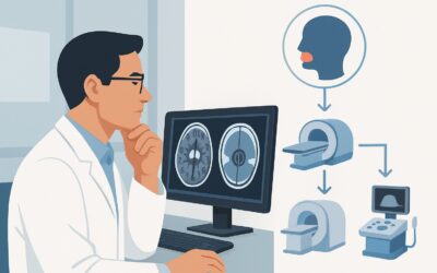
Alternative studies are rated lower for specific reasons. CT neck with IV contrast, rated May be appropriate, can identify large masses or bony abnormalities like an elongated styloid process. However, it offers poor visualization of the nerve itself, the brainstem, and subtle neurovascular conflicts. Its use of ionizing radiation (☢☢☢ 1-10 mSv) makes it a secondary choice. Ultrasound of the neck is rated Usually not appropriate because it cannot penetrate the bony skull base to visualize the intracranial and foraminal segments of the nerve, where the most consequential pathology is typically located.
What’s Next After MRI? Downstream Workflow
The results of the MRI will guide the subsequent clinical pathway. The workflow typically branches based on whether a structural cause is identified.
If the MRI is positive for neurovascular compression:
A definitive finding of an artery compressing the CN IX root exit zone in a patient with classic glossopharyngeal neuralgia confirms the diagnosis. The next step is a referral to Neurology or Neurosurgery. Treatment often begins with medical management (e.g., carbamazepine or gabapentin). For refractory cases, the patient may be a candidate for microvascular decompression (MVD) surgery.
If the MRI is positive for a tumor or mass:
The patient should be referred to Neurosurgery and potentially Neuro-oncology for further evaluation and management. The specific treatment plan will depend on the tumor’s type, size, and location, and may involve surgical resection, radiation therapy, or observation.
If the MRI is negative:
A negative MRI is a common outcome and helps rule out the most dangerous structural causes. The diagnosis shifts to idiopathic glossopharyngeal neuralgia. Management is typically medical, led by a neurologist. If dysphagia is the primary symptom and the MRI is negative, further functional testing, such as a modified barium swallow study or fiberoptic endoscopic evaluation of swallowing (FEES), may be warranted to assess the physiology of the swallow.
Pitfalls to Avoid (and When to Get Help)
Navigating the workup for suspected CN IX neuropathy requires avoiding several common pitfalls to ensure a timely and accurate diagnosis.
- Ordering a Non-Contrast MRI: A non-contrast study is insufficient. It will likely miss enhancing tumors, inflammatory lesions, or signs of perineural spread of malignancy, leading to a false-negative result. Always specify “without and with IV contrast.”
- Defaulting to CT: While CT is faster and more accessible, its low sensitivity for nerve pathology and neurovascular conflict makes it a suboptimal first-line test for this indication. Starting with CT often leads to a non-diagnostic result, requiring a follow-up MRI and delaying care.
- Misinterpreting Incidental Vascular Loops: Not every vessel adjacent to a nerve is causing symptoms. The imaging findings must be tightly correlated with the classic clinical presentation of glossopharyngeal neuralgia.
If red flag symptoms are present—such as progressive deficits, involvement of multiple cranial nerves, or constitutional symptoms like fever and weight loss—escalate care promptly with a referral to a neurologist for a more urgent and comprehensive evaluation.
Related ACR Topics and Tools
This article focuses on a single clinical variant. For a comprehensive overview of imaging for all twelve cranial nerves, consult the parent topic article. For technical details or to explore adjacent scenarios, the following resources are available.
- For breadth across all scenarios in Cranial Neuropathy, see our parent guide: Cranial Neuropathy: ACR Appropriateness Decoded.
- To explore other clinical presentations and their corresponding ACR ratings, use the ACR Appropriateness Criteria Lookup.
- For technical specifications of the recommended MRI, see the Imaging Protocol Library.
- To discuss radiation exposure from alternative studies like CT, use the Radiation Dose Calculator.
Frequently Asked Questions
Why is MRI of the head recommended instead of MRI of the neck for glossopharyngeal nerve pain?
The most critical portion of the glossopharyngeal nerve (CN IX) to evaluate for common pathologies like vascular compression or schwannomas is its origin from the brainstem and its path through the skull base. An ‘MRI head’ protocol is optimized to visualize these intracranial and cisternal spaces with high resolution. While an ‘MRI neck’ covers the lower course of the nerve, it often lacks the specific sequences (like FIESTA/CISS) and resolution needed to assess the nerve root exit zone, which is the most frequent site of pathology.
Is a contrast allergy a contraindication to the recommended MRI?
A severe allergy to gadolinium-based contrast agents is a relative contraindication. In such cases, an ‘MRI head without IV contrast’ is rated as ‘May be appropriate’ and can still identify neurovascular compression. However, it cannot reliably rule out tumors or inflammatory causes. The decision to proceed without contrast, use premedication, or switch to an alternative modality like CT should be made in consultation with the radiologist, weighing the specific clinical suspicion against the risk of an allergic reaction.
What if the patient has a pacemaker or other MRI-incompatible implant?
If a patient cannot undergo an MRI, ‘CT neck with IV contrast’ is the next best option and is rated ‘May be appropriate’ by the ACR. While less sensitive for nerve pathology, it can identify large masses, bony abnormalities like Eagle syndrome, and other significant structural lesions. A CTA (CT Angiography) may also be considered to evaluate the vascular anatomy, though it provides less information about the nerve itself.
Does the imaging protocol change if I suspect Eagle syndrome?
While MRI is the recommended initial study to rule out intracranial pathology, if clinical suspicion for Eagle syndrome is very high (e.g., pain on turning the head, palpable styloid process), a CT with 3D reconstructions is the best test to visualize and measure the elongated styloid process or calcified stylohyoid ligament. Often, clinicians will start with the ACR-recommended MRI to be comprehensive, and if it is negative, may proceed to a dedicated CT if Eagle syndrome remains a strong possibility.
Can MRA (Magnetic Resonance Angiography) be used instead of a standard MRI?
MRA of the head and neck, whether with or without contrast, is rated ‘Usually not appropriate’ as the primary initial study for this scenario. While MRA is excellent for visualizing blood vessels, it does not provide the detailed soft-tissue contrast needed to see the nerve itself or to detect non-vascular pathologies like small tumors or inflammatory changes. The necessary high-resolution nerve-visualizing sequences are part of a standard brain MRI protocol, not a standard MRA protocol.
Reviewed by Pouyan Golshani, MD, Interventional Radiologist — May 29, 2026