How Should You Image Persistent Central Vertigo? An ACR-Guided Workflow
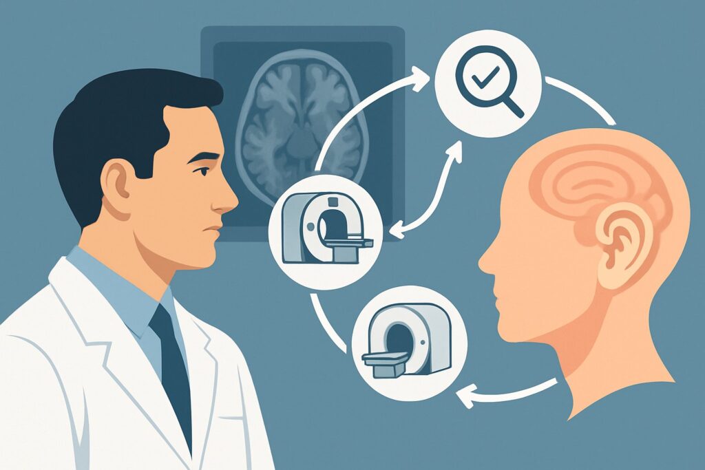
A 58-year-old patient presents to your clinic with six weeks of persistent, low-grade dizziness, describing it as a constant feeling of being “off-balance.” Over the last week, he’s also noticed some mild clumsiness in his right hand and intermittent double vision when looking to the side. The HINTS (Head-Impulse, Nystagmus, Test-of-Skew) exam is equivocal, but your clinical suspicion is high for a central process. You know imaging is necessary, but which study provides the highest diagnostic yield while minimizing unnecessary tests? This is the critical decision point for a patient with suspected central vertigo.
For this specific scenario—persistent vertigo with or without neurological symptoms—the American College of Radiology (ACR) Appropriateness Criteria rate MRI head and internal auditory canal without and with IV contrast as Usually Appropriate. This article details the clinical workflow, differential diagnosis, and imaging rationale for this common and high-stakes presentation.
Who Fits This Clinical Scenario?
This guidance applies to adult patients presenting with persistent vertigo, which is typically described as a constant or near-constant sensation of dizziness, unsteadiness, or motion that lasts for days to weeks. The key differentiator is the chronicity and persistence, as opposed to brief, episodic spells. The presence of associated neurological signs or symptoms—such as diplopia, dysarthria, ataxia, focal weakness, or sensory changes—strongly points toward a central etiology and places the patient squarely in this category.
It is crucial to distinguish this presentation from related but distinct clinical scenarios that follow different imaging pathways:
- Exclusion 1: Episodic Vertigo. If the patient’s symptoms are brief, recurrent, and often triggered by position changes (suggesting Benign Paroxysmal Positional Vertigo) or accompanied by fluctuating hearing loss and aural fullness (suggesting Meniere’s disease), they fit the peripheral vertigo scenario, which has a different workup.
- Exclusion 2: Isolated Hearing Loss. If the primary complaint is hearing loss (conductive, sensorineural, or mixed) without significant or persistent vertigo, the workup should follow the guidelines for those specific scenarios. While there is overlap (e.g., a vestibular schwannoma can cause both), the initial imaging approach is tailored to the dominant symptom.
This workflow is designed for the initial diagnostic imaging of a patient in whom the brainstem, cerebellum, or cranial nerves are the suspected source of pathology.
What Diagnoses Are You Working Up in This Scenario?
When ordering imaging for persistent central vertigo, you are primarily investigating pathologies of the posterior fossa and cranial nerves. The differential is broad, but the most consequential diagnoses are those that require prompt intervention or specialized management.
Posterior Circulation Stroke: An ischemic or hemorrhagic stroke involving the cerebellum or brainstem is a can’t-miss diagnosis. Symptoms can be subtle, and vertigo is a common presentation of vertebral or basilar artery territory infarcts. The clinical urgency is high, as timely diagnosis can impact treatment and secondary prevention.
Vestibular Schwannoma: This benign tumor arises from the vestibulocochlear nerve (CN VIII) in the internal auditory canal or cerebellopontine angle. While often associated with unilateral hearing loss, it can present with persistent vertigo and imbalance as it grows and compresses adjacent structures like the brainstem and cerebellum.
Demyelinating Disease: For younger patients in particular, persistent vertigo can be the initial presentation of Multiple Sclerosis (MS). A demyelinating plaque in the brainstem (e.g., at the root entry zone of CN VIII) or cerebellum can produce these exact symptoms. Identifying inflammatory activity is key to diagnosis and treatment initiation.
Other Posterior Fossa Tumors: Beyond schwannomas, other masses such as meningiomas, metastases, or primary brain tumors (e.g., ependymoma, medulloblastoma) can present with central vertigo due to mass effect on the cerebellum, brainstem, or fourth ventricle. These are less common but critical to identify.
Why Is MRI of the Head and Internal Auditory Canal the Recommended Study?
The ACR designates MRI head and internal auditory canal without and with IV contrast as Usually Appropriate because it offers superior soft-tissue contrast and is exquisitely sensitive for the key pathologies in the differential diagnosis for central vertigo. This single study can effectively evaluate the brain parenchyma, cranial nerves, and surrounding structures without exposing the patient to ionizing radiation (0 mSv).
The rationale for this specific protocol includes:
- Sensitivity for Ischemia: Diffusion-Weighted Imaging (DWI), a standard sequence in a brain MRI, is highly sensitive and specific for detecting acute and subacute ischemic strokes within minutes of onset, far outperforming CT.
- Cranial Nerve and Canal Visualization: High-resolution T2-weighted sequences (like CISS or FIESTA) provide detailed anatomical views of the cranial nerves within the internal auditory canals and cerebellopontine angle, making it the gold standard for detecting small vestibular schwannomas.
- Value of IV Contrast: The administration of gadolinium-based contrast is critical. It causes avid enhancement of vestibular schwannomas and other tumors (meningiomas, metastases) and highlights active inflammatory plaques in Multiple Sclerosis, which would be invisible or inconspicuous on non-contrast imaging.
Alternative studies are rated lower for specific reasons in this clinical context:
- CT head without IV contrast is rated May be appropriate. While it is fast and excellent for detecting acute hemorrhage, its utility is limited. It has poor sensitivity for small posterior fossa infarcts, demyelination, and non-hemorrhagic tumors due to beam-hardening artifact, which obscures detail in this region.
- CT temporal bone without IV contrast is rated Usually not appropriate. This is a high-resolution bone algorithm study designed to evaluate the ossicles, mastoid air cells, and cochlea. It provides very poor visualization of the brain parenchyma and cranial nerves, making it the wrong tool for evaluating suspected central pathology.
In summary, an MRI with and without contrast provides a comprehensive, high-yield evaluation for the most likely and most dangerous causes of persistent central vertigo in a single, non-invasive examination.
What’s Next After MRI? Downstream Workflow
The results of the MRI will guide your next steps, often leading to a subspecialty consultation and further management. The downstream workflow depends directly on the findings.
- If the MRI is positive for an acute/subacute stroke: This is a medical emergency. The patient requires immediate neurology consultation, likely hospital admission for stroke workup, and initiation of secondary prevention. Further vascular imaging, such as an MRA or CTA of the head and neck (both rated May be appropriate as initial studies), may be performed to identify the underlying cause (e.g., dissection, stenosis).
- If the MRI identifies a mass (e.g., vestibular schwannoma): The next step is a referral to Neurosurgery and/or Otolaryngology (ENT). Management options for a schwannoma include observation with serial imaging, stereotactic radiosurgery, or surgical resection, depending on tumor size, patient age, and symptoms.
- If the MRI shows findings suggestive of demyelination: A neurology referral is indicated for a full Multiple Sclerosis workup. This typically includes an MRI of the spine to look for additional lesions and may involve a lumbar puncture for cerebrospinal fluid analysis.
- If the MRI is negative: A negative, high-quality MRI provides strong evidence against a structural cause in the posterior fossa. The workup should pivot back to a thorough clinical re-evaluation. This may include formal vestibular testing, a trial of vestibular rehabilitation therapy, or consideration of less common or non-structural causes of vertigo.
Pitfalls to Avoid (and When to Get Help)
Navigating the workup for central vertigo requires careful attention to clinical detail and imaging choice. Here are a few common pitfalls to avoid:
- Ordering a non-contrast CT as the definitive test. A negative non-contrast head CT does not rule out a posterior circulation stroke or a small tumor. If clinical suspicion for a central cause remains high, an MRI is necessary.
- Forgetting to specify “with and without contrast.” For a suspected tumor or inflammatory process, the “with contrast” portion of the MRI is essential. Omitting it can lead to a non-diagnostic study and delay care.
- Misinterpreting “persistent” vs. “episodic.” Applying this central vertigo imaging pathway to a patient with classic episodic, positional vertigo (BPPV) leads to unnecessary, low-yield imaging. A careful history is paramount.
If a patient presents with acute, severe vertigo accompanied by the “5 D’s” (dizziness, diplopia, dysarthria, dysphagia, dystaxia) or sudden hearing loss, this represents a neurological emergency. Escalate immediately for an urgent neurology evaluation and emergent imaging.
Related ACR Topics and Tools
The ACR Appropriateness Criteria provide evidence-based guidance for a wide range of clinical scenarios. For breadth across all scenarios in Hearing Loss and/or Vertigo, see our parent guide: Hearing Loss and/or Vertigo: ACR Appropriateness Decoded.
For additional decision support and technical details, the following GigHz tools may be helpful:
- Imaging Appropriateness Selector — To review guidelines for adjacent or alternative clinical presentations.
- Imaging Protocol Library — For detailed technical parameters of MRI and CT protocols.
- Radiation Dose Calculator — To help in discussions with patients about cumulative radiation exposure from CT scans.
Frequently Asked Questions
Why is an MRI of the internal auditory canal (IAC) specified instead of just a routine brain MRI?
A routine brain MRI may not use the thin, high-resolution imaging sequences necessary to clearly visualize the small structures within the internal auditory canals and cerebellopontine angle. Specifying ‘and internal auditory canal’ ensures the protocol includes these dedicated sequences (e.g., CISS/FIESTA), which are essential for detecting small vestibular schwannomas or other cranial nerve pathologies that could be missed otherwise.
Is an MRA or CTA ever the first-choice study for persistent vertigo?
While MRA and CTA are rated as ‘May be appropriate,’ they are typically not the first-choice initial study unless there is a very high pre-test probability of a primary vascular etiology, such as vertebrobasilar insufficiency or dissection. An MRI of the brain provides a more comprehensive evaluation of the brain parenchyma itself, which is the primary concern. MRA/CTA is often a downstream study after an initial MRI shows a stroke.
What if my patient has a contraindication to MRI, like a non-compatible pacemaker?
In cases with a hard contraindication to MRI, a CT-based study is the next best option. A CT head with IV contrast is rated ‘May be appropriate’ and can identify larger masses, hemorrhage, or significant strokes. If vascular pathology is the primary concern, a CTA head and neck with IV contrast (‘May be appropriate’) would be the preferred study to evaluate the arteries of the posterior circulation.
Does a non-contrast MRI have any role in this scenario?
Yes, an MRI of the head and IAC without IV contrast is also rated as ‘Usually Appropriate.’ It is an excellent choice if the primary suspicion is an ischemic stroke (DWI is non-contrast) or if the patient has a severe allergy to gadolinium-based contrast agents or very poor renal function. However, it is less sensitive for detecting small enhancing tumors or active demyelinating plaques, which is why the ‘with contrast’ study is often preferred for a complete workup.
The patient’s vertigo is persistent but they have no other neurological symptoms. Does the recommendation change?
No, the recommendation remains the same. The ACR scenario is ‘Persistent vertigo with OR without neurological symptoms.’ While the presence of other neurological signs increases the pre-test probability of a central cause, isolated but persistent vertigo can still be the sole manifestation of a significant posterior fossa pathology, such as a small cerebellar stroke or vestibular schwannoma. The high-sensitivity workup with MRI is still warranted.
Reviewed by Pouyan Golshani, MD, Interventional Radiologist — May 26, 2026