Which Imaging Is Best for Altered Mental Status with a Focal Neurologic Deficit?
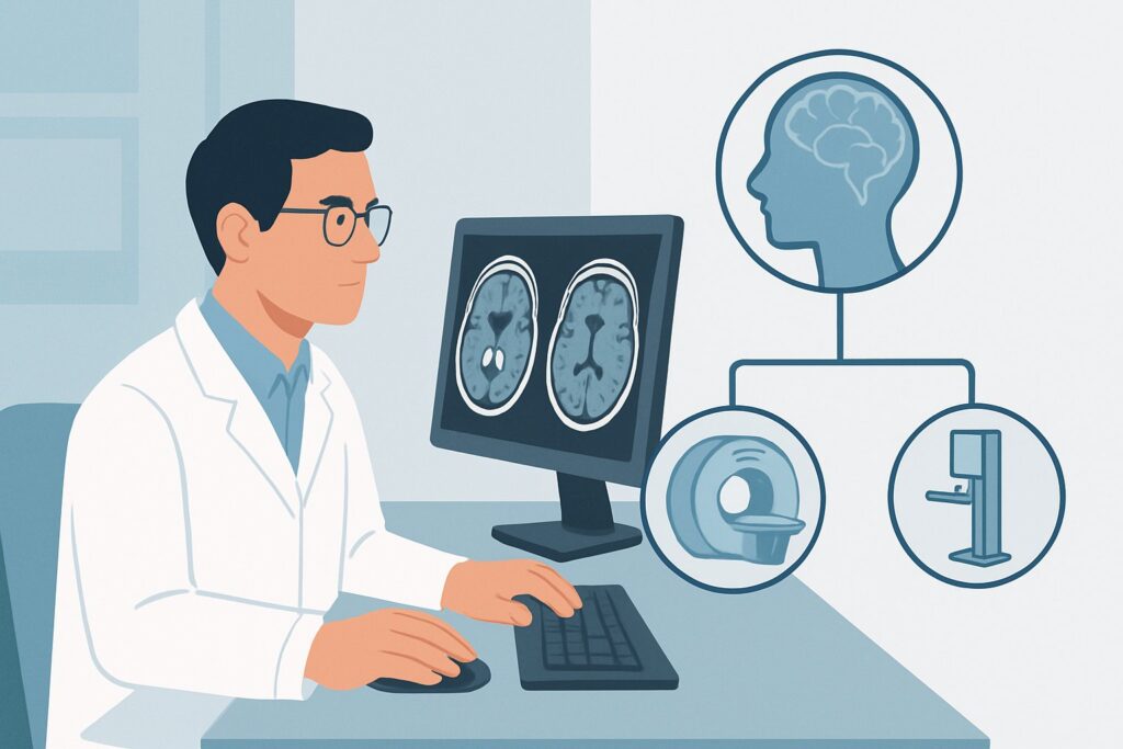
It’s 2 AM in the emergency department, and you’re evaluating a 72-year-old patient brought in by family for acute confusion. During your exam, you discover new-onset left-sided weakness and slurred speech. The presentation screams intracranial pathology, but the differential is broad—ischemic stroke, hemorrhage, or something else. Your immediate decision is which imaging study to order first to get the fastest, most actionable answer. This article provides a deep dive into the American College of Radiology (ACR) workflow for an adult with altered mental status and a suspected focal neurologic deficit, explaining why a specific initial study is recommended. For this scenario, the ACR rates `CT head without IV contrast` as Usually Appropriate.
Who Fits This Clinical Scenario?
This guidance applies to a specific and urgent clinical situation: an adult patient presenting with a change in mental status accompanied by new focal neurologic findings or a high clinical suspicion for a new structural brain lesion. “Altered mental status” can range from confusion and lethargy to delirium or coma. The key differentiator for this scenario is the presence of “focal neurologic deficits,” such as:
- Unilateral weakness (hemiparesis) or paralysis (hemiplegia)
- New-onset aphasia (difficulty with speech production or comprehension)
- Visual field cuts
- Significant new ataxia or vertigo
- Cranial nerve palsies
This workflow is distinct from other similar presentations. If your patient has altered mental status but a non-focal exam, and you suspect a systemic cause like sepsis, uremia, or drug toxicity, the workup follows a different path. Similarly, a patient with a known history of intracranial pathology (e.g., a brain tumor) who presents with worsening symptoms falls under a separate ACR variant. This guidance is for the initial, undifferentiated workup where a new intracranial process is the primary concern.
What Diagnoses Are You Working Up in This Scenario?
When a patient presents with altered mental status and a focal deficit, the differential diagnosis is anchored by time-sensitive and life-threatening intracranial conditions. The initial imaging choice is designed to rapidly differentiate among these possibilities.
Intracranial Hemorrhage: This is a primary “can’t-miss” diagnosis. It includes intraparenchymal hemorrhage (often related to hypertension), subarachnoid hemorrhage (from a ruptured aneurysm), or extra-axial collections like a subdural or epidural hematoma, which may be seen after trauma that was unwitnessed or forgotten by the patient.
Acute Ischemic Stroke: The most common cause of acute focal deficits. While early ischemic changes may not be visible on initial imaging, the study is critical to rule out hemorrhage, which is a contraindication for thrombolytic therapy. The imaging can also sometimes reveal early signs of a large vessel occlusion.
Space-Occupying Lesion: A primary brain tumor, metastasis, or brain abscess can present acutely with focal deficits and altered mental status due to mass effect, edema, or hemorrhage into the lesion. While less common as an initial presentation than stroke, it remains a key consideration.
Herniation Syndromes: A severe consequence of any of the above conditions is increased intracranial pressure leading to brain herniation. This is a neurosurgical emergency, and rapid diagnosis is essential. Initial imaging is the fastest way to identify midline shift, effacement of the basal cisterns, or other signs of impending herniation.
Why CT Head without IV Contrast Is the Recommended Study for This Presentation
For the initial evaluation of an adult with altered mental status and a focal neurologic deficit, the ACR designates `CT head without IV contrast` as Usually Appropriate. The rationale is rooted in speed, accessibility, and diagnostic utility for the most critical differential diagnoses.
The primary strength of a non-contrast CT is its exceptional sensitivity for detecting acute intracranial hemorrhage. Blood appears hyperdense (bright white) on CT, making it readily identifiable. In a patient being considered for thrombolysis for a suspected ischemic stroke, ruling out hemorrhage is the most important first step, and non-contrast CT accomplishes this in minutes. It is also the fastest and most widely available advanced imaging modality in most emergency settings, which is critical when “time is brain.”
Alternative studies are rated lower for this initial step. An `MRI head without IV contrast` is rated as May be appropriate. While MRI is far more sensitive for detecting early ischemic stroke, it is significantly slower to acquire, less available on an emergent basis, and more challenging for monitoring an unstable patient. Its role is often as a follow-up study after the initial CT has excluded hemorrhage.
Studies involving intravenous contrast, such as `CT head with IV contrast` (Usually not appropriate), are not recommended for the initial workup. Contrast does not improve the detection of acute hemorrhage and adds potential risks (allergic reaction, contrast-induced nephropathy) and time to the study. While contrast is essential for evaluating for tumor or abscess, that is typically a secondary question after hemorrhage has been ruled out.
The radiation dose for a non-contrast head CT is moderate (ACR Relative Radiation Level ☢☢☢, 1-10 mSv), a necessary trade-off for the rapid, life-saving information it provides. Once you’ve decided on this study, our protocol guide covers the technical details. For a complete walkthrough of the technique, common findings, and reporting principles, see our guide: CT Brain Without Contrast.
What’s Next After CT Head without IV Contrast? Downstream Workflow
The results of the initial non-contrast head CT will dictate your next steps and dramatically narrow the clinical pathway. The workflow branches based on the findings:
- If the CT is positive for hemorrhage: The immediate next step is an urgent consultation with neurosurgery and neurology. Management will focus on blood pressure control, reversal of any anticoagulation, and determining the need for surgical intervention. Further imaging like CT angiography (CTA) may be ordered to look for an underlying vascular lesion.
- If the CT is negative for hemorrhage but suspicion for ischemic stroke is high: This is a classic “code stroke” pathway. The negative CT “rules in” the patient for consideration of thrombolysis (tPA/TNK) if they are within the treatment window. Further vascular imaging with CTA of the head and neck is typically performed concurrently or immediately after to assess for a large vessel occlusion (LVO) that may be amenable to mechanical thrombectomy.
- If the CT is negative and clinical suspicion for stroke is low or atypical: The workup broadens. If the patient does not improve, an MRI of the brain becomes the next logical step to evaluate for subtle ischemia (especially in the posterior fossa, which is poorly visualized on CT), encephalitis, or other non-hemorrhagic structural lesions. If the MRI is also unrevealing, the focus shifts back toward a comprehensive workup for toxic-metabolic, infectious, or seizure-related causes of altered mental status.
- If the CT shows a mass or other unexpected finding: A follow-up MRI with and without contrast is typically the next step to better characterize the lesion. A neurology or neurosurgery consultation is warranted to guide further management.
Pitfalls to Avoid (and When to Get Help)
In this high-stakes scenario, several common pitfalls can delay diagnosis or lead to misinterpretation. Be mindful of the following:
- Over-reliance on a negative CT: A normal non-contrast head CT does not rule out acute ischemic stroke, particularly in the first few hours or for strokes in the brainstem or cerebellum. Maintain a high index of suspicion based on your clinical exam.
- Delaying the scan: For both hemorrhagic and ischemic stroke, time is critical. Do not delay imaging for routine labs if a stroke is suspected; obtain the scan as quickly as possible.
- Misinterpreting subtle signs: Early signs of a large ischemic stroke on non-contrast CT can be subtle, such as loss of the gray-white matter differentiation, sulcal effacement, or a hyperdense MCA sign. If you are unsure, a formal radiology read or neurology consultation is essential.
If the patient is rapidly deteriorating, has signs of herniation on exam (e.g., a “blown pupil”), or the CT shows a large hemorrhage or mass with significant midline shift, escalate immediately to your senior resident, attending physician, and the on-call neurosurgery team.
Related ACR Topics and Tools
This article covers one specific clinical variant. For a comprehensive overview of imaging for all related presentations, from coma to psychosis, please see our parent guide. For help with adjacent scenarios or understanding the technical aspects of the recommended studies, the following GigHz resources are available:
- For breadth across all scenarios in Altered Mental Status, Coma, Delirium, and Psychosis, see our parent guide: Altered Mental Status, Coma, Delirium, and Psychosis: ACR Appropriateness Decoded.
- ACR Appropriateness Criteria Lookup — for adjacent scenarios
- Imaging Protocol Library — for technique on the recommended study
- Radiation Dose Calculator — for cumulative dose conversations
Frequently Asked Questions
Why not start with an MRI if it’s more sensitive for an ischemic stroke?
While MRI is more sensitive for early ischemia, a non-contrast CT is prioritized because it is much faster, more widely available, and is excellent at answering the most immediate life-threatening question: is there a brain bleed? Ruling out hemorrhage is the critical first step before administering thrombolytic therapy for an ischemic stroke.
Does a negative non-contrast head CT completely rule out a stroke?
No. A non-contrast CT can be normal in the early hours of an ischemic stroke. It is also less sensitive for small strokes or those located in the posterior fossa (cerebellum and brainstem). If clinical suspicion for a stroke remains high despite a negative CT, further imaging with MRI/MRA is often necessary.
In what situation would I add IV contrast to the initial CT scan?
For the initial, undifferentiated workup of suspected stroke or hemorrhage, contrast is rated ‘Usually not appropriate’ because it adds time and risk without improving detection of acute blood. Contrast is added in downstream imaging if the initial CT suggests a mass, abscess, or if there is a concern for meningitis/encephalitis, but it is not part of the primary evaluation in this scenario.
What if the patient has a known brain tumor and presents with worsening confusion?
That presentation falls under a different ACR clinical variant: ‘Altered mental status with known history of intracranial pathology.’ The imaging choice in that case may be different, often involving a contrast-enhanced study (either CT or MRI) to assess for tumor progression, hemorrhage into the tumor, or treatment-related changes.
How does this workflow change if the patient has altered mental status but no focal deficits?
If the neurologic exam is non-focal, the pre-test probability of a primary intracranial catastrophe is lower, and the likelihood of a toxic-metabolic or systemic cause is higher. The ACR scenario ‘Altered mental status. Suspected medical illness or toxic-metabolic cause’ applies, and imaging may not be the first-line investigation. The decision to image is based on the failure to improve after correcting metabolic derangements or if the cause remains elusive.
Reviewed by Pouyan Golshani, MD, Interventional Radiologist — May 30, 2026