What Is the Best Initial Imaging for Recurrent Pregnancy Loss? An ACR-Guided Workflow
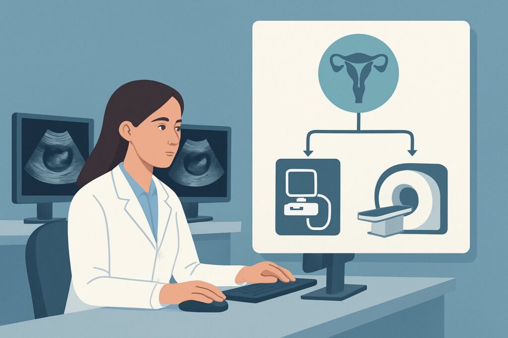
A 34-year-old patient is in your clinic for evaluation after her third consecutive first-trimester pregnancy loss. The genetic and hormonal workups have been unrevealing, and she is anxious for answers about potential anatomic causes. You are now faced with the decision of which imaging study to order first to evaluate her uterine anatomy. This choice is critical for identifying treatable conditions and guiding her future care. For this specific clinical scenario—female infertility due to recurrent pregnancy loss—the American College of Radiology (ACR) provides clear guidance. The ACR Appropriateness Criteria rate US pelvis transabdominal, along with its transvaginal and sonohysterography counterparts, as Usually Appropriate for this initial evaluation.
Who Fits This Clinical Scenario?
This workflow is designed for patients experiencing recurrent pregnancy loss (RPL), which is clinically defined as two or more failed clinical pregnancies. The evaluation discussed here is for the initial imaging workup focused on identifying anatomic abnormalities of the uterus that may contribute to RPL. The patient has typically undergone a preliminary evaluation, including hormonal and genetic testing, without a definitive diagnosis, prompting an investigation into structural causes.
It is crucial to distinguish this scenario from others that may present with infertility but have different primary drivers. This guidance does not apply if:
- The primary clinical suspicion is tubal occlusion. While RPL and tubal factors can coexist, a workup focused specifically on tubal patency follows a different imaging pathway, often prioritizing hysterosalpingography.
- The patient presents with a classic history and clinical features of polycystic ovary syndrome (PCOS), such as oligo-ovulation and hyperandrogenism. That presentation has its own dedicated ACR variant for initial imaging.
- The dominant clinical feature is severe pelvic pain or dysmenorrhea suggesting endometriosis. While endometriosis can be associated with infertility, its dedicated workup prioritizes different imaging features and techniques.
This article focuses squarely on the patient whose history points toward an underlying uterine factor as a potential cause for repeated pregnancy failure.
What Anatomic Causes Are You Working Up in Recurrent Pregnancy Loss?
When ordering initial imaging for recurrent pregnancy loss, the primary goal is to identify and characterize structural abnormalities of the uterus that can interfere with embryo implantation or fetal development. The differential diagnosis for anatomic causes is focused and specific.
Congenital Uterine (Müllerian Duct) Anomalies: These are among the most significant findings. The septate uterus is the most common Müllerian duct anomaly and carries the highest risk of pregnancy loss, thought to be due to implantation on an avascular fibrous septum. Other anomalies include the bicornuate uterus (incomplete fusion of the uterine horns), unicornuate uterus (underdevelopment of one side), and uterus didelphys (a double uterus). Differentiating these is critical, as a septate uterus is often correctable with hysteroscopic resection, whereas a bicornuate uterus is not.
Submucosal Leiomyomas (Fibroids): While fibroids are extremely common, only those that impinge upon or distort the endometrial cavity (submucosal or intracavitary) are strongly associated with RPL. They can disrupt implantation by acting as a foreign body, causing local inflammation, or altering endometrial receptivity. Intramural or subserosal fibroids are less likely to be the primary cause unless they are very large.
Intrauterine Adhesions (Asherman Syndrome): Scar tissue within the endometrial cavity can obliterate the normal uterine architecture, preventing proper implantation and placentation. These adhesions are typically an acquired condition resulting from prior uterine surgery (like a dilation and curettage), infection (endometritis), or other trauma to the uterine lining.
Endometrial Polyps: These benign overgrowths of endometrial tissue can also interfere with implantation. While often asymptomatic, larger or multiple polyps may contribute to both infertility and pregnancy loss and are an important target of the imaging evaluation.
Why Is Pelvic Ultrasound the Recommended Initial Study for This Presentation?
The ACR designates several imaging modalities as Usually Appropriate for the initial workup of recurrent pregnancy loss, with ultrasound-based techniques forming the cornerstone of the evaluation. This recommendation is based on a balance of diagnostic accuracy, safety, accessibility, and cost-effectiveness.
The initial study is typically a combination of US pelvis transabdominal and US pelvis transvaginal. This non-invasive, radiation-free examination provides a comprehensive overview of the pelvic anatomy. The transabdominal view assesses the overall uterine size and contour, while the high-frequency transvaginal approach offers superior resolution of the endometrium, myometrium, and adnexa. This initial step can often identify major uterine anomalies, large fibroids, and ovarian pathology.
For a more detailed evaluation of the endometrial cavity, US sonohysterography is also rated Usually Appropriate and is often the definitive next step if the initial 2D ultrasound is inconclusive or suspicious. This technique involves instilling sterile saline into the endometrial cavity, which distends it and outlines intracavitary structures with remarkable clarity. It is highly effective for detecting subtle polyps, submucosal fibroids, and intrauterine adhesions. Crucially, sonohysterography helps differentiate a septate uterus from a bicornuate uterus by clearly visualizing the external uterine contour—a distinction that has major surgical implications.
While MRI pelvis without and with IV contrast is also rated Usually Appropriate, it is generally reserved as a problem-solving tool rather than a first-line test due to higher cost and lower accessibility. MRI provides excellent soft tissue contrast and is considered the gold standard for classifying complex Müllerian duct anomalies when ultrasound findings are equivocal.
Alternative studies are rated lower for this specific indication. Fluoroscopy hysterosalpingography (HSG) is rated May be appropriate (Disagreement). Although excellent for assessing tubal patency, HSG provides only an indirect, two-dimensional view of the uterine cavity and cannot evaluate the myometrium or external uterine contour. It also involves ionizing radiation (☢☢ 0.1-1mSv) and iodinated contrast, making it less ideal as a primary tool for evaluating uterine structure in RPL.
What’s Next After Pelvic Ultrasound? Downstream Workflow
The results of the initial pelvic ultrasound and sonohysterography will guide the subsequent clinical and diagnostic pathway. The workflow branches based on whether the findings are positive, negative, or indeterminate.
If the study is positive for a significant anatomic abnormality:
- Septate Uterus: The patient should be referred to a reproductive endocrinologist or gynecologic surgeon for consideration of hysteroscopic septoplasty. This minimally invasive procedure can significantly improve live birth rates.
- Submucosal Leiomyoma or Large Polyp: Referral for hysteroscopic myomectomy or polypectomy is the standard next step. Removing lesions that distort the cavity can restore a more favorable environment for implantation.
- Intrauterine Adhesions (Asherman Syndrome): The patient should be referred for operative hysteroscopy to lyse the adhesions (adhesiolysis).
If the study is negative:
A normal ultrasound and sonohysterogram effectively rule out most significant anatomic causes of RPL. The clinical focus should then shift back to non-anatomic factors. This may involve re-evaluating or expanding the workup for thrombophilias, autoimmune conditions (e.g., antiphospholipid syndrome), or parental karyotyping. Further imaging is generally not indicated unless new clinical concerns arise.
If the study is indeterminate or complex:
In cases where ultrasound and sonohysterography cannot definitively characterize a congenital uterine anomaly (e.g., distinguishing a severe bicornuate from a septate uterus), the next step is MRI of the pelvis. As a Usually Appropriate modality, MRI serves as the ultimate arbiter for complex pelvic anatomy, providing clear delineation of both the internal and external uterine contours to guide appropriate management.
Pitfalls to Avoid (and When to Get Help)
Navigating the imaging workup for recurrent pregnancy loss requires attention to detail to avoid common missteps.
- Pitfall 1: Stopping at 2D Ultrasound. A standard transvaginal ultrasound may not adequately distend the endometrial cavity, potentially missing subtle polyps, adhesions, or the true nature of a uterine septum. If suspicion remains, proceeding to sonohysterography is essential.
- Pitfall 2: Misinterpreting a Uterine Anomaly. The most critical error is confusing a septate uterus with a bicornuate uterus. An incorrect diagnosis could lead to an unnecessary or ineffective surgery. If there is any ambiguity on ultrasound, do not hesitate to order an MRI for clarification.
- Pitfall 3: Timing the Study Incorrectly. For premenopausal patients, sonohysterography and HSG should be performed in the early follicular phase of the menstrual cycle (typically days 6-10), after cessation of menses but before ovulation. This timing ensures a thin endometrium for optimal visualization and avoids interrupting a potential early pregnancy.
If a complex Müllerian duct anomaly is identified or suspected, consultation with a radiologist specializing in gynecologic imaging or a reproductive endocrinologist is highly recommended before proceeding with any surgical intervention.
Related ACR Topics and Tools
For a comprehensive overview of imaging for all clinical variants related to female infertility, see our parent guide. For tools to help you navigate other scenarios, select appropriate imaging protocols, or discuss radiation safety with patients, the following resources are available.
- For breadth across all scenarios in Female Infertility, see our parent guide: Female Infertility: ACR Appropriateness Decoded.
- Imaging Appropriateness Selector — for adjacent scenarios
- Imaging Protocol Library — for technique on the recommended study
- Radiation Dose Calculator — for cumulative dose conversations
Frequently Asked Questions
Why is sonohysterography preferred over hysterosalpingography (HSG) for evaluating the uterus in RPL?
Sonohysterography is generally preferred because it provides a more detailed evaluation of the endometrial cavity and uterine walls without using ionizing radiation. It is superior for detecting polyps and submucosal fibroids and, crucially, can assess the external uterine contour to differentiate a septate from a bicornuate uterus. HSG is primarily a test for tubal patency and is less sensitive for intracavitary pathology.
Is a 3D ultrasound necessary for evaluating recurrent pregnancy loss?
While not explicitly rated as a separate procedure by the ACR, 3D ultrasound is an extremely valuable tool in this setting. It can provide coronal plane reconstructions of the uterus, which is the ideal view for classifying Müllerian duct anomalies. Many practices incorporate 3D acquisitions as part of their standard RPL evaluation, often reducing the need for subsequent MRI.
If a patient has a known fibroid, is imaging still necessary for RPL workup?
Yes. Even with a known fibroid, imaging is crucial to determine its exact location and relationship to the endometrial cavity. An intramural fibroid that does not contact the endometrium is an unlikely cause of RPL, whereas a small submucosal fibroid could be highly significant. Sonohysterography or MRI is needed to make this distinction.
When should I order an MRI as the very first imaging test for recurrent pregnancy loss?
Ordering MRI first is rarely necessary. The standard workflow begins with ultrasound and sonohysterography due to their high accuracy, lower cost, and greater accessibility. MRI is best used as a second-line, problem-solving tool when ultrasound findings are complex, indeterminate, or conflict with clinical suspicion, particularly for characterizing complex congenital uterine anomalies.
Does the imaging workup change if the patient has had only two pregnancy losses instead of three?
No, the imaging approach remains the same. The American Society for Reproductive Medicine (ASRM) defines recurrent pregnancy loss as two or more failed clinical pregnancies. Therefore, the workup for an anatomic cause is indicated after two losses, and the ACR guidance for initial imaging with ultrasound would apply.
Reviewed by Pouyan Golshani, MD, Interventional Radiologist — May 26, 2026