What Is the Best Initial Imaging for Suspected Osteomyelitis in a Child Over 5?
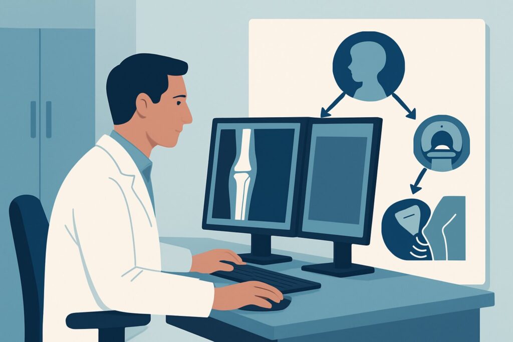
A 7-year-old boy presents to the emergency department on a Friday evening with a three-day history of worsening left knee pain, fever, and refusal to bear weight. His C-reactive protein and erythrocyte sedimentation rate are significantly elevated. You are concerned for a musculoskeletal infection, but the differential is broad. Is this septic arthritis, osteomyelitis, or something else? Deciding on the right initial imaging study is critical for timely diagnosis and management. This article details the clinical workflow for this specific scenario, explaining why the American College of Radiology (ACR) Appropriateness Criteria guide your first step. For this presentation, an ultrasound of the area of interest is rated as Usually appropriate.
Who Fits This Clinical Scenario for Suspected Osteomyelitis?
This guidance applies to a specific and common clinical presentation: a child, five years of age or older, with a clinical concern for osteomyelitis or septic arthritis involving an extremity, who is undergoing their initial imaging workup. The key inclusion criteria are the patient’s age (skeletally maturing but past the infant stage), the location of symptoms (an extremity like a leg, arm, foot, or hand), and the timing (this is the first imaging test being ordered for this episode of care).
It is crucial to distinguish this scenario from similar but distinct clinical situations that follow different diagnostic pathways:
- Children Younger Than 5 Years: Infants and toddlers have a different metaphyseal blood supply (transphyseal vessels) that can alter the presentation and spread of infection. Their workup is covered in a separate ACR variant.
- Axial Skeleton Involvement: If you suspect infection of the spine (diskitis-osteomyelitis) or pelvis (sacroiliitis), the imaging approach differs significantly, often prioritizing Magnetic Resonance Imaging (MRI) earlier.
- Post-Radiograph Workup: This article addresses the initial imaging choice. If plain radiographs have already been obtained and are normal or equivocal, the next step is addressed by a different ACR scenario, which often proceeds directly to more advanced imaging like MRI.
What Diagnoses Are You Working Up in This Scenario?
When a child presents with a focal limb complaint and systemic signs of infection, several diagnoses must be considered. The imaging strategy is designed to differentiate between these possibilities, some of which are orthopedic emergencies.
Septic Arthritis: This is often the most urgent consideration. A bacterial infection within the joint space can rapidly destroy cartilage, leading to long-term disability. The diagnosis requires identifying a joint effusion, which must be aspirated to confirm the presence of infection. Prompt surgical drainage is the standard of care.
Acute Osteomyelitis: An infection of the bone itself, most commonly in the metaphysis of long bones. It can occur alongside septic arthritis or in isolation. Early diagnosis is key to initiating appropriate antibiotic therapy and preventing complications like bone necrosis, abscess formation, or chronic osteomyelitis.
Cellulitis or Pyomyositis: These are infections of the skin/subcutaneous tissue and muscle, respectively. They can present with focal pain, swelling, and fever, closely mimicking bone or joint infection. Imaging helps to localize the infection to the soft tissues and rule out deeper involvement.
Transient Synovitis: A common, benign, and self-limiting inflammation of a joint’s synovial lining, particularly the hip. It is a diagnosis of exclusion after more serious conditions like septic arthritis have been ruled out. It typically presents with pain and a limp but often with less severe systemic symptoms.
Occult Trauma: A non-displaced fracture or significant bone bruise can cause pain, swelling, and refusal to bear weight. While less likely to be associated with high fevers, it remains an important part of the differential diagnosis that initial imaging can clarify.
Why Is Ultrasound the Recommended First Study for Suspected Osteomyelitis or Septic Arthritis?
For a child over five with suspected extremity infection, both ultrasound and radiography are rated as Usually appropriate. However, ultrasound often provides the most immediate, actionable information for the most urgent diagnoses, making it an excellent first-line choice.
The primary strength of US area of interest is its high sensitivity for detecting joint effusions. Identifying an effusion is the critical first step in diagnosing or excluding septic arthritis. Ultrasound can guide a diagnostic aspiration in real-time, providing a definitive sample for analysis. Furthermore, ultrasound excels at evaluating the surrounding soft tissues. It can readily identify cellulitis, fluid collections, and abscesses, including subperiosteal fluid, which is an early sign of osteomyelitis before bony changes are visible on radiographs. As a modality, it is fast, widely available, non-invasive, and uses no ionizing radiation (0 mSv).
Radiography area of interest is also Usually appropriate and is frequently performed alongside or just before ultrasound. While radiographs are insensitive to early osteomyelitis (bony changes may not appear for 10-14 days), they are essential for evaluating for alternative diagnoses like fractures, bone tumors (e.g., Ewing sarcoma), or other osseous abnormalities. They provide a crucial anatomic baseline.
Other powerful modalities are rated lower as initial tests for specific reasons:
- MRI area of interest without and with IV contrast is also rated Usually appropriate and is the most sensitive and specific imaging test for diagnosing osteomyelitis. However, it is more resource-intensive, takes longer to perform, is more expensive, and may require sedation in an anxious or pained child. It is often reserved as a second-line or problem-solving tool if the diagnosis remains unclear after initial studies.
- Bone scan whole body and 3-phase bone scan area of interest is rated Usually not appropriate. While sensitive for detecting bone inflammation, it is non-specific and cannot distinguish infection from trauma or other inflammatory processes. Most importantly, it involves a significant radiation dose for a child (☢☢☢☢ 3-10 mSv).
- CT area of interest is also Usually not appropriate due to its use of ionizing radiation (Varies) and its inferior soft tissue contrast compared to ultrasound and MRI for detecting early inflammatory changes and fluid collections.
What’s Next After Ultrasound? Downstream Workflow
The results of the initial ultrasound and radiographs will guide your subsequent management decisions in a branching pathway.
If the ultrasound is positive for a significant joint effusion: This finding is highly suspicious for septic arthritis. The immediate next step is an urgent orthopedic consultation for joint aspiration. If the aspirate is purulent or meets laboratory criteria for infection, the patient will require surgical irrigation and debridement followed by intravenous antibiotics.
If the ultrasound is negative for effusion but shows a subperiosteal fluid collection or deep soft tissue abscess: This is suggestive of osteomyelitis or pyomyositis. An orthopedic consultation is still warranted for possible surgical drainage. MRI with and without contrast is often the next step to define the full extent of bone marrow and soft tissue involvement, which helps guide the duration of antibiotic therapy and surgical planning.
If the ultrasound and radiographs are both negative or non-specific: If clinical suspicion for osteomyelitis remains high despite negative initial imaging (e.g., persistent point tenderness over bone, worsening inflammatory markers), the next step is typically an MRI. This moves the workup into the ACR variant for a patient with normal or equivocal initial radiographs. MRI can detect early bone marrow edema of osteomyelitis days before other imaging modalities become positive.
If the ultrasound shows only soft tissue edema (cellulitis) and radiographs are normal: The diagnosis is likely a superficial infection. The patient can often be managed with antibiotics, and further advanced imaging may not be necessary unless they fail to improve clinically.
Pitfalls to Avoid (and When to Get Help)
Navigating this workup requires avoiding several common pitfalls. First, do not delay imaging in a child with a high fever and refusal to bear weight; septic arthritis is a time-sensitive diagnosis. Second, do not rely solely on a normal radiograph to rule out acute osteomyelitis, as findings lag behind the clinical course by up to two weeks. Third, remember that ultrasound is operator-dependent; ensure the study is performed by a sonographer experienced in pediatric musculoskeletal imaging. Finally, a small, simple joint effusion can be seen in conditions other than septic arthritis, such as reactive effusions from nearby osteomyelitis or transient synovitis. If the clinical picture and ultrasound findings are equivocal, an orthopedic consultation and consideration for diagnostic aspiration or MRI are the appropriate escalation steps.
Related ACR Topics and Tools
This article covers one specific scenario in depth. For a broader overview of related pediatric musculoskeletal infection workups, or to explore the tools used to develop these guidelines, the following resources are available.
- For breadth across all scenarios in Osteomyelitis or Septic Arthritis-Child (Excluding Axial Skeleton), see our parent guide: Osteomyelitis or Septic Arthritis-Child (Excluding Axial Skeleton): ACR Appropriateness Decoded.
- To explore other clinical presentations, use the ACR Appropriateness Criteria Lookup.
- For details on imaging techniques, consult the Imaging Protocol Library.
- To discuss radiation exposure with families, use the Radiation Dose Calculator.
Frequently Asked Questions
Why not just order an MRI first for every child with suspected osteomyelitis?
While MRI is the most sensitive test for osteomyelitis, it is not always the best initial study. Ultrasound is faster, cheaper, requires no sedation, and is excellent for answering the most urgent question: is there a joint effusion concerning for septic arthritis? Answering that question quickly is paramount. MRI is an outstanding second-line test if the diagnosis remains uncertain after ultrasound and radiographs.
Are radiographs still necessary if I’m ordering an ultrasound?
Yes. Radiographs and ultrasound are complementary. While ultrasound assesses for joint fluid and soft tissue collections, radiographs are essential for providing an osseous baseline and ruling out other important diagnoses like fractures, benign bone lesions, or malignant tumors that can mimic infection.
What if the child has pain in multiple locations?
Multifocal pain changes the differential diagnosis and imaging strategy. While multifocal osteomyelitis can occur, especially in certain patient populations, this presentation should also raise concern for systemic inflammatory conditions or malignancy (e.g., leukemia). In this case, a whole-body imaging modality like whole-body MRI (rated ‘Usually not appropriate’ for focal symptoms) may become more relevant, and a pediatric rheumatology or hematology-oncology consultation may be needed.
Does the choice of imaging change if I’m more concerned about septic arthritis than osteomyelitis?
No, the initial approach is the same. Ultrasound is the best initial test for suspected septic arthritis because of its high sensitivity for detecting joint effusions, which is the key imaging finding. The workup for both conditions begins with the same initial studies (ultrasound and radiographs) because the two diseases often coexist and present similarly.
Is a CT scan ever useful in this scenario?
CT is rated ‘Usually not appropriate’ for the initial diagnosis of osteomyelitis in children. Its primary disadvantages are radiation exposure and poor soft tissue contrast compared to MRI. However, CT may have a niche role later in management, such as for surgical planning in cases of chronic osteomyelitis to evaluate for a sequestrum (a piece of dead bone) or to guide percutaneous drainage of a well-defined abscess.
Reviewed by Pouyan Golshani, MD, Interventional Radiologist — May 29, 2026