Which Imaging Study Is Best for a Child’s Back Pain with Suspected Infection or Neoplasm?
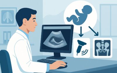
A 9-year-old presents to your clinic with three weeks of worsening thoracic back pain. The pain is now waking him from sleep, and he has had intermittent low-grade fevers and a documented 5-pound weight loss. His C-reactive protein (CRP) and erythrocyte sedimentation rate (ESR) are both elevated. You are concerned about a serious underlying process—infection, inflammation, or neoplasm—and need to decide on the most appropriate initial imaging study. This clinical workflow article details the American College of Radiology (ACR) guidance for this specific scenario. For a child with back pain and suspected inflammation, infection, or neoplasm, the ACR rates MRI complete spine without and with IV contrast as Usually Appropriate.
## Who Fits This Clinical Scenario for Pediatric Back Pain?
This guidance applies to children presenting with back pain accompanied by clinical or laboratory “red flags” that raise suspicion for a serious underlying etiology. The key is the suspicion of infection, inflammation, or neoplasm, which is typically based on a constellation of findings rather than a single symptom.
Inclusion criteria for this workflow include:
- Systemic Symptoms: Persistent fever, night sweats, weight loss, or malaise.
- Pain Characteristics: Constant, progressive, non-mechanical pain, or pain that awakens the child from sleep.
- Neurologic Deficits: New-onset weakness, sensory changes, bowel or bladder dysfunction, or abnormal reflexes.
- Elevated Inflammatory Markers: Unexplained elevation in ESR or CRP.
- Known Predisposing Condition: A child with a known primary malignancy (e.g., leukemia, neuroblastoma) who develops new back pain.
This workflow is distinct from other common pediatric back pain presentations. It does not apply to:
- Back Pain Without Red Flags: A child with back pain but a normal physical exam, no systemic symptoms, and normal inflammatory markers. This presentation follows a different diagnostic pathway.
- Mechanical or Overuse Injury: An adolescent athlete (e.g., a gymnast or football player) with activity-related pain that resolves with rest, suggesting a mechanical cause like spondylolysis.
- Cutaneous Stigmata: A child with back pain and a visible overlying skin abnormality like a hairy patch, deep dimple, or hemangioma, which suggests a congenital spinal anomaly.
Correctly identifying your patient’s clinical cluster is crucial for selecting the right initial imaging test and avoiding unnecessary radiation or diagnostic delays.
## What Diagnoses Are You Working Up in This Scenario?
When a child presents with back pain and red flags, the differential diagnosis is broad and includes several high-consequence conditions. The choice of imaging is driven by the need to effectively evaluate for these possibilities.
Infection: This is a primary concern and includes discitis, vertebral osteomyelitis, and epidural abscess. These conditions often present with fever, localized spinal tenderness, and elevated inflammatory markers. Staphylococcus aureus is a common causative organism. The infection can lead to rapid bone destruction, spinal instability, or neurologic compromise if not diagnosed and treated promptly. Imaging must be sensitive enough to detect early inflammatory changes in the bone marrow and soft tissues.
Neoplasm: Malignancy is a less common but critical consideration. Back pain can be the presenting symptom of both primary bone tumors (like Ewing sarcoma or osteosarcoma) and metastatic disease. Hematologic malignancies, such as leukemia and lymphoma, frequently infiltrate the vertebral marrow, causing pain. In younger children, metastatic neuroblastoma is also a key differential. The imaging study must be able to assess bone marrow, the spinal canal, and adjacent soft tissues for tumor infiltration.
Inflammatory Conditions: Non-infectious inflammatory processes, such as spondyloarthropathies (including juvenile idiopathic arthritis), can cause back pain. While often associated with other joint involvement, sacroiliitis or spondylitis can be the initial manifestation. Langerhans cell histiocytosis, an inflammatory disorder, can also present with lytic bone lesions in the spine causing pain.
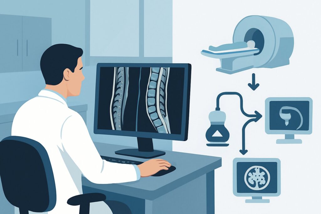
## Why Is MRI of the Complete Spine With and Without Contrast Usually Appropriate?
The ACR designates MRI complete spine without and with IV contrast as Usually Appropriate because it is the single most effective modality for evaluating the full spectrum of potential diagnoses in this scenario. Its superior soft tissue resolution and lack of ionizing radiation make it the clear choice for initial imaging in children with suspected serious spinal pathology.
The rationale for this recommendation is multi-faceted:
- Superior Tissue Characterization: MRI provides unparalleled detail of the bone marrow, intervertebral discs, spinal cord, nerve roots, and paraspinal soft tissues. It can detect bone marrow edema—an early sign of both infection and tumor infiltration—long before bony changes become visible on radiographs or CT.
- Role of IV Contrast: The administration of gadolinium-based contrast is essential. It helps differentiate between phlegmon and a drainable, ring-enhancing abscess. It also highlights areas of neoplastic enhancement, defines tumor margins, and can reveal leptomeningeal disease. Without contrast, the full extent and nature of the pathology can be missed.
- **Importance of the Complete Spine:** Pathologic processes like infection and malignancy can be multifocal. A tumor may have “skip” metastases, or an infection could involve multiple vertebral levels. Imaging only the most symptomatic area risks missing the full disease burden, which is critical for accurate staging and treatment planning.
- Zero Ionizing Radiation: This is a paramount consideration in pediatric imaging. MRI uses no ionizing radiation (0 mSv), unlike CT or radiography. This avoids contributing to the child’s cumulative lifetime radiation exposure, aligning with the As Low As Reasonably Achievable (ALARA) principle.
Comparison to Alternative Studies:
- Radiography complete spine: Rated May be appropriate (Disagreement). While often obtained, plain films are highly insensitive in the early stages of infection or neoplasm. Bony destruction or periosteal reaction may not be visible for weeks after symptom onset, leading to a false sense of security from a “normal” report.
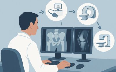
- CT complete spine without IV contrast: Rated May be appropriate. CT is excellent for evaluating cortical bone and is faster than MRI. However, it delivers a significant radiation dose (pediatric RRL ☢☢☢☢, 3-10 mSv) and has far inferior soft tissue contrast for assessing marrow, disc, or neural element involvement. It is not the preferred initial study for this clinical question.
Once you’ve decided on MRI of the complete spine, our protocol guide covers the technique, contrast, and reading principles: MRI Lumbar Spine Without Contrast.
## What’s Next After MRI? Downstream Workflow
The results of the contrast-enhanced spine MRI will guide your subsequent management. The next steps are determined by whether the findings are positive, negative, or indeterminate.
- If the MRI is positive for infection (e.g., discitis, osteomyelitis, abscess): The immediate next step is consultation with pediatric infectious disease and potentially pediatric neurosurgery or orthopedic surgery, especially if there is an epidural abscess causing neurologic compromise. A CT-guided or open biopsy may be necessary to obtain cultures and guide antibiotic therapy.
- If the MRI is positive for a neoplastic process: Urgent consultation with pediatric oncology/hematology is required. Further staging studies (such as a whole-body bone scan or PET/CT) and biopsy will be coordinated by the oncology team to confirm the diagnosis and initiate treatment.
- If the MRI is negative: A negative, high-quality MRI of the entire spine makes a serious occult infection or tumor highly unlikely. The workup should pivot back to considering other causes. If clinical suspicion for an inflammatory spondyloarthropathy remains high, referral to a pediatric rheumatologist is appropriate. This may also route the patient to the ACR variant for “Child. Back pain. With at least one clinical red flag. Negative radiographs. Next imaging study,” though MRI has already been performed.
- If the MRI is indeterminate: In rare cases, the findings may be nonspecific. This may prompt a discussion with the radiologist about the need for short-interval follow-up imaging or alternative modalities. A nuclear medicine bone scan, rated Usually not appropriate for initial imaging, might be considered in specific, complex cases after multidisciplinary discussion to look for multifocal osseous activity.
## Pitfalls to Avoid (and When to Get Help)
Navigating this workup requires vigilance to avoid common diagnostic errors.
- Pitfall 1: Accepting a negative radiograph. Do not be falsely reassured by normal plain films in a child with red flag symptoms. These studies lack the sensitivity to rule out early-stage infection or malignancy.
- Pitfall 2: Ordering a limited MRI. Insisting on an MRI of the complete spine is crucial. A focal study of the lumbar spine could miss a thoracic neuroblastoma or cervical osteomyelitis.
- Pitfall 3: Omitting IV contrast. Ordering a non-contrast MRI can miss or underestimate the extent of an abscess or enhancing tumor, delaying appropriate surgical or medical management. Always specify “without and with IV contrast.”
- Pitfall 4: Delaying the imaging. If there are neurologic deficits or high suspicion of an epidural abscess, imaging should be obtained emergently.
If you identify any new or progressive neurologic deficits, such as weakness or bladder changes, this constitutes a medical emergency. Escalate immediately for an urgent MRI and neurosurgical consultation.
## Related ACR Topics and Tools
This article focuses on a single clinical scenario. For a comprehensive overview of all pediatric back pain variants and for tools to help with ordering and dose management, please see the following resources.
For breadth across all scenarios in Back Pain-Child, see our parent guide: Back Pain-Child: ACR Appropriateness Decoded.
- ACR Appropriateness Criteria Lookup — for adjacent scenarios
- Imaging Protocol Library — for technique on the recommended study
- Radiation Dose Calculator — for cumulative dose conversations
Frequently Asked Questions
Why is a ‘complete spine’ MRI necessary instead of just imaging the painful area?
Imaging the complete spine is critical because serious conditions like malignancy (e.g., leukemia, metastatic neuroblastoma) and infection can be multifocal. A tumor or infection might be present at a different spinal level than the area of maximal pain. A limited study risks missing ‘skip lesions’ or the primary site of disease, leading to an incomplete diagnosis and improper treatment planning.
Is an MRI without contrast ever sufficient in this scenario?
According to the ACR, an MRI of the complete spine without IV contrast is rated ‘May be appropriate.’ However, omitting contrast significantly limits the study’s diagnostic power. Contrast is essential for identifying and characterizing abscesses (which typically show ring enhancement), delineating the extent of tumors, and detecting leptomeningeal disease. For the initial workup of suspected infection or neoplasm, a contrast-enhanced study is strongly preferred and considered ‘Usually Appropriate’.
My hospital has a long wait time for MRI. Should I order a CT scan instead?
While a CT scan is faster, it is rated ‘May be appropriate’ and has significant drawbacks, including substantial ionizing radiation (3-10 mSv in children) and inferior soft tissue detail compared to MRI. If the child has neurologic deficits or signs of spinal cord compression, the MRI should be performed emergently. In a stable patient, it is generally better to wait for the superior diagnostic information from an MRI than to proceed with a lower-yield, higher-radiation CT scan.
What if the child has a contraindication to MRI, like an incompatible implanted device?
In the rare case of an absolute contraindication to MRI, a CT of the complete spine without and with IV contrast would be the next best option, despite its limitations. This is rated ‘May be appropriate (Disagreement)’. A nuclear medicine bone scan with SPECT/CT could also be considered in consultation with a radiologist, though it is rated ‘Usually not appropriate’ as a first-line test.
Do I need to check renal function before ordering an MRI with gadolinium contrast in a child?
Yes, it is standard practice to assess renal function (e.g., with a serum creatinine/eGFR) before administering gadolinium-based contrast agents, especially in patients with known or suspected kidney disease. While the risk of nephrogenic systemic fibrosis (NSF) is very low with modern agents in patients with normal renal function, institutional protocols for screening should always be followed.
Reviewed by Pouyan Golshani, MD, Interventional Radiologist — May 30, 2026