What Is the Right Imaging for a Child with Congenital or Developmental Vision Loss?
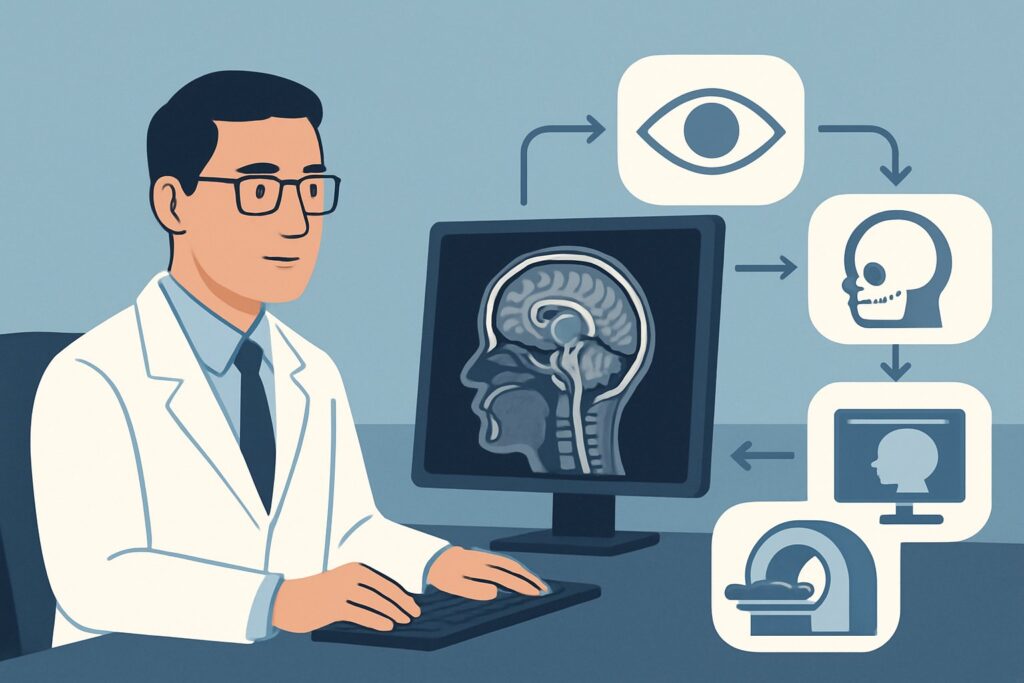
A four-year-old is referred to your clinic by a pediatric ophthalmologist after failing a routine vision screening. The exam confirms decreased visual acuity in the left eye that has likely been present since birth. There is no history of trauma, no signs of infection, and critically, no leukocoria on fundoscopy. You suspect a congenital or developmental cause for the vision loss, but the etiology is unclear. To investigate a potential structural abnormality along the visual pathway, you need to select the most appropriate initial imaging study. This decision requires balancing diagnostic yield with the unique safety considerations of pediatric imaging.
For a child with suspected congenital or developmental vision loss, the American College of Radiology (ACR) Appropriateness Criteria rates MRI head and orbits without IV contrast as Usually appropriate. This article details the clinical workflow for this specific scenario, explaining the rationale behind this recommendation and outlining the downstream steps based on imaging results.
Who Fits This Clinical Scenario?
This guidance applies to a specific pediatric population: a child presenting with decreased visual acuity or vision loss believed to be from a congenital or developmental cause. The presentation can be unilateral or bilateral. A key negative finding for this scenario is the absence of leukocoria (a white pupillary reflex), which would trigger a different workup focused on retinoblastoma.
This workflow is intended for the initial imaging of these patients. It is crucial to distinguish this presentation from similar, but distinct, clinical situations that follow different diagnostic pathways:
- Acute Traumatic Vision Loss: If the vision loss occurred after an injury, the primary concern is orbital fracture or intracranial hemorrhage. This follows the Child. Traumatic visual loss. Suspected orbital injury ACR variant.
- Acute Nontraumatic Vision Loss: A sudden onset of vision loss without trauma suggests etiologies like optic neuritis or ischemia, which may require different imaging protocols (often including contrast) as detailed in the Child. Nontraumatic acute vision loss without papilledema variant.
- Suspected Optic Pathway Tumor: If there is a strong clinical suspicion for a tumor, such as in a child with neurofibromatosis type 1 (NF1), the imaging approach is tailored to tumor detection and characterization, often requiring contrast.
Correctly identifying your patient within this specific scenario ensures the most appropriate and highest-yield initial study is ordered.
What Diagnoses Are You Working Up in This Scenario?
When a child presents with congenital or developmental vision loss, the differential diagnosis spans a range of structural abnormalities affecting the eyes, optic nerves, and brain. The choice of imaging is driven by the need to visualize these structures with high fidelity.
A primary consideration is optic nerve hypoplasia, a condition where the optic nerve is underdeveloped. This can occur in isolation or as part of a broader syndrome. MRI is exceptionally sensitive for measuring optic nerve caliber and can readily identify this anomaly.
Closely related is septo-optic dysplasia (de Morsier syndrome), a clinical spectrum that includes optic nerve hypoplasia, pituitary hormone deficiencies, and midline brain defects such as the absence of the septum pellucidum or corpus callosum abnormalities. Identifying these associated brain findings on MRI is critical, as it mandates an endocrine evaluation that would otherwise be missed.
Other congenital brain malformations can also lead to vision loss, particularly those affecting the posterior visual pathways. This category includes cortical dysplasia, schizencephaly (clefts in the cerebral hemispheres), and lissencephaly (a “smooth brain” appearance). These conditions result in cortical visual impairment, where the eyes are structurally normal but the brain cannot process visual information correctly. MRI is the definitive modality for characterizing these complex brain abnormalities.
Less commonly, an indolent, slow-growing tumor like an optic pathway glioma can manifest as progressive, seemingly developmental vision loss. While a dedicated tumor workup is a separate scenario, a high-resolution non-contrast MRI can often detect mass-like expansion of the optic nerve or chiasm, prompting further investigation.
Why Is MRI Head and Orbits Without IV Contrast the Recommended Study?
The ACR designates MRI head and orbits without IV contrast as Usually appropriate because it provides the most comprehensive and safest evaluation for the leading differential diagnoses in this scenario.
The rationale is threefold: superior soft-tissue visualization, avoidance of ionizing radiation, and the targeted scope of a non-contrast study for this indication.
- Unmatched Anatomic Detail: MRI offers unparalleled soft-tissue contrast, which is essential for evaluating the small, intricate structures of the visual pathway. It can precisely measure the diameter of the optic nerves, assess the optic chiasm and tracts, and characterize the brain parenchyma for associated developmental anomalies.
- No Ionizing Radiation: This is a paramount consideration in pediatric imaging. MRI uses magnetic fields and radio waves, not ionizing radiation. It carries a pediatric relative radiation level of O (0 mSv). In contrast, all CT variants are rated Usually not appropriate for this indication, in large part due to the associated radiation dose (pediatric RRL of ☢☢☢ to ☢☢☢☢, or 0.3-10 mSv), which should be avoided unless absolutely necessary.
- Rationale for Omitting Contrast: For the initial evaluation of a congenital or developmental abnormality, the primary goal is to assess anatomy and morphology. Conditions like optic nerve hypoplasia and midline brain defects are structural and are well-visualized without gadolinium-based contrast. While an MRI with contrast is rated May be appropriate (Disagreement), the lack of consensus suggests it is not required for the initial step. Omitting contrast avoids potential risks associated with gadolinium administration and shortens scan time, which is beneficial for young children who may require sedation.
An MRI of the orbits alone or MRI of the head alone are both rated as May be appropriate. However, ordering them separately risks missing the full picture. An orbits-only study could miss a central cause like septo-optic dysplasia, while a head-only study might lack the dedicated high-resolution sequences needed to optimally assess the intraorbital optic nerves. The combined MRI head and orbits is the most robust initial examination.
What’s Next After MRI Head and Orbits Without IV Contrast? Downstream Workflow
The results of the MRI will guide the subsequent clinical pathway, which often involves multidisciplinary care.
- If the MRI is positive for optic nerve hypoplasia or midline brain anomalies (e.g., absent septum pellucidum): This finding is highly suggestive of septo-optic dysplasia. The immediate next step is a referral to a pediatric endocrinologist to screen for pituitary hormone deficiencies, which can be life-threatening if untreated. A pediatric neurology consultation is also warranted to manage any associated developmental or seizure-related issues.
- If the MRI is positive for other brain malformations (e.g., cortical dysplasia): The patient should be referred to pediatric neurology for long-term management of developmental delays and potential epilepsy. Vision therapy and early intervention services are also key components of care.
- If the MRI is negative: A normal MRI of the brain and orbits effectively rules out a structural cause along the central visual pathways. The etiology of the vision loss is likely located within the retina or is functional. The next step is a referral back to the pediatric ophthalmologist for further specialized testing, such as an electroretinogram (ERG), to assess retinal function and investigate conditions like Leber congenital amaurosis or other inherited retinal dystrophies.
- If the MRI is indeterminate or shows a suspected mass: If a finding is unclear or suspicious for a neoplasm (e.g., subtle thickening of an optic nerve), a follow-up MRI head and orbits without and with IV contrast (May be appropriate (Disagreement)) may be necessary to better characterize the lesion. A consultation with pediatric neuro-oncology would be the next step if a tumor is confirmed.
Pitfalls to Avoid (and When to Get Help)
Navigating this workup requires avoiding several common pitfalls to ensure a timely and accurate diagnosis.
- Pitfall 1: Ordering CT instead of MRI. Due to its speed and availability, CT is sometimes ordered reflexively. For this indication, CT provides poor visualization of the optic nerves and brain parenchyma while exposing the child to unnecessary ionizing radiation.
- Pitfall 2: Incomplete imaging. Ordering an MRI of only the orbits or only the head can miss crucial findings. The pathology can exist anywhere from the globe to the occipital cortex, making a combined study essential.
- Pitfall 3: Underestimating the need for sedation. Young children cannot remain still for the duration of an MRI scan. A motion-degraded, non-diagnostic study is a waste of resources and delays care. Plan for anesthesia or sedation in collaboration with the radiology department to ensure high-quality images are obtained.
- Pitfall 4: Missing the endocrine connection. If the MRI reveals findings consistent with septo-optic dysplasia, the workup is not complete. Failure to refer for an endocrine evaluation can lead to missed diagnoses of adrenal insufficiency or hypothyroidism.
If you are uncertain about the imaging findings or the appropriate next clinical step, a consultation with a pediatric radiologist or pediatric neurologist is always recommended.
Related ACR Topics and Tools
For a comprehensive understanding of pediatric orbital imaging and to explore related clinical scenarios, the following resources are available. For breadth across all scenarios in Orbital Imaging and Vision Loss-Child, see our parent guide: Orbital Imaging and Vision Loss-Child: ACR Appropriateness Decoded.
- ACR Appropriateness Criteria Lookup: Access the full guidelines for thousands of clinical variants.
- Imaging Protocol Library: Review detailed imaging techniques for recommended studies.
- Radiation Dose Calculator: Model and discuss cumulative radiation exposure with patients and families when considering CT.
Frequently Asked Questions
Why is intravenous contrast not routinely recommended for the initial imaging in this scenario?
For congenital or developmental abnormalities like optic nerve hypoplasia or midline brain defects, the primary goal is to assess structure and anatomy. These are typically well-visualized on non-contrast MRI sequences. Omitting contrast avoids the risks associated with gadolinium administration and shortens the scan time, which is a significant benefit for young children who may require sedation.
What if the child cannot tolerate an MRI or if sedation is contraindicated?
This is a challenging situation. If MRI is not feasible, the options are limited. A high-resolution CT of the head and orbits could provide some information about gross structural abnormalities but is significantly less sensitive for optic nerve or subtle brain malformations and involves ionizing radiation. The decision should be made in consultation with a pediatric radiologist to weigh the low diagnostic yield and radiation risk of CT against the risks of sedation for MRI.
Does this imaging guidance change if the vision loss is bilateral versus unilateral?
No, the initial imaging recommendation of MRI head and orbits without IV contrast remains the same regardless of whether the vision loss is unilateral or bilateral. Bilateral findings, such as bilateral optic nerve hypoplasia, may increase the clinical suspicion for a systemic or syndromic condition like septo-optic dysplasia, making the comprehensive evaluation of the brain even more critical.
My patient has nystagmus along with developmental vision loss. Does that change the imaging choice?
Nystagmus is a common co-occurring feature in children with congenital vision loss and does not change the primary imaging recommendation. Its presence reinforces the need to investigate for an underlying structural cause, such as optic nerve hypoplasia or foveal hypoplasia (which may be better seen on optical coherence tomography). The ACR has a separate variant for isolated nystagmus, but when it accompanies vision loss in this context, this workflow applies.
If the MRI of the head and orbits is completely normal, what is the most likely next step?
A normal MRI effectively rules out a structural cause in the brain, optic nerves, or orbits. The diagnostic focus then shifts to the retina. The patient should be referred back to their pediatric ophthalmologist for further evaluation, which often includes an electroretinogram (ERG) to test retinal function. This can help diagnose conditions like Leber congenital amaurosis or other inherited retinal dystrophies that are not visible on MRI.
Reviewed by Pouyan Golshani, MD, Interventional Radiologist — May 30, 2026