What Is the Best Initial Imaging for Suspected Pulmonary Hypertension? An ACR-Guided Workflow
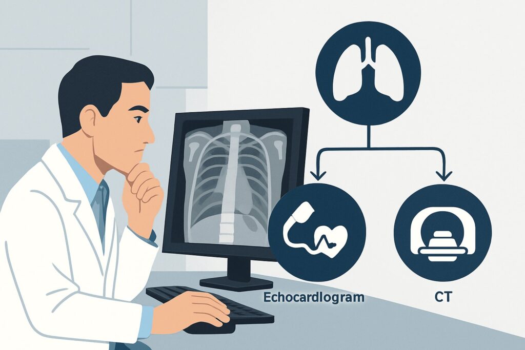
A 55-year-old woman presents to your clinic with six months of progressive dyspnea on exertion and new bilateral lower extremity edema. Her past medical history is notable only for well-controlled hypothyroidism. On exam, you note an elevated jugular venous pressure, a loud P2 component of the second heart sound, and 2+ pitting edema to the mid-calves. You suspect pulmonary hypertension, a diagnosis with significant morbidity if missed. You know imaging is the next step, but which study provides the most diagnostic value upfront without unnecessary radiation or risk? This clinical workflow article details the initial imaging pathway for a patient just like this one. For this specific scenario, the American College of Radiology (ACR) rates a resting transthoracic echocardiogram as Usually Appropriate, making it the clear starting point for your workup.
Who Fits This Clinical Scenario for Suspected Pulmonary Hypertension?
This guidance applies to adult and pediatric patients undergoing an initial evaluation for suspected pulmonary hypertension (PH). These are typically individuals presenting with unexplained signs and symptoms such as progressive dyspnea, fatigue, exertional chest pain, syncope, or peripheral edema. Physical exam findings may heighten suspicion, including a loud pulmonic valve closure sound (P2), a right ventricular heave, elevated jugular venous pressure, hepatomegaly, or ascites. This workflow is intended for the first diagnostic imaging step, before a definitive diagnosis of PH has been established and before its specific etiology is known.
This article does not cover patients with an already-confirmed diagnosis of PH who are undergoing follow-up imaging to monitor disease progression or response to therapy. It also does not apply to patients presenting with acute, severe cardiorespiratory distress where a massive pulmonary embolism (PE) is the leading diagnosis; that scenario requires an emergent and distinct imaging protocol, typically a dedicated CTA for PE. Finally, while patients with known severe left-sided heart disease can develop secondary PH, this guidance is focused on the undifferentiated patient where PH itself is the primary question to be answered.
What Diagnoses Are You Working Up in This Scenario?
When you order the initial imaging for suspected PH, you are evaluating a broad differential organized by the World Health Organization (WHO) classification system. The goal is to confirm the presence of PH and find clues to its underlying cause.
Pulmonary Hypertension due to Left Heart Disease (WHO Group 2): This is the most common cause of elevated pulmonary pressures. Conditions like left ventricular systolic or diastolic dysfunction, or significant mitral or aortic valve disease, lead to a “backup” of pressure into the pulmonary circulation. The initial imaging study must be able to thoroughly evaluate left heart structures and function to identify or exclude this group.
Pulmonary Arterial Hypertension (PAH) (WHO Group 1): This is a rarer but serious condition involving proliferative changes in the small pulmonary arteries. It can be idiopathic, heritable, or associated with other conditions like connective tissue disease (e.g., scleroderma), congenital heart disease, or HIV infection. Early detection is critical as specific therapies are available.
Pulmonary Hypertension due to Lung Disease or Hypoxia (WHO Group 3): Chronic lung diseases such as Chronic Obstructive Pulmonary Disease (COPD) or interstitial lung disease are common causes of PH. The imaging workup should provide information about the lung parenchyma, though this is often better characterized by subsequent tests like CT.
Chronic Thromboembolic Pulmonary Hypertension (CTEPH) (WHO Group 4): A potentially curable form of PH, CTEPH results from unresolved pulmonary emboli that organize into fibrous blockages. Identifying this etiology is crucial because it can be treated with pulmonary thromboendarterectomy or balloon pulmonary angioplasty. The initial imaging may show signs of chronic right heart strain that raise suspicion for this diagnosis.
Why Is Transthoracic Echocardiography the Recommended First Step for Suspected Pulmonary Hypertension?
The ACR Appropriateness Criteria panel designates US echocardiography transthoracic resting as Usually Appropriate for the initial imaging of suspected pulmonary hypertension. This recommendation is based on the study’s high diagnostic yield, widespread availability, and excellent safety profile.
A transthoracic echocardiogram (TTE) is the ideal first-line test because it provides a wealth of information in a single, non-invasive session. It uses no ionizing radiation (O 0 mSv) and does not require IV contrast. Most importantly, it allows for the estimation of the pulmonary artery systolic pressure (PASP) via Doppler assessment of the tricuspid regurgitation jet. Beyond just a pressure estimate, the TTE provides a comprehensive assessment of the consequences of elevated pressures, including the size, thickness, and function of the right ventricle and right atrium. It can also reveal secondary findings like paradoxical septal motion. Furthermore, a TTE is essential for evaluating for the most common causes of PH (WHO Group 2) by directly visualizing left ventricular function, chamber sizes, and valvular integrity.
Other imaging studies are rated lower for the initial step for specific reasons:
- Right Heart Catheterization: While this is the gold standard for confirming and quantifying PH, the ACR rates it as Usually Not Appropriate for the initial workup. It is an invasive procedure with associated risks and is properly reserved for confirming the diagnosis after non-invasive testing is suggestive and for guiding specific therapeutic decisions.
- CTA Chest with IV Contrast: This study is also rated Usually Appropriate but is not typically the first choice. While it provides excellent detail of the lung parenchyma (for WHO Group 3) and can identify chronic thromboemboli (for WHO Group 4), it involves both radiation (☢☢☢ 1-10 mSv) and IV contrast. It cannot provide the direct hemodynamic estimates or assess cardiac function with the same detail as an echocardiogram. It is often a critical second step in the workup after an abnormal TTE.
When ordering the TTE, it is helpful to specify that you are evaluating for pulmonary hypertension and request a full assessment of right heart size and function, along with an estimation of the PASP.
What Is the Downstream Workflow After an Initial Echocardiogram?
The results of the transthoracic echocardiogram will guide your subsequent management and diagnostic strategy.
If the study is positive or suggests a high probability of PH: When the TTE shows an elevated estimated PASP, right ventricular dilation or dysfunction, or other signs of right heart pressure overload, the next step is a comprehensive workup to determine the underlying cause (the WHO Group). This typically involves referral to a pulmonologist or cardiologist with expertise in PH. Further testing will likely include pulmonary function tests, a ventilation/perfusion (V/Q) scan to screen for CTEPH, and serologic testing for associated conditions like connective tissue disease. Ultimately, a right heart catheterization will be needed to confirm the diagnosis, measure hemodynamics precisely, and guide therapy.
If the study is negative or shows a low probability of PH: A normal echocardiogram with no signs of right heart strain makes significant pulmonary hypertension highly unlikely. The diagnostic focus should then shift to other potential causes of the patient’s symptoms, such as primary pulmonary disease not causing PH (e.g., asthma, mild COPD), occult coronary artery disease, or deconditioning.
If the study is indeterminate or borderline: In cases where the estimated PASP is borderline or the images are technically limited, the clinical context is paramount. If suspicion remains high, a different imaging modality may be considered. A cardiac MRI (rated May be Appropriate) can provide a more accurate and reproducible assessment of right ventricular size and function. In some cases, a transesophageal echocardiogram (TEE) may be used to obtain better acoustic windows.
Common Pitfalls to Avoid in the Initial Workup of Pulmonary Hypertension
Navigating the initial workup for suspected PH requires careful interpretation and avoiding common missteps. One major pitfall is over-relying on the estimated PASP value from the echocardiogram as a single diagnostic number. This value is an estimate, and its accuracy can be affected by technical factors; it must be interpreted in the context of the complete structural and functional assessment of the right heart. Another common error is prematurely anchoring on a more common diagnosis like asthma or anxiety for a patient’s dyspnea, thereby delaying the PH workup, especially in a high-risk individual. Finally, do not stop the workup if a chest radiograph is normal; a plain film can be completely unremarkable even in the presence of significant pulmonary hypertension. If a patient presents with syncope, chest pain, or signs of severe right heart failure like ascites, escalate immediately for specialist consultation and likely inpatient evaluation.
Related ACR Topics and Tools
This article covers a single, common scenario in depth. For a broader view of all clinical variants related to this condition, please consult the parent topic article. For additional decision support, the following tools can help you navigate imaging choices, technical specifications, and patient communication.
- For breadth across all scenarios in Suspected Pulmonary Hypertension, see our parent guide: Suspected Pulmonary Hypertension: ACR Appropriateness Decoded.
- To explore other clinical presentations, use the Imaging Appropriateness Selector.
- For technical details on performing the recommended study, see the Imaging Protocol Library.
- To discuss radiation exposure with your patients, use the Radiation Dose Calculator.
Frequently Asked Questions
Why isn’t a chest CT the first test? It can see the lungs and pulmonary arteries.
While a CT scan is excellent for evaluating lung disease and the pulmonary arteries, a transthoracic echocardiogram is the recommended first step because it provides essential functional and hemodynamic information non-invasively and without radiation. It can estimate pulmonary pressures and directly assess right heart function, which are critical initial data points. A CT is often an important second step after an abnormal echo to help determine the underlying cause.
My patient cannot lie flat for an echo. What are my options?
Skilled sonographers can often obtain adequate images with the patient in other positions, such as semi-upright or the left lateral decubitus position. If a standard transthoracic echo is truly non-diagnostic due to patient factors like body habitus or lung disease, the ACR lists a transesophageal echocardiogram (TEE) or a cardiac MRI as ‘May be appropriate’ alternatives to better visualize the heart.
The echo report gives a wide range for the estimated pulmonary pressure. How do I interpret this?
The pulmonary artery systolic pressure (PASP) on an echo is an estimate derived from the tricuspid regurgitation jet velocity and an estimated right atrial pressure. It is not a direct measurement. It’s crucial to interpret this number in the context of all other echocardiographic findings, such as the size and function of the right ventricle, the position of the interventricular septum, and the size of the inferior vena cava. An equivocal pressure with other signs of right heart strain is more concerning than an isolated borderline value.
When is a right heart catheterization necessary if it’s ‘Usually Not Appropriate’ for initial imaging?
Right heart catheterization (RHC) is the gold standard for confirming a diagnosis of pulmonary hypertension, not for initial screening. It is performed after non-invasive tests like an echocardiogram suggest a high probability of PH. The RHC provides precise, direct hemodynamic measurements that are essential for confirming the diagnosis, classifying the specific type and severity of PH, and determining the most appropriate therapy, particularly for patients with suspected Pulmonary Arterial Hypertension (WHO Group 1).
Reviewed by Pouyan Golshani, MD, Interventional Radiologist — May 26, 2026