What Is the Best Initial Procedure for Malignant Hilar Biliary Obstruction?
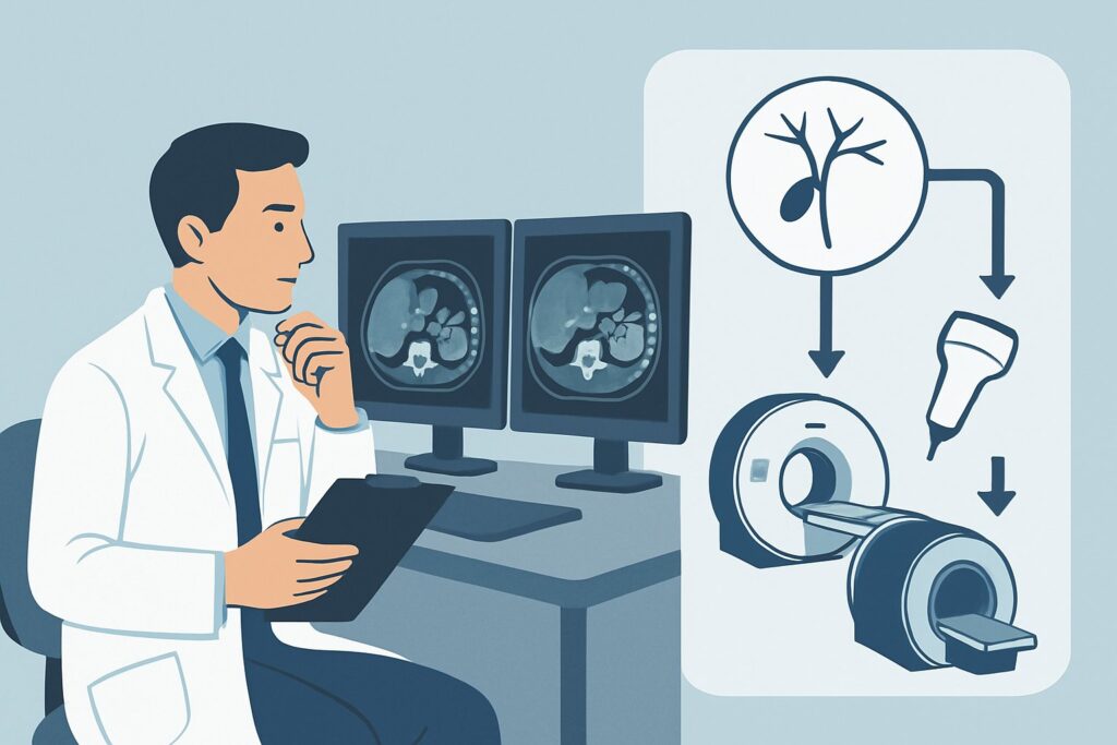
A 68-year-old man presents with two months of progressive, painless jaundice, pruritus, and unintentional weight loss. An abdominal Magnetic Resonance Cholangiopancreatography (MRCP) reveals a mass at the confluence of the right and left hepatic ducts, highly suspicious for a perihilar cholangiocarcinoma, also known as a Klatskin tumor. His total bilirubin is 18 mg/dL. The multidisciplinary tumor board recommends biliary decompression to improve his liver function before considering neoadjuvant chemotherapy. As the consulting physician, you must decide on the most appropriate initial therapeutic procedure to relieve the obstruction. This clinical decision is critical, as the chosen method directly impacts the patient’s eligibility for further treatment and overall prognosis. According to the American College of Radiology (ACR) Appropriateness Criteria, the initial placement of a `Percutaneous internal/external biliary catheter` is considered Usually Appropriate for this specific scenario.
Who Fits This Clinical Scenario?
This guidance applies specifically to patients with biliary obstruction located at the hepatic hilum (the confluence of the right and left hepatic ducts) that is caused by a suspected or confirmed malignancy. The quintessential example is a perihilar cholangiocarcinoma (Klatskin tumor), but this scenario also includes advanced gallbladder cancer invading the hilum or extrinsic compression from metastatic lymphadenopathy in the porta hepatis.
The key elements defining this scenario are:
- Location: The obstruction is high in the biliary tree, at or above the insertion of the cystic duct, often separating the right and left ductal systems (Bismuth-Corlette type II, III, or IV).
- Etiology: The cause is malignant, not benign.
- Goal: The procedure is the initial therapeutic intervention for biliary drainage, intended as a bridge to surgery, to enable systemic therapy, or for palliation.
It is crucial to distinguish this presentation from similar but distinct clinical problems. This guidance does not apply to patients with distal common bile duct obstruction (often from pancreatic carcinoma), benign strictures (like primary sclerosing cholangitis), or obstruction caused by choledocholithiasis. Each of those situations has a different recommended management pathway.
What Diagnoses Are You Working Up in This Scenario?
When a patient presents with a malignant-appearing hilar biliary obstruction, several diagnoses are on the differential, though one is far more common than the others. The primary goal of the initial intervention is decompression, but confirming the specific histology is a critical secondary objective.
Perihilar Cholangiocarcinoma (Klatskin Tumor)
This is the most common cause of malignant hilar obstruction. These tumors arise from the biliary epithelium at the hepatic duct confluence. They are typically slow-growing but infiltrate locally, encasing vascular structures and causing progressive biliary obstruction. Cross-sectional imaging often shows a poorly defined, infiltrating mass at the hilum with associated upstream ductal dilation that may be isolated to one side of the liver.
Gallbladder Carcinoma
An advanced gallbladder cancer can grow beyond the gallbladder fossa and directly invade the biliary confluence and porta hepatis. On imaging, it can be difficult to distinguish from a primary cholangiocarcinoma if the tumor is large, but identifying a primary mass centered in the gallbladder fossa is a key clue.
Metastatic Disease
Lymph node metastases in the porta hepatis can extrinsically compress the bile ducts, leading to a hilar obstruction. Primary cancers that commonly metastasize to these nodes include colorectal, gastric, pancreatic, and breast cancer. In these cases, the biliary ducts are typically compressed rather than infiltrated, and a history of a known primary malignancy is often present.
Hepatocellular Carcinoma (HCC)
Less commonly, a primary liver tumor (HCC) located near the hilum can either invade the biliary ducts or cause extrinsic compression. This is more common in patients with underlying cirrhosis. Serum tumor markers like Alpha-Fetoprotein (AFP) can help differentiate this from cholangiocarcinoma.
Why Is a Percutaneous Biliary Catheter the Recommended Initial Procedure?
For malignant hilar obstruction, the ACR designates `Percutaneous internal/external biliary catheter` placement as Usually Appropriate. This interventional radiology procedure involves accessing the biliary tree through the liver under imaging guidance (ultrasound and fluoroscopy). The rationale for this recommendation is rooted in the unique anatomical challenge posed by hilar tumors.
Hilar malignancies frequently isolate the right and left hepatic ductal systems, and may even obstruct secondary branches. Effective drainage requires decompressing a sufficient volume of the liver (typically >30-50% of the total volume) to lower bilirubin levels. The percutaneous approach provides direct, selective access to the desired ductal system. An interventional radiologist can target specific, dilated ducts—often in the future liver remnant if a major resection is planned—ensuring controlled and effective drainage. This method has a high technical success rate for accessing obstructed systems that may be impossible to reach from below.
In contrast, other options are rated lower for this specific initial intervention:
- Endoscopic internal biliary catheter (ERCP): Rated as May be appropriate, this approach is the standard for distal obstructions but is less effective for complex hilar lesions. Cannulating separated right and left systems from the duodenum can be technically difficult or impossible, leading to failed procedures or, more dangerously, incomplete drainage. Draining one segment while leaving another obstructed can seed the undrained segment with bacteria, precipitating severe cholangitis.
- Surgery: Also rated May be appropriate, major surgery is generally not the initial step for decompression. Patients with deep jaundice have impaired synthetic function and are at high risk for perioperative complications. Biliary drainage is performed first to optimize the patient’s condition before a potential curative resection is attempted.
- Endoscopic US-guided biliary drainage (EUS-BD): This is rated Usually not appropriate as a first-line therapy for this scenario. EUS-BD is a valuable but highly specialized salvage technique used when both percutaneous and ERCP approaches have failed or are not feasible.
The percutaneous catheter provides both internal drainage (into the duodenum) and external drainage (into a bag), offering flexibility in management. It also establishes a mature access tract that can be used later for tumor biopsy or for converting the catheter to a permanent internal metallic stent.
What’s Next After Percutaneous Biliary Catheter Placement? Downstream Workflow
The placement of a percutaneous biliary drain is the start, not the end, of the management pathway. The subsequent steps depend on the clinical response and the overall treatment plan.
If the procedure is successful and bilirubin levels decrease: The patient’s clinical status improves, relieving pruritus and reducing the risk of cholangitis. With improved liver function, they can now safely proceed with the planned oncologic therapy. This may involve neoadjuvant chemotherapy or radiation, followed by reassessment for surgical resectability. The percutaneous access tract can also be used to obtain tissue samples via intraductal biopsy or brushings to confirm the diagnosis if it remains uncertain.
If bilirubin levels fail to decrease or cholangitis develops: This often indicates incomplete drainage of the functional liver volume. Because hilar tumors can isolate multiple segments, draining only one system may be insufficient. The next step is typically to perform cholangiography through the existing catheter to map the biliary anatomy and identify other obstructed, undrained segments. A second (or even third) percutaneous catheter may be required to achieve adequate drainage.
If the patient is deemed non-surgical or has a palliative care plan: Once the biliary system is decompressed and any infection is controlled, the external catheter can be managed long-term. In many cases, after a tract has matured over several weeks, the external drain can be exchanged for a fully internalized, self-expanding metallic stent, which improves quality of life by removing the external tube and bag.
Pitfalls to Avoid (and When to Get Help)
Navigating the management of malignant hilar obstruction requires careful planning and multidisciplinary coordination. Here are a few common pitfalls to avoid:
- The “ERCP-first” reflex: While ERCP is dominant for distal obstructions, reflexively sending a patient with a high, complex hilar block for ERCP can lead to procedural failure and delay appropriate treatment. Always review cross-sectional imaging first to define the level of obstruction.
- Incomplete drainage: Placing a single drain may not be enough. It is critical to drain the segments that constitute the bulk of the functional liver volume. Draining an atrophic lobe while leaving the hypertrophied lobe obstructed is a common error.
- Delaying tissue diagnosis: While decompression is the priority, obtaining a tissue diagnosis is essential for guiding chemotherapy and radiation. The percutaneous access provides an excellent opportunity for this.
- Not involving a multidisciplinary team: Management of Klatskin tumors is complex and should always involve a team of interventional radiologists, gastroenterologists, surgical oncologists, medical oncologists, and radiation oncologists from the outset.
If a patient develops fever, chills, or new abdominal pain after a drainage procedure, escalate immediately. This may signal cholangitis in an undrained segment, a medical emergency requiring urgent imaging and potentially another drainage procedure.
Related ACR Topics and Tools
For a comprehensive overview of managing different types of biliary obstruction and to explore related clinical scenarios, please consult our parent guide. Additional GigHz resources can help you apply these guidelines in your daily practice.
- For breadth across all scenarios in Radiologic Management of Biliary Obstruction, see our parent guide: Radiologic Management of Biliary Obstruction: ACR Appropriateness Decoded.
- To explore other clinical presentations, use the Imaging Appropriateness Selector.
- For details on imaging techniques for biliary pathology, see the Imaging Protocol Library.
- To discuss radiation exposure with patients, the Radiation Dose Calculator can help frame the conversation.
Frequently Asked Questions
Why not just place a permanent metallic stent during the first procedure?
Placing a permanent metallic stent is rated ‘May be appropriate’ but is often avoided as the initial step. A primary stent placement can obscure the stricture, making future surgery more difficult. It also eliminates the possibility of using the tract for biopsy. Furthermore, if the stent is misplaced or fails to drain the correct segments, it is very difficult to reposition or remove. An external catheter provides more flexibility to adjust the drainage strategy before committing to a permanent internal device.
What is the role of MRCP or CT before the drainage procedure?
Pre-procedural cross-sectional imaging, preferably with MRCP, is essential. It provides the roadmap for the interventional radiologist by defining the level of obstruction (Bismuth-Corlette classification), showing which lobes or segments are dilated, identifying any liver lobe atrophy, and delineating the relationship of the tumor to the portal vein and hepatic artery. This information is critical for planning the safest and most effective access route for percutaneous drainage.
If a patient has a Klatskin tumor, how many drains are typically needed?
The number of drains depends on the anatomy of the obstruction. For a Bismuth type I or II tumor (involving the confluence), a single drain that allows communication between the right and left systems may suffice. For type III (involving the confluence and right or left secondary confluence) or type IV (involving both secondary confluences), two or more separate drains are often required to decompress isolated systems and achieve an adequate clinical response.
Is there a role for medical management alone for malignant hilar obstruction?
Medical management alone is rated ‘Usually not appropriate’ by the ACR. While medications can manage symptoms like pruritus (e.g., cholestyramine), they do not relieve the physical obstruction. Untreated, progressive biliary obstruction leads to hepatic failure, coagulopathy, and recurrent cholangitis, precluding any possibility of oncologic treatment and leading to a poor outcome.
What if the patient is not a candidate for a percutaneous approach due to ascites or coagulopathy?
Significant ascites can make a percutaneous approach risky due to the inability to tamponade bleeding and the risk of bile leakage into the peritoneum (bile peritonitis). Severe, uncorrectable coagulopathy is also a relative contraindication. In these challenging cases, an endoscopic approach (ERCP or EUS-guided drainage) may be attempted, accepting the lower success rate for hilar lesions. This decision requires careful discussion between the interventional radiologist and the endoscopist.
Reviewed by Pouyan Golshani, MD, Interventional Radiologist — May 26, 2026