What Is the Best Management for a Subperiosteal Abscess of the Ankle?
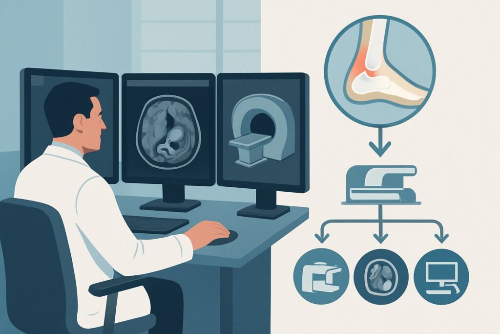
A 24-year-old male arrives in the emergency department on a busy Friday night. He reports five days of worsening right ankle swelling after a minor scrape while playing basketball, and for the past 48 hours, he has felt feverish and unwell. The ankle is exquisitely tender, erythematous, and diffusely swollen, limiting his ability to bear weight. Given the concern for a deep soft-tissue infection, a point-of-care ultrasound was performed, revealing a hypoechoic fluid collection beneath the periosteum of the distal tibia. With a diagnosis of a subperiosteal abscess confirmed and IV antibiotics initiated, the critical question becomes one of management: what is the definitive next step to control the source of infection? This article details the clinical workflow for this specific scenario, guided by the American College of Radiology (ACR) Appropriateness Criteria, which rates Surgical drainage as Usually appropriate.
Who Fits This Clinical Scenario?
This guidance applies to patients who present with signs of a localized musculoskeletal infection (e.g., progressive swelling, pain, erythema) and systemic symptoms (e.g., fever), where an initial imaging study, typically ultrasound, has definitively identified a subperiosteal abscess. The key inclusion criteria are the confirmed presence of a fluid collection located between the cortical bone and the overlying periosteum, coupled with a clinical picture of an active infection requiring antibiotic therapy.
This workflow is distinct from several related but different clinical situations. It does not apply to patients with:
- Simple cellulitis or a superficial soft-tissue abscess without evidence of subperiosteal or bone involvement. These conditions often have different management pathways that may not require aggressive surgical intervention.
- Septic arthritis without a subperiosteal collection. While also a surgical emergency, the focus of drainage is the joint space itself, not the periosteum.
- Chronic osteomyelitis without an acute, drainable fluid collection. The management of chronic bone infection is complex and follows a different diagnostic and therapeutic algorithm.
- An infected post-operative fluid collection in the abdomen. As detailed in a neighboring ACR scenario, these collections often arise in a different clinical context and may be more amenable to percutaneous drainage techniques.
Correctly identifying the patient as having a discrete, infected subperiosteal collection is crucial for applying the following management rationale.
What Diagnoses Are You Working Up in This Scenario?
While the ultrasound has identified a subperiosteal abscess, this finding is a consequence of an underlying pathology. The management strategy is designed to address both the abscess and its cause. The primary diagnostic considerations in this context include:
Acute Osteomyelitis: This is the most common underlying cause. An infection within the bone (metaphysis in children, often epiphysis in adults) can rupture through the cortex and accumulate in the potential space beneath the tightly adherent periosteum. This creates a high-pressure, contained purulent collection that further compromises blood supply to the bone, risking bone necrosis (sequestrum formation). The source can be hematogenous (spread through the blood) or from direct contiguous spread from an overlying wound or soft-tissue infection.
Primary Soft-Tissue Abscess with Secondary Bone Involvement: Less commonly, a deep abscess originating in the muscle or subcutaneous tissue can extend inward to involve the periosteum and underlying bone. This is more frequent in cases of penetrating trauma, where bacteria are directly inoculated into deep tissues.
Septic Arthritis with Contiguous Spread: An infection originating within the ankle joint can, in some cases, erode through the joint capsule and spread along the bone surface, leading to a secondary subperiosteal abscess. This highlights the importance of a thorough physical examination of joint range of motion and stability.
Why Is Surgical Drainage the Recommended Study for This Presentation?
For a confirmed subperiosteal abscess of the ankle, the ACR designates Surgical drainage as Usually appropriate. The rationale is rooted in the pathophysiology of the condition: a subperiosteal abscess is a closed-space infection that requires definitive source control. The collection of pus strips the periosteum from the bone, disrupting its blood supply and creating an ideal environment for bacterial proliferation, which is largely inaccessible to systemic antibiotics.
Surgical intervention directly addresses this by providing complete evacuation of purulent material and allowing for thorough debridement of any underlying necrotic bone or soft tissue. This mechanical removal of the infectious burden is critical for infection control, pain relief, and preventing the progression to chronic osteomyelitis. Cultures obtained intraoperatively are essential for tailoring antibiotic therapy to the specific pathogen.
Alternative management strategies are rated lower for this specific scenario for clear reasons:
- Needle aspiration and Percutaneous catheter drainage only are both rated as May be appropriate. While less invasive, these methods carry a higher risk of incomplete drainage, particularly with the thick, viscous pus often found in musculoskeletal abscesses. A needle may not fully evacuate the collection, and a catheter can easily become clogged. These approaches may be considered in patients who are extremely poor surgical candidates or as a temporizing measure, but they are not the definitive first-line treatment.
- Continued conservative management (i.e., antibiotics alone) is rated Usually not appropriate. Relying solely on antibiotics without draining the abscess is associated with a high failure rate. It risks systemic sepsis, extensive bone necrosis, and the development of chronic, difficult-to-treat osteomyelitis.
The principle is clear: once a contained, purulent subperiosteal collection is identified, prompt and complete drainage is the cornerstone of effective treatment.
What’s Next After Surgical Drainage? Downstream Workflow
The management of a subperiosteal abscess does not end in the operating room. The post-procedure workflow is critical for ensuring a successful outcome and preventing recurrence. The path forward depends on the intraoperative findings and the patient’s clinical response.
If the procedure is successful and cultures are positive: The patient should show clinical improvement, with resolution of fever and decreasing inflammatory markers (e.g., C-reactive protein) within 48-72 hours. The initial broad-spectrum IV antibiotics should be narrowed based on culture sensitivities. A prolonged course of antibiotics is typically required, often 4 to 6 weeks, to fully eradicate the underlying osteomyelitis. The transition from IV to a highly bioavailable oral antibiotic regimen is guided by the patient’s clinical status and infectious disease consultation.
If symptoms persist or worsen post-operatively: This suggests inadequate source control. The next step is to obtain further imaging to assess for residual or recurrent collections, or unaddressed necrotic bone. Cross-sectional imaging, particularly MRI with contrast, is highly sensitive for evaluating the extent of bone marrow edema, identifying sequestra, and delineating any remaining fluid collections that may require a second-look debridement.
If intraoperative cultures are negative: This can occur in about 20-30% of cases, sometimes due to prior antibiotic administration. In this situation, broad-spectrum IV antibiotics are typically continued. Consultation with an infectious disease specialist is recommended to guide the duration and choice of empiric therapy. If the clinical picture is atypical, non-infectious inflammatory conditions should be considered.
Pitfalls to Avoid (and When to Get Help)
Navigating the care of a patient with a subperiosteal abscess requires vigilance to avoid common missteps that can lead to poor outcomes. Key pitfalls include:
- Delaying Drainage: A subperiosteal abscess is a surgical emergency. Delays in definitive source control increase the risk of bone necrosis and sepsis. Once the diagnosis is confirmed on imaging, an urgent surgical or interventional radiology consultation is warranted.
- Underestimating the Underlying Osteomyelitis: Draining the abscess is only one part of the treatment. Failing to complete a full, prolonged course of appropriate antibiotics is a primary cause of treatment failure and progression to chronic osteomyelitis.
- Inadequate Debridement: Simply draining the pus may not be enough. If necrotic bone (a sequestrum) is present, it must be surgically removed as it acts as a nidus for persistent infection.
- Relying on Incomplete Drainage: Choosing a less-invasive drainage method like needle aspiration must be done with caution and close follow-up, as the risk of leaving behind infected material is higher.
Escalate care immediately if the patient develops signs of sepsis, such as hypotension, persistent tachycardia, or altered mental status. This requires aggressive resuscitation and emergent source control.
Related ACR Topics and Tools
This article focuses on a single, specific clinical scenario. For a comprehensive overview of the management of infected fluid collections in other anatomical locations and clinical contexts, refer to the parent topic guide. The following GigHz tools can also support clinical decision-making:
- For breadth across all scenarios in Radiologic Management of Infected Fluid Collections, see our parent guide: Radiologic Management of Infected Fluid Collections: ACR Appropriateness Decoded.
- ACR Appropriateness Criteria Lookup — for quickly finding guidance on adjacent or alternative clinical presentations.
- Imaging Protocol Library — for technical details on imaging studies used to evaluate musculoskeletal infections.
- Radiation Dose Calculator — for discussing cumulative radiation exposure with patients who may require serial imaging.
Frequently Asked Questions
Why can’t a subperiosteal abscess be treated with antibiotics alone?
A subperiosteal abscess is a contained collection of pus under high pressure, located in a space with poor blood supply. Systemic antibiotics struggle to penetrate this collection in sufficient concentrations to be effective. Furthermore, the pressure from the abscess can compromise blood flow to the underlying bone, leading to bone death (necrosis). Drainage is essential for source control—to remove the bulk of the bacteria and inflammatory debris—allowing antibiotics to treat the remaining infection in the bone and surrounding tissues effectively.
What is the role of MRI if ultrasound has already found the abscess?
While ultrasound is excellent for identifying a fluid collection and guiding initial diagnosis, MRI provides superior detail of the bone and soft tissues. An MRI can more accurately define the full extent of the abscess, assess the degree of underlying osteomyelitis (bone marrow infection), identify any necrotic bone fragments (sequestra) that need to be removed, and evaluate for complications like septic arthritis. It is often used for pre-operative planning or to investigate treatment failure.
How is surgical drainage different from percutaneous catheter drainage for this condition?
Surgical drainage involves an open incision, allowing the surgeon to directly visualize the abscess cavity, completely evacuate the pus, and, crucially, debride (remove) any unhealthy or dead tissue and bone. Percutaneous catheter drainage is a less invasive technique where a radiologist uses imaging guidance to place a drain into the abscess. While effective for some abscesses, it can be less successful for thick musculoskeletal pus and does not allow for tissue debridement, leading to a higher risk of recurrence or incomplete treatment for subperiosteal abscesses.
What kind of follow-up is needed after the drainage procedure?
Post-procedure follow-up involves both clinical and laboratory monitoring. Clinically, the patient’s fever, pain, and swelling should resolve. Laboratory follow-up includes tracking inflammatory markers like C-reactive protein (CRP) and erythrocyte sedimentation rate (ESR) to ensure they are trending down. After completing a prolonged course of antibiotics (typically 4-6 weeks), follow-up imaging may be considered in some cases to confirm resolution, though treatment decisions are often guided primarily by the patient’s clinical status.
Is a subperiosteal abscess always caused by a direct injury to the ankle?
Not always. While direct trauma or a skin breach (like the scrape in the vignette) can introduce bacteria and lead to a contiguous infection, subperiosteal abscesses can also result from hematogenous spread. This occurs when bacteria from an infection elsewhere in the body (e.g., a skin boil, urinary tract infection, or even transient bacteremia) travel through the bloodstream and seed the bone, leading to osteomyelitis and subsequent abscess formation without any local injury.
Reviewed by Pouyan Golshani, MD, Interventional Radiologist — May 29, 2026